Recognition: unknown
CTseg: A Tool for Brain CT Segmentation, Spatial Normalisation, and Volumetrics
Pith reviewed 2026-05-08 15:18 UTC · model grok-4.3
The pith
CTseg tool produces accurate brain tissue maps and volumes from routine CT scans
A machine-rendered reading of the paper's core claim, the machinery that carries it, and where it could break.
Core claim
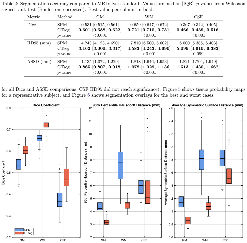
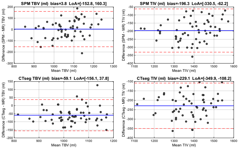
CTseg applies a generative modelling approach to CT images to generate tissue probability maps, deformation fields for spatial normalisation, and estimates of total brain and intracranial volume in a format compatible with established MRI analysis chains, and validation on paired MR/CT scans shows significantly higher segmentation accuracy, sharper group-average normalisation, lower voxelwise variability, and stronger agreement on total brain volume than direct application of the MRI pipeline to CT.
What carries the argument
The CTseg pipeline that generates CT-specific tissue maps and deformation fields from raw head CT images without preprocessing or resampling
If this is right
- Volumetric measurements of total brain volume from CT become more reliable and can be compared directly with MRI-derived values.
- Group-level spatial normalisation of CT images reaches consistency levels previously available only for MRI cohorts.
- Tissue maps produced by CTseg support downstream classification tasks such as sex prediction at levels comparable to MRI-based maps.
- Routine clinical CT scans can be fed into the same analysis workflows used for MRI without additional image preparation steps.
Where Pith is reading between the lines
- Hospital CT databases could be mined retrospectively for brain morphometry at scales not feasible with MRI alone.
- The same modelling strategy might be adapted to produce quantitative outputs from other CT body regions or modalities that currently lack dedicated segmentation tools.
- Integration of CTseg into existing hospital PACS systems would allow automated volumetrics to appear alongside standard radiological reports.
Load-bearing premise
The MRI-derived reference labels accurately mark the true tissue boundaries visible on CT, and the paired scans used for testing represent ordinary clinical CT data.
What would settle it
Independent manual tracings of tissue boundaries on the same CT scans that show lower overlap with CTseg outputs than with the MRI silver standard, or a drop in performance when the tool is run on unpaired CT scans from varied scanners and protocols.
Figures


read the original abstract
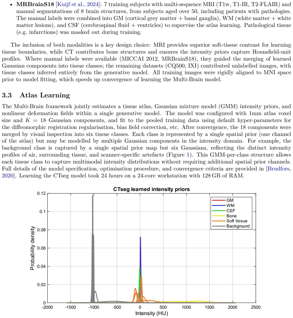
This paper presents and validates CTseg, a freely available software for brain CT segmentation, spatial normalisation, and volumetrics. CTseg builds on the Multi-Brain generative modelling framework, providing a CT-specific pipeline that produces tissue maps, deformation fields, and brain volume estimates in the same format as SPM's unified segmentation, thereby extending SPM's established analysis chain from MRI to CT. CTseg is designed for routine hospital CT scans without requiring preprocessing or resampling in deployment. Although CTseg has been adopted in clinical research spanning, among other things, stroke, dementia, and brain morphometry, a systematic validation against an independent reference standard has been lacking. Using paired MR/CT head scans, we evaluate CTseg across four dimensions: segmentation accuracy against an MRI-derived silver standard; spatial normalisation consistency through group-average sharpness and voxelwise coefficient of variation; brain volume agreement via intraclass correlation and Bland-Altman analysis; and downstream sex classification performance from normalised tissue maps. As a baseline, we apply SPM's MRI-based unified segmentation directly to the CT images. CTseg significantly outperformed this baseline for segmentation and normalisation, showed stronger TBV agreement, and achieved comparable TIV agreement. CTseg is freely available at https://github.com/WCHN/CTseg, and all experiment code is included in the repository for full reproducibility.
Editorial analysis
A structured set of objections, weighed in public.
Referee Report
Summary. The manuscript presents CTseg, a freely available tool extending the Multi-Brain generative modelling framework to provide CT-specific brain segmentation, spatial normalisation, and volumetrics in SPM-compatible format. It is designed for routine clinical CT scans without preprocessing. Validation uses paired MR/CT head scans to compare against SPM's MRI-based unified segmentation baseline across segmentation accuracy (vs. MRI-derived silver standard), normalisation consistency (group-average sharpness and voxelwise CoV), brain volume agreement (ICC and Bland-Altman for TBV/TIV), and downstream sex classification from normalised maps. The abstract reports that CTseg significantly outperformed the baseline on segmentation and normalisation, showed stronger TBV agreement, and comparable TIV agreement.
Significance. If the central claims hold after addressing validation details, the work is significant for extending established MRI analysis pipelines to CT, which is widely used in clinical settings for stroke, dementia, and morphometry research. The provision of all experiment code in the public repository is a clear strength supporting reproducibility and adoption.
major comments (2)
- [Abstract and Methods (validation)] Abstract and Methods (validation section): Segmentation accuracy is assessed against an MRI-derived silver standard transferred to paired CT images. CT has substantially lower soft-tissue contrast than MRI (particularly GM/WM boundaries and partial-volume effects), so the transferred labels may not reflect CT-visible anatomy; without reported quantification of MR-CT registration error, label uncertainty specific to CT, or sensitivity analysis, the reported significant outperformance over SPM on Dice/accuracy metrics is not yet secured.
- [Results (volume agreement and normalisation)] Results (volume agreement and normalisation): The claims of stronger TBV agreement and superior normalisation rest on ICC, Bland-Altman, sharpness, and CoV metrics, but the manuscript provides no details on statistical testing (e.g., p-values for outperformance), sample size after exclusions, or handling of outliers in the paired scans; these omissions are load-bearing for the superiority conclusions.
minor comments (2)
- [Abstract] The abstract states that CTseg 'significantly outperformed' the baseline but does not include effect sizes or exact p-values in the summary, which would improve clarity for readers.
- [Availability and Reproducibility] The GitHub repository link is given, but the manuscript could more explicitly state which validation datasets (if any) are included or how users can reproduce the exact paired-scan experiments.
Simulated Author's Rebuttal
We thank the referee for their detailed and constructive comments. The points raised identify opportunities to strengthen the reporting of our validation procedures. We address each major comment below and will incorporate the requested details into a revised manuscript.
read point-by-point responses
-
Referee: [Abstract and Methods (validation)] Abstract and Methods (validation section): Segmentation accuracy is assessed against an MRI-derived silver standard transferred to paired CT images. CT has substantially lower soft-tissue contrast than MRI (particularly GM/WM boundaries and partial-volume effects), so the transferred labels may not reflect CT-visible anatomy; without reported quantification of MR-CT registration error, label uncertainty specific to CT, or sensitivity analysis, the reported significant outperformance over SPM on Dice/accuracy metrics is not yet secured.
Authors: We agree that the MRI-derived silver standard is an imperfect reference for CT due to modality-specific contrast differences and potential registration inaccuracies. The paired MR/CT design was chosen precisely to enable this cross-modal comparison, with the silver standard generated via a multi-modal registration pipeline. To secure the outperformance claims, the revised Methods section will include: (i) quantitative assessment of MR-CT registration accuracy (e.g., landmark-based errors or overlap metrics on a subset of cases), (ii) explicit discussion of label uncertainty arising from partial-volume effects in CT, and (iii) a sensitivity analysis showing how variations in registration parameters affect the reported Dice and accuracy differences. These additions will allow readers to evaluate the robustness of the superiority findings. revision: yes
-
Referee: [Results (volume agreement and normalisation)] Results (volume agreement and normalisation): The claims of stronger TBV agreement and superior normalisation rest on ICC, Bland-Altman, sharpness, and CoV metrics, but the manuscript provides no details on statistical testing (e.g., p-values for outperformance), sample size after exclusions, or handling of outliers in the paired scans; these omissions are load-bearing for the superiority conclusions.
Authors: We acknowledge that the current Results section omits key statistical details supporting the superiority claims. In the revision we will add: (i) p-values from appropriate paired statistical tests (e.g., Wilcoxon signed-rank or paired t-tests) comparing CTseg versus baseline metrics, (ii) the exact sample size after any exclusions (with reasons for exclusion such as motion or failed registration), and (iii) a description of outlier handling together with sensitivity results obtained with and without outliers. These additions will make the evidence for stronger TBV agreement and superior normalisation fully transparent and reproducible. revision: yes
Circularity Check
Minor self-citation to Multi-Brain framework; validation chain remains independent
full rationale
The paper extends an existing generative modelling framework to CT images and validates performance on paired MR/CT scans using external metrics (Dice against MRI silver standard, group-average sharpness, ICC/Bland-Altman for volumes). No derivation reduces by construction to fitted parameters or self-defined quantities; the baseline (SPM applied to CT) and silver standard are independent references. Self-citation to the prior framework is present but not load-bearing for the reported superiority claims.
Axiom & Free-Parameter Ledger
Reference graph
Works this paper leans on
-
[1]
V Adduru, S A Baum, C Zhang, M Helguera, R Zand, M Lichtenstein, C J Griessenauer, and A M Michael. A method to estimate brain volume from head CT images and application to detect brain atrophy in Alzheimer disease. American Journal of Neuroradiology, 41 0 (2): 0 224--230, 2020. doi:10.3174/ajnr.A6402
-
[2]
Voxel-based morphometry---the methods
John Ashburner and Karl J Friston. Voxel-based morphometry---the methods. NeuroImage, 11 0 (6): 0 805--821, 2000
2000
-
[3]
Unified segmentation
John Ashburner and Karl J Friston. Unified segmentation. NeuroImage, 26 0 (3): 0 839--851, 2005
2005
-
[4]
Voxel-based lesion--symptom mapping
Elizabeth Bates, Stephen M Wilson, Ayse Pinar Saygin, Frederic Dick, Martin I Sereno, Robert T Knight, and Nina F Dronkers. Voxel-based lesion--symptom mapping. Nature Neuroscience, 6 0 (5): 0 448--450, 2003. doi:10.1038/nn1050
-
[5]
Statistical methods for assessing agreement between two methods of clinical measurement
J Martin Bland and Douglas G Altman. Statistical methods for assessing agreement between two methods of clinical measurement. The Lancet, 327 0 (8476): 0 307--310, 1986
1986
-
[6]
Generative Models for Preprocessing of Hospital Brain Scans
Mikael Brudfors. Generative Models for Preprocessing of Hospital Brain Scans. PhD thesis, UCL (University College London), 2020
2020
-
[7]
A tool for super-resolving multimodal clinical MRI
Mikael Brudfors, Ya \"e l Balbastre, Parashkev Nachev, and John Ashburner. A tool for super-resolving multimodal clinical MRI . arXiv preprint arXiv:1909.01140, 2019
-
[8]
Flexible B ayesian modelling for nonlinear image registration
Mikael Brudfors, Ya \"e l Balbastre, Guillaume Flandin, Parashkev Nachev, and John Ashburner. Flexible B ayesian modelling for nonlinear image registration. In International Conference on Medical Image Computing and Computer Assisted Intervention, pages 253--263. Springer, 2020
2020
-
[9]
Fully automated segmentation of head CT neuroanatomy using deep learning
Jason C Cai, Zeynettin Akkus, Kenneth A Philbrick, Arunnit Boonrod, Safa Hoodeshenas, Alexander D Weston, Pouria Rouzrokh, Gian Marco Conte, Atefeh Zeinoddini, David C Vogelsang, Qiao Huang, and Bradley J Erickson. Fully automated segmentation of head CT neuroanatomy using deep learning. Radiology: Artificial Intelligence, 2 0 (5): 0 e190183, 2020. doi:10...
-
[10]
Deep learning algorithms for detection of critical findings in head CT scans: a retrospective study
Sasank Chilamkurthy et al. Deep learning algorithms for detection of critical findings in head CT scans: a retrospective study. The Lancet, 392 0 (10162): 0 2388--2396, 2018
2018
-
[11]
Kuaikuai Duan, Enrico Premi, Andrea Pilotto, Viviana Cristillo, Alberto Benussi, Ilenia Libri, Marcello Giunta, H. Jeremy Bockholt, Jingyu Liu, Riccardo Campora, Alessandro Pezzini, Roberto Gasparotti, Mauro Magoni, Alessandro Padovani, and Vince D. Calhoun. Alterations of frontal-temporal gray matter volume associate with clinical measures of older adult...
-
[12]
SW Fielden, D Beiler, KA Cauley, and V Troiani. A comparison of global brain volumetrics obtained from CT versus MRI using 2 publicly available software packages. American Journal of Neuroradiology, 43 0 (2): 0 245--250, 2022. doi:10.3174/ajnr.A7403
-
[13]
FreeSurfer
Bruce Fischl. FreeSurfer . NeuroImage, 62 0 (2): 0 774--781, 2012
2012
-
[14]
Spatial registration and normalization of images
Karl J Friston, John Ashburner, Christopher D Frith, Jean-Baptiste Poline, John D Heather, and Richard SJ Frackowiak. Spatial registration and normalization of images. Human Brain Mapping, 3 0 (3): 0 165--189, 1995. doi:10.1002/hbm.460030303
-
[15]
Annika Gerken, Sina Walluscheck, Peter Kohlmann, Ivana Galinovic, Kersten Villringer, Jochen B Fiebach, Jan Klein, and Stefan Heldmann. Deep learning-based segmentation of brain parenchyma and ventricular system in CT scans in the presence of anomalies. Frontiers in Neuroimaging, 2: 0 1228255, 2023. doi:10.3389/fnimg.2023.1228255
-
[16]
Hannes Gramespacher, Maximilian HT Schmieschek, Clemens Warnke, Christoph Adler, et al. Analysis of cerebral CT based on supervised machine learning as a predictor of outcome after out-of-hospital cardiac arrest. Neurology, 103 0 (1): 0 e209583, 2024. doi:10.1212/WNL.0000000000209583
-
[17]
Varsha Gupta, Wojciech Ambrosius, Guoyu Qian, Anna Blazejewska, Radoslaw Kazmierski, Andrzej Urbanik, and Wieslaw L Nowinski. Automatic segmentation of cerebrospinal fluid, white and gray matter in unenhanced computed tomography images. Academic Radiology, 17 0 (11): 0 1350--1358, 2010. doi:10.1016/j.acra.2010.06.005
-
[18]
Impact of intracranial volume and brain volume on the prognostic value of computed tomography perfusion core volume in acute ischemic stroke
Jan W Hoving, Praneeta R Konduri, Manon L Tolhuisen, et al. Impact of intracranial volume and brain volume on the prognostic value of computed tomography perfusion core volume in acute ischemic stroke. Journal of Cardiovascular Development and Disease, 11 0 (3): 0 80, 2024
2024
-
[19]
Validation of SynthSeg segmentation performance on CT using paired MRI from radiotherapy patients
Selena Huisman, Matteo Maspero, Marielle Philippens, Joost Verhoeff, and Szabolcs David. Validation of SynthSeg segmentation performance on CT using paired MRI from radiotherapy patients. NeuroImage, 303: 0 120922, 2024. doi:10.1016/j.neuroimage.2024.120922
-
[20]
Mark Jenkinson, Christian F Beckmann, Timothy EJ Behrens, Mark W Woolrich, and Stephen M Smith. FSL . NeuroImage, 62 0 (2): 0 782--790, 2012
2012
-
[21]
Polona Kalc, Felix Hoffstaedter, Eileen Luders, Christian Gaser, and Robert Dahnke. Approximation of bone mineral density and subcutaneous adiposity using T1 -weighted images of the human head. Imaging Neuroscience, 2: 0 1--22, 2024. doi:10.1162/imag_a_00390
-
[22]
Decomposing the Hounsfield unit: probabilistic segmentation of brain tissue in computed tomography
A Kemmling, H Wersching, K Berger, S Knecht, C Groden, and I N \"o lte. Decomposing the Hounsfield unit: probabilistic segmentation of brain tissue in computed tomography. Clinical Neuroradiology, 22 0 (1): 0 79--91, 2012. doi:10.1007/s00062-011-0123-0
-
[23]
Elastix: a toolbox for intensity-based medical image registration
Stefan Klein, Marius Staring, Keelin Murphy, Max A Viergever, and Josien PW Pluim. Elastix: a toolbox for intensity-based medical image registration. IEEE Transactions on Medical Imaging, 29 0 (1): 0 196--205, 2010
2010
-
[24]
MR brain segmentation challenge 2018 data, 2024
Hugo J Kuijf, Edwin Bennink, Koen L Vincken, Nick Weaver, Geert Jan Biessels, and Max A Viergever. MR brain segmentation challenge 2018 data, 2024. Version 1.0
2018
-
[25]
MICCAI 2012 workshop on multi-atlas labeling
Bennett Landman and Simon Warfield. MICCAI 2012 workshop on multi-atlas labeling. In Proc. MICCAI Grand Challenge on Multi-Atlas Labeling, 2012
2012
-
[26]
Chang Liu, Hansheng Liu, Deping Wu, Zhiming Zhou, WenGuo Huang, Zhilin Wu, Wenjie Zi, and Qingwu Yang. Severe brain atrophy predicts poor clinical outcome after endovascular treatment of acute basilar artery occlusion. Frontiers in Aging Neuroscience, 13: 0 720061, 2021. doi:10.3389/fnagi.2021.720061
-
[27]
Sven P R Luijten, Aravind Ganesh, Adam P Marcus, et al. A CT imaging-based prediction model of functional outcome and benefit of endovascular thrombectomy for ischemic stroke. European Radiology, 2026. doi:10.1007/s00330-025-12207-7
-
[28]
White matter and gray matter segmentation in 4D computed tomography
Rashindra Manniesing, Marcel T H Oei, Luuk J Oostveen, Jaime Melendez, Ewoud J Smit, Bram Platel, Clara I S \'a nchez, Frederick J A Meijer, Mathias Prokop, and Bram van Ginneken. White matter and gray matter segmentation in 4D computed tomography. Scientific Reports, 7: 0 119, 2017
2017
-
[29]
A publicly available, high resolution, unbiased CT brain template
John Muschelli. A publicly available, high resolution, unbiased CT brain template. In International Conference on Information Processing and Management of Uncertainty in Knowledge-Based Systems, pages 358--366. Springer, 2020
2020
-
[30]
Age-specific CT and MRI templates for spatial normalization
Christopher Rorden, Leonardo Bonilha, Julius Fridriksson, Benjamin Bender, and Hans-Otto Karnath. Age-specific CT and MRI templates for spatial normalization. NeuroImage, 61 0 (4): 0 957--965, 2012
2012
-
[31]
Voxel-based dysconnectomic brain morphometry with computed tomography in Down syndrome
Beatriz S \'a nchez-Moreno, Linda Zhang, Gloria Mateo, et al. Voxel-based dysconnectomic brain morphometry with computed tomography in Down syndrome. Annals of Clinical and Translational Neurology, 11 0 (1): 0 143--155, 2024. doi:10.1002/acn3.51940
-
[32]
Beatriz S \'a nchez-Moreno, Linda Zhang, Ana Carril Salaberry, et al. Performance of computed tomography and magnetic resonance morphometry in evaluating brain atrophy in Down syndrome. Alzheimer's & Dementia, 21: 0 e70296, 2025. doi:10.1002/alz.70296
-
[33]
PRoNTo : pattern recognition for neuroimaging toolbox
Jessica Schrouff, Maria J Rosa, Jane M Rondina, Andre F Marquand, Carlton Chu, John Ashburner, Christophe Phillips, Jonas Richiardi, and Janaina Mour \ a o-Miranda. PRoNTo : pattern recognition for neuroimaging toolbox. Neuroinformatics, 11 0 (3): 0 319--337, 2013
2013
-
[34]
Intraclass correlations: uses in assessing rater reliability
Patrick E Shrout and Joseph L Fleiss. Intraclass correlations: uses in assessing rater reliability. Psychological Bulletin, 86 0 (2): 0 420--428, 1979
1979
-
[35]
Trends in use of medical imaging in US health care systems and in Ontario , Canada , 2000-2016
Rebecca Smith-Bindman, Marilyn L Kwan, Emily C Marlow, Mary Kay Theis, Wesley Bolch, Stephanie Y Cheng, Erin J A Bowles, James R Duncan, Robert T Greenlee, Lawrence H Kushi, Jason D Pole, Alanna K Rahm, Natasha K Stout, Sheila Weinmann, and Diana L Miglioretti. Trends in use of medical imaging in US health care systems and in Ontario , Canada , 2000-2016....
-
[36]
Won Jun Son, Sung Jun Ahn, Ji Young Lee, and Hyunyeol Lee. Automated brain segmentation on computed tomographic images using perceptual loss based convolutional neural networks. Investigative Magnetic Resonance Imaging, 28 0 (4): 0 193--201, 2024. doi:10.13104/imri.2024.0023
-
[37]
Deep learning from MRI -derived labels enables automatic brain tissue classification on human brain CT
Meera Srikrishna, Joana B Pereira, Rolf A Heckemann, Giovanni Volpe, Danielle van Westen, Anna Zettergren, Silke Kern, Lars-Olof Wahlund, Eric Westman, Ingmar Skoog, and Michael Sch \"o ll. Deep learning from MRI -derived labels enables automatic brain tissue classification on human brain CT . NeuroImage, 244: 0 118606, 2021
2021
-
[38]
Deep generative computed perfusion-deficit mapping of ischaemic stroke
Chayanin Tangwiriyasakul, Pedro Borges, Guilherme Pombo, Stefano Moriconi, Michael S Elmalem, et al. Deep generative computed perfusion-deficit mapping of ischaemic stroke. Communications Biology, 9 0 (1): 0 219, 2026. doi:10.1038/s42003-025-09495-6
-
[39]
Medical Physics52(7), e17981 (2025)
Adrian Thummerer, Erik van der Bijl, Arthur Jr Galapon, Florian Kamp, Mark Savenije, Christina Muijs, Shafak Aluwini, Roel J H M Steenbakkers, Stephanie Beuel, Martijn P W Intven, Johannes A Langendijk, Stefan Both, Stefanie Corradini, Viktor Rogowski, Maarten Terpstra, Niklas Wahl, Christopher Kurz, Guillaume Landry, and Matteo Maspero. SynthRAD2025 gran...
-
[40]
Tim M. Tierney, Nicholas A. Alexander, Nicole Labra Avila, Yael Balbastre, Gareth Barnes, Yulia Bezsudnova, Mikael Brudfors, Korbinian Eckstein, Guillaume Flandin, Karl Friston, Amirhossein Jafarian, Olivia S. Kowalczyk, Vladimir Litvak, Johan Medrano, Stephanie Mellor, George O'Neill, Thomas Parr, Adeel Razi, Ryan Timms, and Peter Zeidman. SPM 25: open s...
-
[41]
Stephens, Merc \`e Boada, Gregory Klein, and Marta Marqui \'e
Matteo Tonietto, Oscar Sotolongo-Grau, N \'u ria Ro \'e -Vellv \'e , Santiago Bullich, Juan Pablo Tartari, \'A ngela Sanabria, Ainhoa Garc \'i a-S \'a nchez, Edilio Borroni, Christopher Galli, Esther P \'e rez-Mart \'i nez, Joan Castell-Conesa, Isabel Roca, Llu \'i s T \'a rraga, Agust \'i n Ruiz, Andrew W. Stephens, Merc \`e Boada, Gregory Klein, and Mar...
-
[42]
Predicting mortality in acutely hospitalised older patients: the impact of model dimensionality
Alex Tsui, Petru-Daniel Tudosiu, Mikael Brudfors, et al. Predicting mortality in acutely hospitalised older patients: the impact of model dimensionality. BMC Medicine, 21: 0 10, 2023. doi:10.1186/s12916-022-02698-2
-
[43]
Nathalie Tzourio-Mazoyer, Brigitte Landeau, Dimitri Papathanassiou, Fabrice Crivello, Olivier Etard, Nicolas Delcroix, Bernard Mazoyer, and Marc Joliot. Automated anatomical labeling of activations in SPM using a macroscopic anatomical parcellation of the MNI MRI single-subject brain. NeuroImage, 15 0 (1): 0 273--289, 2002. doi:10.1006/nimg.2001.0978
-
[44]
Gemma Urbanos, Ana M Casta \ n o-Le \'o n, et al. Comprehensive predictive modeling in subarachnoid hemorrhage: integrating radiomics and clinical variables. Neurosurgical Review, 2025. doi:10.1007/s10143-025-03679-8
-
[45]
The diagnostic accuracy of CTseg segmentation software for dementia in a New Zealand memory service
Mukish Yelanchezian, Cristian Gonzalez-Prieto, et al. The diagnostic accuracy of CTseg segmentation software for dementia in a New Zealand memory service. Journal of Alzheimer's Disease Reports, 9: 0 25424823251332448, 2025. doi:10.1177/25424823251332448
discussion (0)
Sign in with ORCID, Apple, or X to comment. Anyone can read and Pith papers without signing in.