Recognition: 2 theorem links
· Lean TheoremWhen Brains Disagree: Biological Ambiguity Underlies the Challenge of Amyloid PET Synthesis from Structural MRI
Pith reviewed 2026-05-13 07:17 UTC · model grok-4.3
The pith
Biological ambiguity from decoupled brain processes makes MRI-to-amyloid PET synthesis inherently inconsistent.
A machine-rendered reading of the paper's core claim, the machinery that carries it, and where it could break.
Core claim
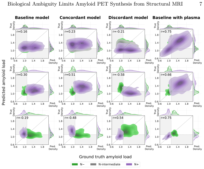
MRI-to-amyloid PET synthesis is intrinsically ill-posed because similar MRI patterns can correspond to different amyloid states due to the temporal decoupling of neurodegeneration and amyloid pathology. When paired data are stratified by amyloid and neurodegeneration status, standard synthesis models learn unambiguous mappings with high performance; introducing ambiguous data causes performance to collapse regardless of model architecture. Incorporating orthogonal information from plasma biomarkers resolves the ambiguity, yielding improved and more stable synthesis results.
What carries the argument
Stratification of paired MRI-PET data by amyloid and neurodegeneration status to isolate unambiguous one-to-one mappings, with multimodal plasma biomarkers serving as disambiguating inputs.
If this is right
- Unambiguous MRI-PET mappings become learnable once data ambiguity is removed through stratification.
- Performance collapse occurs specifically when biologically ambiguous cases enter the training distribution.
- Multimodal inputs such as plasma biomarkers restore accuracy and consistency by resolving one-to-many mappings.
- Architectural complexity alone cannot overcome the limits imposed by data ambiguity.
- Meaningful advances require combining imaging with additional biological signals rather than MRI synthesis in isolation.
Where Pith is reading between the lines
- Purely image-based synthesis methods may reach a performance ceiling in Alzheimer's research unless fluid biomarkers or other modalities are routinely included.
- Similar ambiguity problems are likely in other cross-modal medical imaging tasks where the underlying biological processes evolve on different timescales.
- Future models could benefit from mechanisms that detect or flag ambiguous inputs instead of always producing a single output.
- Clinical use of synthesized PET images would need careful validation across populations with varying degrees of process decoupling.
Load-bearing premise
That grouping the data by amyloid and neurodegeneration status cleanly separates unambiguous mappings without introducing selection bias or hidden confounders.
What would settle it
Demonstrating high and stable synthesis accuracy on mixed ambiguous datasets using only MRI inputs without any multimodal additions.
Figures

read the original abstract
Structural MRI-to-amyloid PET synthesis has been proposed as a non-invasive alternative for amyloid assessment in Alzheimer's disease (AD). However, reported performance of identical models varies widely across studies, and increasingly complex architectures have not led to consistent gains. This inconsistency is thought to be caused by a fundamental biological ambiguity: MRI captures neurodegeneration, while PET measures amyloid pathology - two processes that are often temporally decoupled in AD. As a result, similar MRI patterns may correspond to different amyloid states, creating ambiguous one-to-many mappings. MRI-to-amyloid PET synthesis may therefore be intrinsically ill-posed; however, this idea has yet to be tested scientifically. The aim of this work is to test this hypothesis through two controlled experiments. We first control the training distribution by stratifying paired MRI-PET data by amyloid and neurodegeneration status. Using two standard synthesis models under a controlled design, we show that biologically unambiguous mappings are learnable in isolation, but performance collapses when data ambiguity is introduced. This demonstrates that ambiguity in the data distribution, rather than architectural capacity, constrains performance. Second, we show that introducing orthogonal biological information in the form of plasma biomarkers resolves this ambiguity. When multimodal inputs are incorporated, performance improves and stability is restored. Together, these findings suggest that limited and inconsistent performance in MRI-to-amyloid PET synthesis is explained by intrinsic biological ambiguity, and that stable, meaningful progress requires multimodal integration rather than architectural complexity.
Editorial analysis
A structured set of objections, weighed in public.
Referee Report
Summary. The paper claims that inconsistent performance in structural MRI-to-amyloid PET synthesis arises from intrinsic biological ambiguity caused by the temporal decoupling of neurodegeneration (MRI) and amyloid pathology (PET) in Alzheimer's disease, creating one-to-many mappings. It tests this via two controlled experiments: stratifying paired MRI-PET data by amyloid and neurodegeneration status to show learnable mappings in unambiguous strata but collapse when mixed, and demonstrating that multimodal plasma biomarkers resolve the ambiguity and restore performance. The conclusion is that progress requires multimodal integration rather than architectural complexity.
Significance. If the central claim holds, the work would be significant for shifting focus in neuroimaging synthesis from model complexity to addressing data ambiguity through multimodal inputs, potentially explaining cross-study inconsistencies and guiding more stable non-invasive amyloid assessment. The use of controlled stratification with standard models and the explicit test of the ambiguity hypothesis are strengths that provide a falsifiable framework.
major comments (2)
- [First experiment] §3 (first experiment): Stratification by amyloid status relies on PET-derived labels that are the synthesis target, risking selection bias since subgroups almost certainly differ in size, demographics, disease stage, and comorbidities; without explicit matching, covariate adjustment, or ablation on stratification criteria, the performance collapse cannot be cleanly attributed to mapping ambiguity rather than these confounders.
- [Results] Results (performance contrasts): The described experiments lack reported quantitative metrics (e.g., exact correlation coefficients, Dice scores, or p-values for strata comparisons) and statistical controls for confounders, weakening the load-bearing claim that ambiguity—not architecture or selection effects—explains the collapse.
minor comments (2)
- [Abstract] Abstract: Does not name the two standard synthesis models or provide any numerical performance values, reducing clarity for readers assessing the magnitude of effects.
- [Methods] Methods: Neurodegeneration status definition and any procedures for balancing subgroup sizes or demographics are not detailed, affecting reproducibility.
Simulated Author's Rebuttal
We thank the referee for the detailed and constructive comments, which have helped us strengthen the manuscript. We address each major point below and have revised the paper to incorporate additional controls, quantitative reporting, and supporting analyses where feasible.
read point-by-point responses
-
Referee: [First experiment] §3 (first experiment): Stratification by amyloid status relies on PET-derived labels that are the synthesis target, risking selection bias since subgroups almost certainly differ in size, demographics, disease stage, and comorbidities; without explicit matching, covariate adjustment, or ablation on stratification criteria, the performance collapse cannot be cleanly attributed to mapping ambiguity rather than these confounders.
Authors: We acknowledge that defining strata using the PET target introduces potential confounders, as the resulting subgroups differ in size, demographics, disease stage, and comorbidities. Our core design isolates the effect of ambiguity by holding the model architecture and data source fixed while varying only the presence of mixed vs. unambiguous mappings. Nevertheless, to rule out selection effects more rigorously, the revised manuscript now includes: (1) full demographic and clinical tables for each stratum, (2) covariate-adjusted regression analyses, and (3) a matched ablation in which strata are balanced on age, sex, and clinical stage (where sample size permits). These additions support that the performance drop is attributable to the introduction of biologically ambiguous pairs rather than unbalanced confounders alone. revision: partial
-
Referee: [Results] Results (performance contrasts): The described experiments lack reported quantitative metrics (e.g., exact correlation coefficients, Dice scores, or p-values for strata comparisons) and statistical controls for confounders, weakening the load-bearing claim that ambiguity—not architecture or selection effects—explains the collapse.
Authors: We have added the requested quantitative detail. The revised results section now reports exact Pearson correlation coefficients, MAE, and SSIM values for every stratum and condition, together with p-values from paired statistical tests comparing unambiguous vs. mixed strata. As noted in the response to the first comment, we also include covariate-adjusted models and the matched ablation results. These metrics and controls directly quantify the performance collapse and its attribution to data ambiguity. revision: yes
Circularity Check
No circularity: empirical experiments test hypothesis without self-referential derivations
full rationale
The paper advances its central claim through two controlled empirical experiments on paired MRI-PET data: stratifying by amyloid/neurodegeneration status to compare learnable mappings in unambiguous vs. mixed strata, and testing multimodal plasma biomarkers for performance recovery. No equations, fitted parameters renamed as predictions, or self-definitional constructs appear. The performance contrasts are measured outcomes on held-out data rather than quantities forced by construction from the stratification criteria or model inputs. No load-bearing self-citations, uniqueness theorems, or ansatzes are invoked; the hypothesis test is self-contained against external benchmarks of synthesis accuracy.
Axiom & Free-Parameter Ledger
axioms (1)
- domain assumption MRI captures neurodegeneration while PET measures amyloid pathology and these processes are often temporally decoupled in AD
Lean theorems connected to this paper
-
IndisputableMonolith/Cost/FunctionalEquation.leanwashburn_uniqueness_aczel unclear?
unclearRelation between the paper passage and the cited Recognition theorem.
performance collapses when data ambiguity is introduced... introducing orthogonal biological information in the form of plasma biomarkers resolves this ambiguity
-
IndisputableMonolith/Foundation/RealityFromDistinction.leanreality_from_one_distinction unclear?
unclearRelation between the paper passage and the cited Recognition theorem.
MRI does not uniquely determine amyloid PET... one-to-many mapping
What do these tags mean?
- matches
- The paper's claim is directly supported by a theorem in the formal canon.
- supports
- The theorem supports part of the paper's argument, but the paper may add assumptions or extra steps.
- extends
- The paper goes beyond the formal theorem; the theorem is a base layer rather than the whole result.
- uses
- The paper appears to rely on the theorem as machinery.
- contradicts
- The paper's claim conflicts with a theorem or certificate in the canon.
- unclear
- Pith found a possible connection, but the passage is too broad, indirect, or ambiguous to say the theorem truly supports the claim.
Reference graph
Works this paper leans on
-
[1]
Journal of the American Geriatrics Society69, 1774–1783 (7 2021)
Aranda, M.P., Kremer, I.N., Hinton, L., Zissimopoulos, J., Whitmer, R.A., Hum- mel, C.H., Trejo, L., Fabius, C.: Impact of dementia: Health disparities, population trends, care interventions, and economic costs. Journal of the American Geriatrics Society69, 1774–1783 (7 2021). https://doi.org/10.1111/jgs.17345
-
[2]
Journal of Nuclear Medicine63, 13S–19S (6 2022)
Chapleau, M., Iaccarino, L., Soleimani-Meigooni, D., Rabinovici, G.D.: The role of amyloid PET in imaging neurodegenerative disorders: A review. Journal of Nuclear Medicine63, 13S–19S (6 2022). https://doi.org/10.2967/jnumed.121.263195
-
[3]
Chen, Y., Su, Y., Dumitrascu, C., Chen, K., Weidman, D., Caselli, R.J., Ashton, N., Reiman, E.M., Wang, Y.: Plasma-CycleGAN: Plasma biomarker-guided MRI to PET cross-modality translation using conditional CycleGAN. pp. 1–5. IEEE (4 2025). https://doi.org/10.1109/ISBI60581.2025.10980900
-
[4]
Chen, Z., Bi, S., Shan, Y., Wang, F., Wang, Y., Qi, Z., Wang, T., Li, X., Li, S., Xiao, H., et al.: MRI-to-PET synthesis via deep learning for amyloid-βquantifi- cation in Alzheimer’s disease. European Radiology (1 2026). https://doi.org/10. 1007/s00330-025-12251-3, https://link.springer.com/10.1007/s00330-025-12251-3
-
[5]
New England Journal of Medicine388, 9–21 (1 2023)
van Dyck, C.H., Swanson, C.J., Aisen, P., Bateman, R.J., Chen, C., Gee, M., Kanekiyo, M., Li, D., Reyderman, L., Cohen, S., et al.: Lecanemab in early Alzheimer’s disease. New England Journal of Medicine388, 9–21 (1 2023). https: //doi.org/10.1056/NEJMoa2212948
-
[6]
Isola, P., Zhu, J.Y., Zhou, T., Efros, A.A.: Image-to-image translation with con- ditional adversarial networks. pp. 5967–5976. IEEE (7 2017). https://doi.org/10. 1109/CVPR.2017.632
work page 2017
-
[7]
Alzheimer’s and Dementia14, 535–562 (4 2018)
Jack, C.R., Bennett, D.A., Blennow, K., Carrillo, M.C., Dunn, B., Haeberlein, S.B., Holtzman, D.M., Jagust, W., Jessen, F., Karlawish, J., et al.: NIA-AA research framework: Toward a biological definition of Alzheimer’s disease. Alzheimer’s and Dementia14, 535–562 (4 2018). https://doi.org/10.1016/j.jalz.2018.02.018
-
[8]
Jin, Y., DuBois, J., Zhao, C., Zhan, L., Gabelle, A., Jahanshad, N., Thompson, P.M., Gafson, A., Belachew, S.: Brain MRI to PET Synthesis and Amyloid Esti- mation in Alzheimer’s Disease via 3D Multimodal Contrastive GAN, pp. 94–103 (2024). https://doi.org/10.1007/978-3-031-45673-2_10
-
[9]
Ageing Re- search Reviews101, 102481 (11 2024)
Kamatham, P.T., Shukla, R., Khatri, D.K., Vora, L.K.: Pathogenesis, diagnostics, and therapeutics for Alzheimer’s disease: Breaking the memory barrier. Ageing Re- search Reviews101, 102481 (11 2024). https://doi.org/10.1016/j.arr.2024.102481
-
[10]
Annals of Neurology72, 578–586 (10 2012)
Landau, S.M., Mintun, M.A., Joshi, A.D., Koeppe, R.A., Petersen, R.C., Aisen, P.S., Weiner, M.W., Jagust, W.J.: Amyloid deposition, hypometabolism, and lon- gitudinal cognitive decline. Annals of Neurology72, 578–586 (10 2012). https: //doi.org/10.1002/ana.23650 10 LEG Baron et al
-
[11]
NeuroImage: Clinical23, 101872 (2019)
Mårtensson,G.,Ferreira,D.,Cavallin,L.,Muehlboeck,J.S.,Wahlund,L.O.,Wang, C., Westman, E.: AVRA: Automatic visual ratings of atrophy from mri images using recurrent convolutional neural networks. NeuroImage: Clinical23, 101872 (2019). https://doi.org/10.1016/j.nicl.2019.101872
-
[12]
IEEE Journal of Biomedical and Health Informatics29, 1221–1231 (2 2025)
Ou, Z., Pan, Y., Xie, F., Guo, Q., Shen, D.: Image-and-label conditioning la- tent diffusion model: Synthesizing aβ-PET from MRI for detecting amyloid sta- tus. IEEE Journal of Biomedical and Health Informatics29, 1221–1231 (2 2025). https://doi.org/10.1109/JBHI.2024.3492020
-
[13]
https://doi.org/10.1609/aaai.v32i1
Perez, E., Strub, F., Vries, H.D., Dumoulin, V., Courville, A.: Film: Visual reason- ing with a general conditioning layer (4 2018). https://doi.org/10.1609/aaai.v32i1. 11671
-
[14]
Rombach, R., Blattmann, A., Lorenz, D., Esser, P., Ommer, B.: High-resolution image synthesis with latent diffusion models. pp. 10674–10685. IEEE (6 2022). https://doi.org/10.1109/CVPR52688.2022.01042
-
[15]
EMBO Molecular Medicine15(5 2023)
Salvadó, G., Ossenkoppele, R., Ashton, N.J., Beach, T.G., Serrano, G.E., Reiman, E.M., Zetterberg, H., Mattsson-Carlgren, N., Janelidze, S., Blennow, K., Hans- son, O.: Specific associations between plasma biomarkers and postmortem amy- loid plaque and tau tangle loads. EMBO Molecular Medicine15(5 2023). https: //doi.org/10.15252/emmm.202217123
-
[16]
arXiv preprint arXiv:2508.01292 (2025)
Sargood, A., Puglisi, L., Cole, J.H., Oxtoby, N.P., Ravì, D., Alexander, D.C.: Co- CoLIT: ControlNet-conditioned latent image translation for MRI to amyloid PET synthesis. arXiv preprint arXiv:2508.01292 (2025)
-
[17]
Sims, J.R., Zimmer, J.A., Evans, C.D., Lu, M., Ardayfio, P., Sparks, J., Wessels, A.M., Shcherbinin, S., Wang, H., Nery, E.S.M., et al.: Donanemab in early symp- tomatic Alzheimer disease: the TRAILBLAZER-ALZ 2 randomized clinical trial. JAMA330, 512 (8 2023). https://doi.org/10.1001/jama.2023.13239
-
[18]
https://doi.org/10.1101/2025.04.23.25326302
Theodorou, B., Dadu, A., Avants, B., Nalls, M., Sun, J., Faghri, F.: MRI2PET: Realistic PET image synthesis from MRI for automated inference of brain atrophy and Alzheimer’s (4 2025). https://doi.org/10.1101/2025.04.23.25326302
-
[19]
JAMA Neurology80, 188 (2 2023)
Therriault, J., Vermeiren, M., Servaes, S., Tissot, C., Ashton, N.J., Benedet, A.L., Karikari, T.K., Lantero-Rodriguez, J., Brum, W.S., Lussier, F.Z., et al.: Associa- tion of phosphorylated tau biomarkers with amyloid positron emission tomogra- phy vs tau positron emission tomography. JAMA Neurology80, 188 (2 2023). https://doi.org/10.1001/jamaneurol.2022.4485
-
[20]
Alzheimer’s and Dementia21(1 2025)
VandeVrede, L., Schindler, S.E.: Clinical use of biomarkers in the era of Alzheimer’s disease treatments. Alzheimer’s and Dementia21(1 2025). https://doi.org/10. 1002/alz.14201
work page 2025
-
[21]
Varesi, A., Carrara, A., Pires, V.G., Floris, V., Pierella, E., Savioli, G., Prasad, S., Esposito, C., Ricevuti, G., Chirumbolo, S., Pascale, A.: Blood-based biomarkers for Alzheimer’s disease diagnosis and progression: An overview. Cells11, 1367 (4 2022). https://doi.org/10.3390/cells11081367
-
[22]
Journal of Magnetic Resonance Imaging59, 1021–1031 (3 2024)
Vega, F., Addeh, A., Ganesh, A., Smith, E.E., MacDonald, M.E.: Image trans- lation for estimating two-dimensional axial amyloid-beta PET from structural MRI. Journal of Magnetic Resonance Imaging59, 1021–1031 (3 2024). https: //doi.org/10.1002/jmri.29070
-
[23]
World Health Organization: Global status report on the public health response to dementia. World Health Organization (2021), https://www.who.int/publications/ i/item/9789240033245
- [24]
discussion (0)
Sign in with ORCID, Apple, or X to comment. Anyone can read and Pith papers without signing in.