Recognition: 2 theorem links
· Lean TheoremLongitudinal Localized Kick Driven Fast Extraction Method and Rapid Cycling Synchrotron Design for 3D PBS Proton FLASH Delivery
Pith reviewed 2026-05-14 01:52 UTC · model grok-4.3
The pith
A rapid cycling synchrotron using localized longitudinal kicks enables 3D pencil beam scanning for proton FLASH delivery.
A machine-rendered reading of the paper's core claim, the machinery that carries it, and where it could break.
Core claim
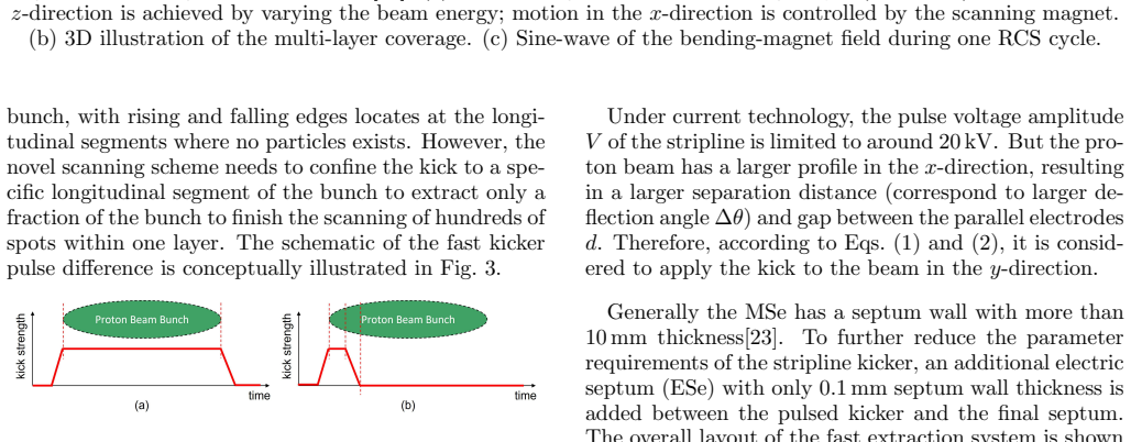
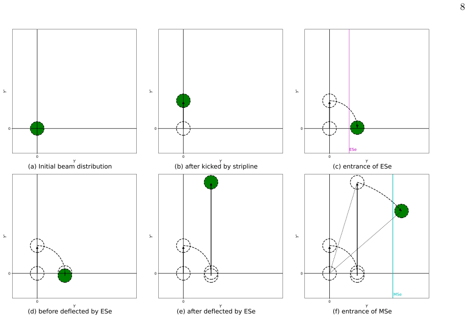
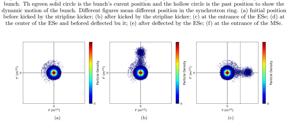
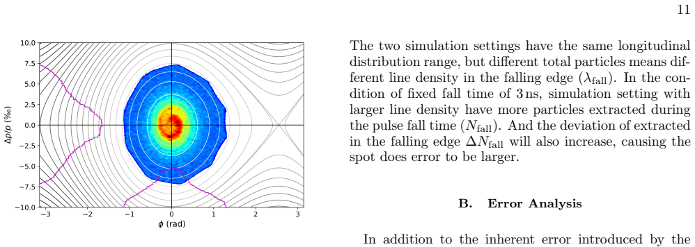
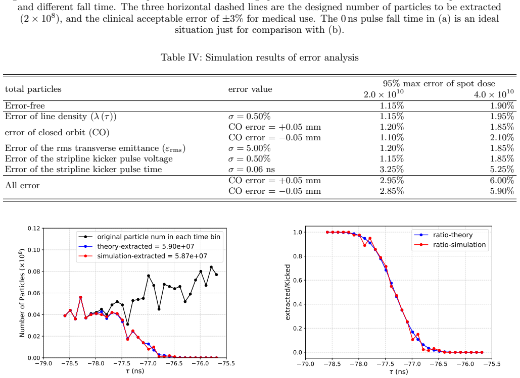
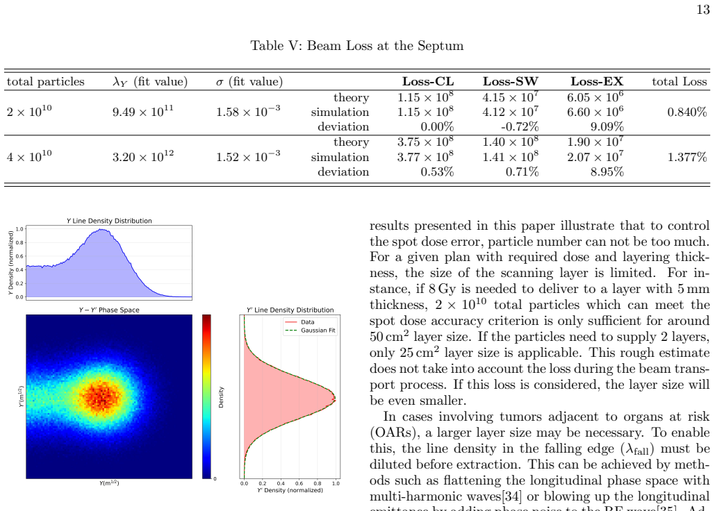
The central claim is that the longitudinal localized kick driven fast extraction method, combined with a novel parallel-layer scanning scheme inside a purpose-built rapid cycling synchrotron, makes 3D PBS proton FLASH delivery practical. The kicker pulse is restricted to specific longitudinal slices of the bunch, with the active slice chosen dynamically from beam-current-monitor data. At 2×10^10 particles the design satisfies spot-dose accuracy requirements and limits septum beam loss to less than 1 percent. The extraction hardware and RCS lattice parameters are chosen to meet these performance targets.
What carries the argument
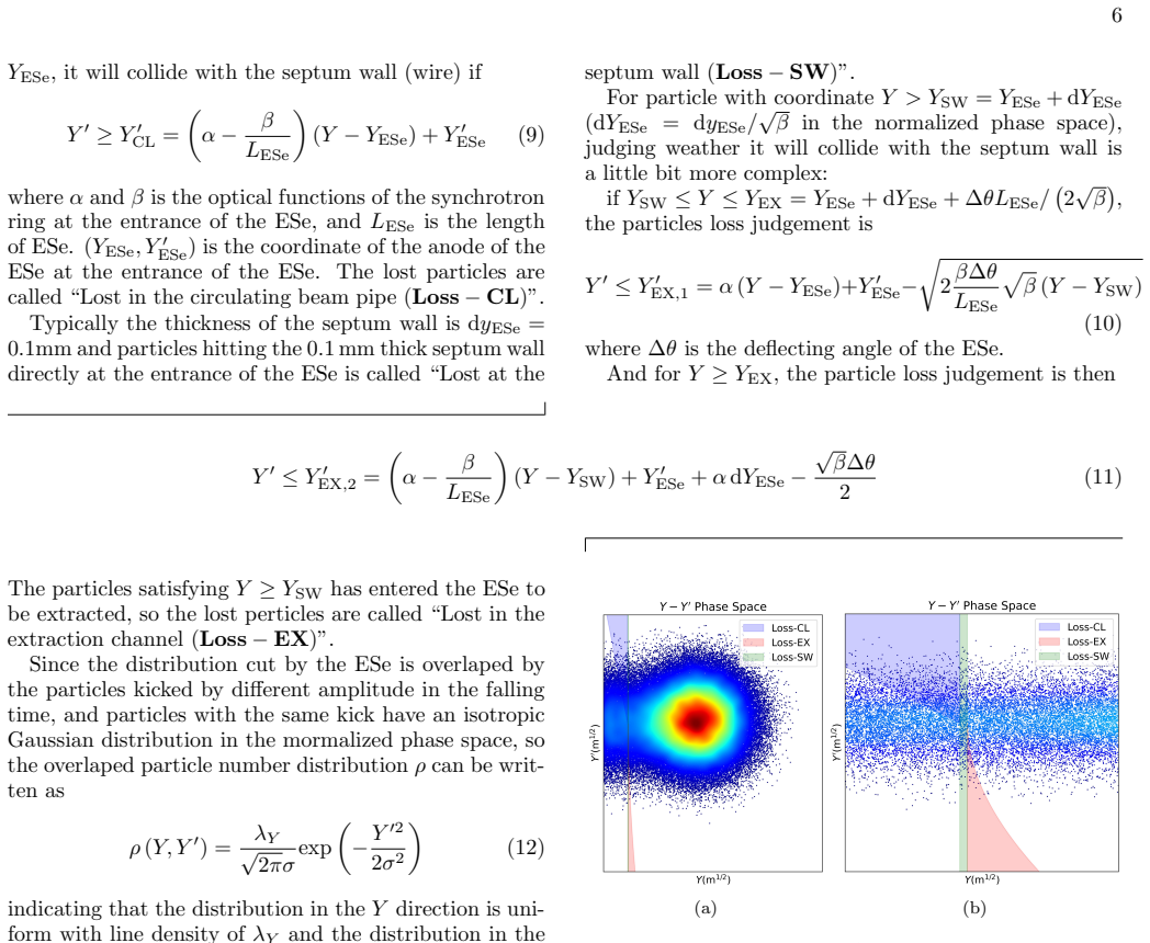
The longitudinal localized kick driven fast extraction system, which applies the kicker waveform only to chosen longitudinal segments of the bunch and adjusts those segments in real time from measured line density.
If this is right
- Spot dose accuracy meets clinical requirements at an intensity of 2×10^10 particles.
- Beam loss at the septum stays below 1 percent.
- The RCS lattice can be tuned to house the stripline kicker together with the electric and magnetic septa.
- The parallel-to-beam scanning scheme cuts overall delivery time compared with standard layer-by-layer methods.
Where Pith is reading between the lines
- The same intensity and timing controls could be tested on existing or scaled-down synchrotrons to check whether the loss budget holds under real beam jitter.
- If the dynamic kicker adjustment proves reliable, the method could be paired with existing FLASH dose-rate monitors to verify ultra-high dose rates in three dimensions.
- Reducing the particle number slightly while tightening kicker rise times might further lower septum losses without sacrificing accuracy.
Load-bearing premise
Real-time longitudinal line-density measurements can set the kicker's active region precisely enough that spot dose accuracy stays acceptable and septum losses remain under 1 percent during actual operation.
What would settle it
A beam test or end-to-end simulation at 2×10^10 particles that records either spot dose errors exceeding tolerance or septum losses above 1 percent when the kicker region is adjusted according to the measured line density.
Figures


read the original abstract
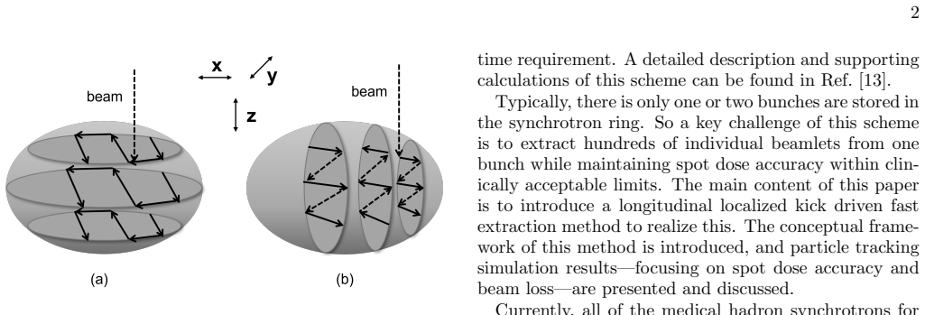
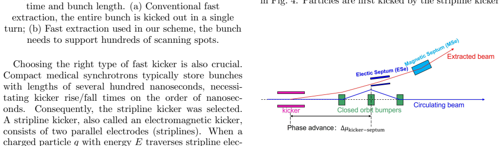
This paper presents the design of a rapid cycling synchrotron (RCS) featuring a longitudinal localized kick driven fast extraction system for three-dimensional (3D) pencil beam scanning (PBS) proton FLASH delivery. The extraction method is designed to accommodate a novel scanning scheme that addresses the stringent requirement for substantially shorter delivery time compared to current solutions, where the scanning layer is parallel to the proton beam direction. In this method, the kicker pulse waveform is applied selectively to specific longitudinal segments of the proton bunch. For each scanning spot, the functional region of the kicker along the longitudinal direction is dynamically adjusted based on real-time beam longitudinal line density measured by a beam current monitor. The corresponding region-determination algorithm is provided. We analyze the spot dose accuracy and the beam loss at the septum, indentifying increased particle longitudinal line density will reduce spot dose accuracy and increase beam loss. A total number of particles of $2\times10^{10}$ can satisfy the requirements of spot dose accuracy and the beam loss due to the septum is less than 1%. The extraction system comprises a stripline kicker, an electric septum (ESe), and a magnetic septum (MSe), imposing specific requirements on the RCS lattice design. The RCS is carefully designed to meet these constraints, and the parameters of the extraction elements are detailed. By integrating a novel scanning scheme with a specially designed RCS and fast extraction method, this work demonstrates the feasibility of achieving 3D PBS proton FLASH delivery.
Editorial analysis
A structured set of objections, weighed in public.
Referee Report
Summary. The manuscript proposes a rapid cycling synchrotron (RCS) design incorporating a longitudinal localized kick driven fast extraction system to enable three-dimensional pencil beam scanning (PBS) proton FLASH delivery. A novel scanning scheme applies the kicker waveform selectively to longitudinal segments of the bunch, with the functional region dynamically determined from real-time beam longitudinal line density measured by a beam current monitor. The authors analyze spot dose accuracy and septum beam loss, concluding that 2×10^{10} particles per spot meets the required accuracy while keeping septum loss below 1%. The extraction hardware (stripline kicker, electric and magnetic septa) imposes lattice constraints that the RCS is designed to satisfy.
Significance. If the performance numbers hold under realistic operating conditions, the approach could enable substantially shorter delivery times for proton FLASH by allowing scanning layers parallel to the beam direction, addressing a central technical barrier in translating FLASH radiotherapy to clinical use. The integration of the extraction method with the RCS lattice and the explicit region-determination algorithm represent concrete engineering contributions that could be tested in future beam studies.
major comments (2)
- [Performance analysis section] The performance analysis (abstract and the section presenting spot dose accuracy and beam loss results): the claim that 2×10^{10} particles satisfies both spot dose accuracy and <1% septum loss is derived from simulations that omit measurement noise, timing jitter, beam-position jitter, and finite kicker rise/fall times in the region-determination algorithm. Because the text already states that higher longitudinal density degrades both metrics, the absence of an error-propagation study leaves the margin under actual beam conditions unquantified and makes the feasibility conclusion only partially supported.
- [Method section on region-determination algorithm] The description of the region-determination algorithm (the paragraph following the scanning scheme introduction): the algorithm selects kicker functional segments from real-time line-density data, yet no quantitative bounds are given on monitor resolution, latency between measurement and pulse shaping, or how these affect the resulting particle distribution delivered to each spot. This directly impacts the central claim that the dynamic adjustment preserves the quoted accuracy and loss figures.
minor comments (2)
- [Abstract] Typo in the abstract: 'indentifying' should read 'identifying'.
- [Introduction and method sections] The longitudinal line density is referenced repeatedly but never given an explicit defining equation or symbol at first use; adding this would improve readability.
Simulated Author's Rebuttal
We thank the referee for the constructive comments that highlight important aspects of our performance analysis and algorithm description. We address each point below and will revise the manuscript to strengthen the supporting evidence.
read point-by-point responses
-
Referee: [Performance analysis section] The performance analysis (abstract and the section presenting spot dose accuracy and beam loss results): the claim that 2×10^{10} particles satisfies both spot dose accuracy and <1% septum loss is derived from simulations that omit measurement noise, timing jitter, beam-position jitter, and finite kicker rise/fall times in the region-determination algorithm. Because the text already states that higher longitudinal density degrades both metrics, the absence of an error-propagation study leaves the margin under actual beam conditions unquantified and makes the feasibility conclusion only partially supported.
Authors: We agree that the presented simulations provide an idealized baseline and omit realistic effects including measurement noise, timing jitter, beam-position jitter, and finite kicker rise/fall times. In the revised manuscript we will add a new subsection on error propagation and sensitivity analysis. Using representative hardware parameters (beam current monitor resolution of 0.5 %, timing jitter of 5 ns, and kicker rise/fall time of 20 ns), we will quantify the resulting degradation in spot dose accuracy and septum loss. The updated analysis will show that 2×10^{10} particles per spot retains adequate margin under these conditions while keeping loss below 1 % and dose accuracy within the stated tolerance. revision: yes
-
Referee: [Method section on region-determination algorithm] The description of the region-determination algorithm (the paragraph following the scanning scheme introduction): the algorithm selects kicker functional segments from real-time line-density data, yet no quantitative bounds are given on monitor resolution, latency between measurement and pulse shaping, or how these affect the resulting particle distribution delivered to each spot. This directly impacts the central claim that the dynamic adjustment preserves the quoted accuracy and loss figures.
Authors: We acknowledge that the current description lacks quantitative bounds on monitor resolution and latency. In the revised manuscript we will expand the algorithm section to specify monitor performance (temporal resolution 1 ns, measurement-to-pulse latency < 50 ns) and include a short sensitivity study showing how these parameters propagate into the extracted particle distribution. The study will confirm that the dynamic region selection preserves the reported spot dose accuracy and keeps septum loss below 1 % for the chosen particle number. revision: yes
Circularity Check
No significant circularity; particle count and extraction parameters chosen via forward analysis of dose accuracy and septum loss
full rationale
The derivation selects 2×10^{10} particles to meet stated spot-dose and <1% loss targets after analyzing the effect of longitudinal line density on those metrics. This is a conventional design-threshold calculation rather than a self-referential loop in which the output is presupposed by the input definitions or by a fitted parameter renamed as a prediction. The region-determination algorithm is described as a novel scheme that uses real-time beam-current-monitor data, but no equations or steps reduce the claimed performance numbers to the algorithm by construction. Lattice constraints for the stripline kicker, electric septum, and magnetic septum are imposed externally by extraction requirements and satisfied by standard RCS design choices. No load-bearing self-citations, uniqueness theorems imported from the authors' prior work, or ansatzes smuggled via citation appear in the provided chain. The feasibility demonstration therefore remains self-contained against external benchmarks such as conventional synchrotron extraction performance.
Axiom & Free-Parameter Ledger
free parameters (1)
- Total particles per spot =
2e10
axioms (1)
- standard math Established principles of beam extraction, kicker waveform control, and synchrotron lattice design hold under the proposed operating conditions.
Lean theorems connected to this paper
-
IndisputableMonolith/Cost/FunctionalEquation.leanwashburn_uniqueness_aczel unclearFor each scanning spot, the functional region of the kicker along the longitudinal direction is dynamically adjusted based on real-time beam longitudinal line density measured by a beam current monitor. The corresponding region-determination algorithm is provided.
-
IndisputableMonolith/Foundation/DimensionForcing.leanalexander_duality_circle_linking unclearThe RCS is carefully designed to meet these constraints, and the parameters of the extraction elements are detailed.
Reference graph
Works this paper leans on
-
[1]
V. Favaudon, L. Caplier, V. Monceau, F. Pouzoulet, M. Sayarath, C. Fouillade, M.-F. Poupon, I. Brito, P. Hup´ e, J. Bourhis, J. Hall, J.-J. Fontaine, and M.- 15 C. Vozenin, Science Translational Medicine6, 245ra93 (2014)
work page 2014
- [2]
-
[3]
M.-C. Vozenin, B. W. Loo, S. Tantawi, P. G. Maxim, D. R. Spitz, C. Bailat, and C. L. Limoli, Rev. Mod. Phys.96, 035002 (2024)
work page 2024
-
[4]
A. E. Mascia, E. C. Daugherty, Y. Zhang, E. Lee, Z. Xiao, M. Sertorio, J. Woo, L. R. Backus, J. M. McDon- ald, C. McCann, K. Russell, L. Levine, R. A. Sharma, D. Khuntia, J. D. Bradley, I. Simone, Charles B., J. P. Perentesis, and J. C. Breneman, JAMA Oncology9, 62 (2023)
work page 2023
-
[5]
A. Darafsheh and A. Bey, Physics in Medicine & Biology 70, 105008 (2025)
work page 2025
-
[6]
E. S. Diffenderfer, I. I. Verginadis, M. M. Kim, K. Shoniy- ozov, A. Velalopoulou, D. Goia, M. Putt, S. Ha- gan, S. Avery, K. Teo, W. Zou, A. Lin, S. Swisher- McClure, C. Koch, A. R. Kennedy, A. Minn, A. Maity, T. M. Busch, L. Dong, C. Koumenis, J. Metz, and K. A. Cengel, International Journal of Radiation Oncol- ogy*Biology*Physics106, 440 (2020)
work page 2020
-
[7]
G. Klimpki, Y. Zhang, G. Fattori, S. Psoroulas, D. C. Weber, A. Lomax, and D. Meer, Physics in Medicine & Biology63, 145006 (2018)
work page 2018
- [8]
-
[9]
J. E. Younkin, M. Bues, T. T. Sio, W. Liu, X. Ding, S. R. Keole, J. B. Stoker, and J. Shen, Advances in Radiation Oncology3, 412 (2018)
work page 2018
-
[10]
B. Roberfroid, M. S. Chocan Vera, C. Draguet, J. A. Lee, A. M. Barrag´ an-Montero, and E. Sterpin, Physics in Medicine & Biology70, 065005 (2025)
work page 2025
- [11]
-
[12]
Flanz,Particle Therapy Technology for Safe Treatment (CRC Press, Boca Raton, 2022) p
J. Flanz,Particle Therapy Technology for Safe Treatment (CRC Press, Boca Raton, 2022) p. 163
work page 2022
- [13]
-
[14]
M. M. Folkerts, E. Abel, S. Busold, J. R. Perez, V. Kr- ishnamurthi, and C. C. Ling, Medical Physics47, 6396 (2020)
work page 2020
- [15]
-
[16]
H. Chen, Y. Chen, S. Fu, L. Ma, S. Wang, F. Chen, Y. Chen, H. Dong, L. Dong, G. Feng, J. Gu, L. He, K. He, W. He, C. Hu, J. Huang, Q. Ji, X. Jia, D. Jin, L. Kang, W. Kang, T. Liang, G. Lin, H. Liu, J. Li, H. Ouyang, F. Qi, X. Qi, H. Qu, H. Sun, Z. Sun, L. Shen, J. Tang, J. Tao, F. Wang, L. Wang, P. Wang, Q. Wang, Y. Wu, J. Xi, T. Xu, W. Yin, B. Zhang, J. ...
work page 2025
- [17]
-
[18]
S. E. Combs, O. J¨ akel, T. Haberer, and J. Debus, Ra- diotherapy and Oncology95, 41 (2010)
work page 2010
-
[19]
J. Cardona, S. Peggs, and J. Kewisch, IEEE Transac- tions on Nuclear Science50, 1147 (2003)
work page 2003
-
[20]
A. Yamaguchi, K. Nakayama, T. Rizawa, S. Sukenobu, K. Satoh, Y. Morii, Y. Tanabe, and Y. Chiba, inProceed- ings of the 1997 Particle Accelerator Conference (Cat. No.97CH36167), Vol. 3 (1997) pp. 3828–3830 vol.3
work page 1997
-
[21]
M. A. Fraser, CERN Yellow Rep. School Proc.5, 285 (2018)
work page 2018
-
[22]
M. P. Sangroula, C. J. Liaw, C. Liu, J. Sandberg, N. Tsoupas, B. Xiao, and X. Sun, Phys. Rev. Accel. Beams28, 020401 (2025)
work page 2025
-
[23]
Y. Wu, W. Kang, Y. Chen, X. Wu, S. Li, L. Wang, C. Deng, L. Li, J. Zhou, and Y. Liu, Nuclear Instruments and Methods in Physics Research Section A: Acceler- ators, Spectrometers, Detectors and Associated Equip- ment882, 45 (2018). [24]CAS - CERN Accelerator School: Beam Diagnostics, CERN (CERN, Geneva, 2009) in collaboration with Syn- chrotron SOLEIL, Gif...
work page 2018
- [24]
-
[25]
W. Kang, C. D. Deng, Q. Li, L. Li, W. Chen, B. G. Yin, J. X. Zhou, X. J. Sun, F. S. Chen, and C. T. Shi, IEEE Transactions on Applied Superconductivity22, 4001204 (2012)
work page 2012
-
[26]
H. Yaoet al., inProc. 10th International Particle Accel- erator Conference (IPAC’19), Melbourne, Australia, 19- 24 May 2019, International Particle Accelerator Confer- ence No. 10 (JACoW Publishing, Geneva, Switzerland,
work page 2019
-
[28]
W. Liu, L. Shang, W. Song, F. Shang, and Z. Sun, Nuclear Instruments and Methods in Physics Research Section A: Accelerators, Spectrometers, Detectors and Associated Equipment961, 163670 (2020)
work page 2020
-
[29]
B. Arjomandy, P. Taylor, C. Ainsley, S. Safai, N. Sahoo, M. Pankuch, J. B. Farr, S. Yong Park, E. Klein, J. Flanz, E. D. Yorke, D. Followill, and Y. Kase, Medical Physics 46, e678 (2019)
work page 2019
-
[31]
C. Yaoet al., inProc. of North American Particle Ac- celerator Conference (NAPAC’16), Chicago, IL, USA, October 9-14, 2016, North American Particle Acceler- ator Conference No. 3 (JACoW, Geneva, Switzerland,
work page 2016
-
[32]
950–952, https://doi.org/10.18429/JACoW- NAPAC2016-WEPOB24
pp. 950–952, https://doi.org/10.18429/JACoW- NAPAC2016-WEPOB24
-
[33]
C. Mitsuda, T. Honiden, K. Kobayashi, T. Kobayashi, S. Sasaki, and N. Sekine, inProc. 13th Annual Meeting of Particle Accelerator Society of Japan, August 8-10, 2016, Chiba, Japan, Annual Meeting of Particle Acceler- ator Society of Japan (Chiba, Japan, 2016) pp. 694–698
work page 2016
- [34]
- [35]
-
[36]
P. Baudrenghien and T. Mastoridis, Nuclear Instruments and Methods in Physics Research Section A: Acceler- ators, Spectrometers, Detectors and Associated Equip- ment726, 181 (2013)
work page 2013
-
[37]
Y. Prezado, M. Grams, E. Jouglar, I. Mart´ ınez-Rovira, R. Ortiz, J. Seco, and S. Chang, Physics in Medicine & Biology69, 10TR02 (2024)
work page 2024
-
[38]
H. Li, N. A. Mayr, R. J. Griffin, H. Zhang, D. Pokhrel, M. Grams, J. Penagaricano, S. Chang, M. B. Spraker, J. Kavanaugh, L. Lin, K. Sheikh, S. Mossahebi, C. B. Si- mone, D. Roberge, J. W. Snider, P. Sabouri, A. Molineu, Y. Xiao, and S. H. Benedict, International Journal of Radiation Oncology*Biology*Physics119, 737 (2024)
work page 2024
discussion (0)
Sign in with ORCID, Apple, or X to comment. Anyone can read and Pith papers without signing in.