WoundFormer: Multi-Scale Spatial Feature Fusion for Multi-Class Wound Tissue Segmentation
Pith reviewed 2026-05-20 05:37 UTC · model grok-4.3
The pith
WoundFormer replaces the SegFormer decoder with a spatially-preserving multi-scale aggregation head to improve multi-class wound tissue segmentation.
A machine-rendered reading of the paper's core claim, the machinery that carries it, and where it could break.
Core claim
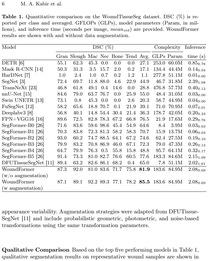
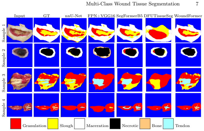
WoundFormer enhances hierarchical spatial feature fusion by replacing the standard SegFormer decoder with a spatially-preserving multi-scale aggregation head that maintains feature topology during cross-scale integration and strengthens contextual interactions through convolutional fusion, achieving 81.9 percent overall Dice on the WoundTissueSeg dataset with consistent gains on minority classes.
What carries the argument
Spatially-preserving multi-scale aggregation head that maintains feature topology during cross-scale integration and strengthens contextual interactions through convolutional fusion.
If this is right
- Boundary localization improves for all wound tissue classes.
- Discrimination increases between visually similar tissue categories.
- Performance rises consistently on minority tissue classes.
- Transformer computational efficiency remains unchanged.
- Quantitative wound assessment becomes more reliable for clinical use.
Where Pith is reading between the lines
- The same head design could transfer to other medical segmentation problems that involve high intra-class variability and limited labels.
- Combining the head with different transformer backbones might produce further accuracy lifts in heterogeneous scene parsing.
- Validation on larger wound image collections would test whether the spatial-preservation benefit scales to real-world deployment.
Load-bearing premise
The observed gains come from the new aggregation head rather than from differences in training procedure, data augmentation, or baseline re-implementations.
What would settle it
Retraining the original SegFormer decoder on the same WoundTissueSeg data with identical augmentation and optimization settings and obtaining comparable Dice scores would show the head is not the source of improvement.
Figures

read the original abstract
Chronic wounds such as diabetic foot ulcers and pressure injuries require accurate tissue-level assessment to guide treatment planning and monitor healing progression. While deep learning methods have advanced automated wound analysis, most existing approaches focus on binary segmentation and inadequately model heterogeneous tissue composition due to high intra-class variability and limited annotated data. Multi-class wound tissue segmentation, therefore, remains a challenging and clinically relevant problem. We propose WoundFormer, a transformer-based framework that enhances hierarchical spatial feature fusion for multi-class wound tissue segmentation. Specifically, we replace the standard SegFormer decoder with a spatially-preserving multi-scale aggregation head that maintains feature topology during cross-scale integration and strengthens contextual interactions through convolutional fusion. This design improves boundary localization and discrimination between visually similar tissue categories while preserving transformer efficiency. We evaluate WoundFormer on the WoundTissueSeg dataset (147 images, six tissue classes) and a second benchmark (DFUTissue dataset). The proposed method achieves an overall Dice score of 81.9%, outperforming strong CNN- and transformer-based baselines by up to 4.3 Dice points on the WoundTissueSeg benchmark, with consistent improvements across minority tissue classes. These results indicate that explicit modeling of hierarchical spatial interactions enhances transformer representations for heterogeneous wound tissue segmentation and supports more reliable quantitative wound assessment.
Editorial analysis
A structured set of objections, weighed in public.
Referee Report
Summary. The paper proposes WoundFormer, a transformer-based framework for multi-class wound tissue segmentation. It replaces the standard SegFormer decoder with a spatially-preserving multi-scale aggregation head that maintains feature topology during cross-scale integration and strengthens contextual interactions via convolutional fusion. The model is evaluated on the WoundTissueSeg dataset (147 images, six tissue classes) and the DFUTissue dataset, reporting an overall Dice score of 81.9% that outperforms CNN- and transformer-based baselines by up to 4.3 points, with gains on minority classes.
Significance. If the reported gains are shown to stem specifically from the proposed decoder head rather than training or implementation differences, the work would offer a practical enhancement to hierarchical feature fusion in transformer segmentation models for heterogeneous medical images. This could support more reliable quantitative assessment of chronic wounds, addressing intra-class variability and limited data in a clinically relevant domain.
major comments (2)
- [Abstract / Experiments] Abstract and Experiments section: The central claim attributes the +4.3 Dice improvement and better minority-class results to the spatially-preserving multi-scale aggregation head, yet no ablation isolating this component (e.g., replacing only the decoder while holding all other factors fixed) is described. On a 147-image dataset, even modest unstated differences in augmentation, loss weighting, or baseline re-implementation can produce Dice shifts of this size, undermining attribution.
- [Experiments] Experiments section: No statistical significance tests, standard deviations across multiple runs, or hyper-parameter matching details for the reported baselines are provided. This leaves open whether the observed improvements on WoundTissueSeg and DFUTissue are robust or sensitive to implementation choices.
minor comments (2)
- [Method] The description of how the convolutional fusion preserves topology during cross-scale integration would benefit from an accompanying diagram or explicit equations showing the aggregation operation.
- [Experiments] Dataset details such as train/validation/test splits, class imbalance handling, and annotation protocol for the six tissue classes should be expanded for reproducibility.
Simulated Author's Rebuttal
We thank the referee for the constructive comments, which help clarify the attribution of our results and the robustness of the reported gains. We address each major comment below and will revise the manuscript to incorporate the suggested analyses.
read point-by-point responses
-
Referee: [Abstract / Experiments] Abstract and Experiments section: The central claim attributes the +4.3 Dice improvement and better minority-class results to the spatially-preserving multi-scale aggregation head, yet no ablation isolating this component (e.g., replacing only the decoder while holding all other factors fixed) is described. On a 147-image dataset, even modest unstated differences in augmentation, loss weighting, or baseline re-implementation can produce Dice shifts of this size, undermining attribution.
Authors: We agree that a controlled ablation isolating the decoder head is necessary to strengthen attribution of the gains, particularly on the small WoundTissueSeg dataset. The manuscript currently compares the full WoundFormer against external baselines but does not report an internal variant that replaces only the proposed head with the standard SegFormer decoder under otherwise identical training conditions. In the revised version we will add this ablation, training the SegFormer decoder variant with the same backbone, augmentations, loss, optimizer, and hyper-parameters, and report both overall and per-class Dice scores to isolate the contribution of the spatially-preserving multi-scale aggregation head. revision: yes
-
Referee: [Experiments] Experiments section: No statistical significance tests, standard deviations across multiple runs, or hyper-parameter matching details for the reported baselines are provided. This leaves open whether the observed improvements on WoundTissueSeg and DFUTissue are robust or sensitive to implementation choices.
Authors: We acknowledge that single-run results and limited implementation details leave the robustness open to question. In the revision we will re-train all models (including baselines) across multiple random seeds, report mean Dice scores together with standard deviations, and add an explicit section detailing hyper-parameter selection and matching procedures used for fair comparison. Where appropriate we will also include statistical significance testing (e.g., paired Wilcoxon test) between WoundFormer and the strongest baseline to quantify whether the observed improvements are statistically reliable. revision: yes
Circularity Check
No circularity: empirical architecture proposal with external benchmarks
full rationale
The paper proposes WoundFormer by describing an architectural replacement of SegFormer's decoder with a spatially-preserving multi-scale aggregation head, then reports empirical Dice scores (81.9% overall) on the external WoundTissueSeg (147 images) and DFUTissue datasets against CNN and transformer baselines. No equations, first-principles derivations, or fitted parameters are presented that reduce to the model's own inputs by construction. Performance claims rest on standard training and evaluation rather than self-referential predictions or load-bearing self-citations. The derivation chain is therefore self-contained as an empirical contribution.
Axiom & Free-Parameter Ledger
axioms (1)
- domain assumption Transformer encoders from SegFormer produce features suitable for the proposed cross-scale fusion head in medical images
invented entities (1)
-
spatially-preserving multi-scale aggregation head
no independent evidence
Lean theorems connected to this paper
-
IndisputableMonolith/Cost/FunctionalEquation.leanwashburn_uniqueness_aczel unclear?
unclearRelation between the paper passage and the cited Recognition theorem.
we replace the standard SegFormer decoder with a spatially-preserving multi-scale aggregation head that maintains feature topology during cross-scale integration and strengthens contextual interactions through convolutional fusion
-
IndisputableMonolith/Foundation/AlexanderDuality.leanalexander_duality_circle_linking unclear?
unclearRelation between the paper passage and the cited Recognition theorem.
Coarse-to-Fine Fusion. Fusion begins from the lowest-resolution feature: X = F̂4. At each stage i=3,2,1 ... X ← σ(BN(Conv1×1([F̂i, X↑])))
What do these tags mean?
- matches
- The paper's claim is directly supported by a theorem in the formal canon.
- supports
- The theorem supports part of the paper's argument, but the paper may add assumptions or extra steps.
- extends
- The paper goes beyond the formal theorem; the theorem is a base layer rather than the whole result.
- uses
- The paper appears to rely on the theorem as machinery.
- contradicts
- The paper's claim conflicts with a theorem or certificate in the canon.
- unclear
- Pith found a possible connection, but the passage is too broad, indirect, or ambiguous to say the theorem truly supports the claim.
Reference graph
Works this paper leans on
-
[1]
Journal of Vascular Nursing (2023)
Alcântara, S.B.C., de Araújo, J.G., Santos, D.F., da Silva, T.R., Goulart, I.M.B., da Silva, A.M.B., Antunes, D.E.: Identification of types of wound bed tissue as a percentage and total wound area by planimetry in neuropathic and venous ulcers. Journal of Vascular Nursing (2023)
work page 2023
-
[2]
Advances in Wound Care11(12), 687–709 (2022)
Anisuzzaman, D., Wang, C., Rostami, B., Gopalakrishnan, S., Niezgoda, J., Yu, Z.: Image-based artificial intelligence in wound assessment: a systematic review. Advances in Wound Care11(12), 687–709 (2022)
work page 2022
-
[3]
IEEE transactions on pat- tern analysis and machine intelligence39(12), 2481–2495 (2017)
Badrinarayanan, V., Kendall, A., Cipolla, R.: Segnet: A deep convolutional encoder-decoder architecture for image segmentation. IEEE transactions on pat- tern analysis and machine intelligence39(12), 2481–2495 (2017)
work page 2017
-
[4]
In: Seminars in vascular surgery
Bandyk, D.F.: The diabetic foot: Pathophysiology, evaluation, and treatment. In: Seminars in vascular surgery. vol. 31, pp. 43–48. Elsevier (2018)
work page 2018
-
[5]
In: Joint Eu- ropean Conference on Machine Learning and Knowledge Discovery in Databases
Borst, V., Dittus, T., Dege, T., Schmieder, A., Kounev, S.: Woundambit: Bridging state-of-the-art semantic segmentation and real-world wound care. In: Joint Eu- ropean Conference on Machine Learning and Knowledge Discovery in Databases. pp. 285–303. Springer (2025)
work page 2025
-
[6]
In: European conference on computer vision
Carion, N., Massa, F., Synnaeve, G., Usunier, N., Kirillov, A., Zagoruyko, S.: End- to-end object detection with transformers. In: European conference on computer vision. pp. 213–229. Springer (2020)
work page 2020
-
[7]
In: Proceedings of the IEEE/CVF international conference on computer vision
Chao, P., Kao, C.Y., Ruan, Y.S., Huang, C.H., Lin, Y.L.: Hardnet: A low mem- ory traffic network. In: Proceedings of the IEEE/CVF international conference on computer vision. pp. 3552–3561 (2019)
work page 2019
-
[8]
Rethinking Atrous Convolution for Semantic Image Segmentation
Chen, L.C., Papandreou, G., Schroff, F., Adam, H.: Rethinking atrous convolution for semantic image segmentation. arXiv preprint arXiv:1706.05587 (2017)
work page internal anchor Pith review Pith/arXiv arXiv 2017
-
[9]
International Wound Journal7(3), 176–183 (2010)
Clerici, G., Caminiti, M., Curci, V., Quarantiello, A., Faglia, E.: The use of a dermal substitute to preserve maximal foot length in diabetic foot wounds with tendon and bone exposure following urgent surgical debridement for acute infec- tion. International Wound Journal7(3), 176–183 (2010)
work page 2010
-
[10]
Journal of wound care11(7), 275–278 (2002)
Cutting, K.F., White, R.J.: Maceration of the skin and wound bed 1: its nature and causes. Journal of wound care11(7), 275–278 (2002)
work page 2002
-
[11]
arXiv preprint arXiv:2406.16012 (2024)
Dhar, M.K., Wang, C., Patel, Y., Zhang, T., Niezgoda, J., Gopalakrishnan, S., Chen, K., Yu, Z.: Wound tissue segmentation in diabetic foot ulcer images using deep learning: a pilot study. arXiv preprint arXiv:2406.16012 (2024)
-
[12]
Biomedical Signal Pro- cessing and Control92, 106057 (2024)
Dhar, M.K., Zhang, T., Patel, Y., Gopalakrishnan, S., Yu, Z.: Fusegnet: A deep convolutional neural network for foot ulcer segmentation. Biomedical Signal Pro- cessing and Control92, 106057 (2024)
work page 2024
-
[13]
In: International MICCAI brainlesion workshop
Hatamizadeh, A., Nath, V., Tang, Y., Yang, D., Roth, H.R., Xu, D.: Swin unetr: Swin transformers for semantic segmentation of brain tumors in mri images. In: International MICCAI brainlesion workshop. pp. 272–284. Springer (2021)
work page 2021
-
[14]
In: Proceedings of the IEEE international conference on computer vision
He, K., Gkioxari, G., Dollár, P., Girshick, R.: Mask r-cnn. In: Proceedings of the IEEE international conference on computer vision. pp. 2961–2969 (2017) 10 M. A. Kabir et al
work page 2017
-
[15]
Nature methods18(2), 203–211 (2021)
Isensee, F., Jaeger, P.F., Kohl, S.A., Petersen, J., Maier-Hein, K.H.: nnu-net: a self-configuring method for deep learning-based biomedical image segmentation. Nature methods18(2), 203–211 (2021)
work page 2021
-
[16]
arXiv preprint arXiv:2502.10652 (2025)
Kabir, M.A., Roy, N., Hossain, M.E., Featherston, J., Ahmed, S.: Deep learning for wound tissue segmentation: A comprehensive evaluation using a novel dataset. arXiv preprint arXiv:2502.10652 (2025)
-
[17]
In: Interna- tional Conference on Pattern Recognition
Niri, R., Douzi, H., Lucas, Y., Treuillet, S.: A superpixel-wise fully convolutional neural network approach for diabetic foot ulcer tissue classification. In: Interna- tional Conference on Pattern Recognition. pp. 308–320. Springer (2021)
work page 2021
-
[18]
Wound repair and regeneration27(1), 114–125 (2019)
Olsson, M., Järbrink, K., Divakar, U., Bajpai, R., Upton, Z., Schmidtchen, A., Car, J.: The humanistic and economic burden of chronic wounds: a systematic review. Wound repair and regeneration27(1), 114–125 (2019)
work page 2019
-
[19]
Rajathi,V.,Chinnasamy,A.,Selvakumari,P.:DUTCNet:Anoveldeepulcertissue classification network with stage prediction and treatment plan recommendation. Biomedical Signal Processing and Control90, 105855 (2024).https://doi.org/ 10.1016/j.bspc.2023.105855
-
[20]
Informatics in Medicine Unlocked37, 101185 (2023)
Reifs, D., Casanova-Lozano, L., Reig-Bolano, R., Grau-Carrion, S.: Clinical vali- dation of computer vision and artificial intelligence algorithms for wound measure- ment and tissue classification in wound care. Informatics in Medicine Unlocked37, 101185 (2023)
work page 2023
-
[21]
Journal of Diabetes Science and Technology4(4), 799–802 (2010)
Rogers, L.C., Bevilacqua, N.J., Armstrong, D.G., Andros, G.: Digital planimetry results in more accurate wound measurements: A comparison to standard ruler measurements. Journal of Diabetes Science and Technology4(4), 799–802 (2010). https://doi.org/10.1177/193229681000400405
-
[22]
Journal of Engineering2021(3) (2021)
Sarp, S., Kuzlu, M., Pipattanasomporn, M., Guler, O.: Simultaneous wound border segmentation and tissue classification using a conditional generative adversarial network. Journal of Engineering2021(3) (2021)
work page 2021
-
[23]
In: Pro- ceedings of the IEEE/CVF conference on computer vision and pattern recognition
Shi, D.: Transnext: Robust foveal visual perception for vision transformers. In: Pro- ceedings of the IEEE/CVF conference on computer vision and pattern recognition. pp. 17773–17783 (2024)
work page 2024
-
[24]
Wang, C., Shirzaei Sani, E., Shih, C.D., Lim, C.T., Wang, J., Armstrong, D.G., Gao, W.: Wound management materials and technologies from bench to bedside and beyond. Nature Reviews Materials pp. 1–17 (2024)
work page 2024
-
[25]
Wang, C., Mahbod, A., Ellinger, I., Galdran, A., Gopalakrishnan, S., Niezgoda, J., Yu, Z.: Fuseg: The foot ulcer segmentation challenge. Information15(3), 140 (2024)
work page 2024
-
[26]
Advances in neural information processing systems34, 12077–12090 (2021)
Xie, E., Wang, W., Yu, Z., Anandkumar, A., Alvarez, J.M., Luo, P.: Segformer: Simple and efficient design for semantic segmentation with transformers. Advances in neural information processing systems34, 12077–12090 (2021)
work page 2021
-
[27]
Medical Image Analysis94, 103153 (2024).https://doi.org/10.1016/j.media.2024.103153
Yap, M.H., Cassidy, B., Byra, M., yu Liao, T., Yi, H., Galdran, A., Chen, Y.H., Brüngel, R., Koitka, S., Friedrich, C.M., wen Lo, Y., hui Yang, C., Li, K., Lao, Q., Ballester, M.A.G., Carneiro, G., Ju, Y.J., Huang, J.D., Pappachan, J.M., Reeves, N.D., Chandrabalan, V., Dancey, D., Kendrick, C.: Diabetic foot ulcers segmenta- tion challenge report: Benchma...
discussion (0)
Sign in with ORCID, Apple, or X to comment. Anyone can read and Pith papers without signing in.