Label-free SERS Discrimination of Native Proline Hydroxylation at Single-molecule peptide by Deep Learning-assisted plasmonic nanopore
Pith reviewed 2026-05-21 01:38 UTC · model grok-4.3
The pith
A particle-in-pore SERS platform with peak-frequency analysis and a 1D convolutional network discriminates hydroxylated from non-hydroxylated HIF peptides at the single-molecule level.
A machine-rendered reading of the paper's core claim, the machinery that carries it, and where it could break.
Core claim
The central claim is that the particle-in-pore SERS sensor, when paired with peak occurrence frequency analysis and a one-dimensional convolutional neural network, captures reproducible hydroxylation-dependent changes in adsorption conformation and surface interactions, enabling reliable single-molecule discrimination of hydroxylated versus non-hydroxylated HIF peptide fragments. For the 7-, 9-, and 15-amino-acid pairs the post-evaluation accuracies reach 72.98 percent, 78.55 percent, and 89.74 percent respectively, each with AUC above 0.80. Gradient-weighted visualization shows that the network attends to the same recurrent spectral features identified by the peak-frequency method, and an增强
What carries the argument
The particle-in-pore single-molecule SERS platform that records Raman spectra while peptides adsorb to gold nanoparticles inside a nanopore, processed by peak occurrence frequency statistics and 1D-CNN classification.
If this is right
- Hydroxylation alters how longer peptides adsorb to the gold surface, amplifying spectral differences and raising classification accuracy.
- The same platform can register weak PTM signatures without labels or ensemble averaging.
- Gradient-weighted maps confirm that the neural network learns chemically meaningful features that match the peak-occurrence statistics.
- The approach works across peptide lengths from 7 to 15 amino acids, suggesting scalability to other HIF-derived fragments.
Where Pith is reading between the lines
- If the adsorption change proves general for other polar PTMs, the same hardware could be used to screen multiple modification types without redesign.
- Integration with microfluidic delivery might allow the sensor to monitor dynamic hydroxylation states in real time rather than in static snapshots.
- The citrate-band enhancement observed for the longest peptide hints that surface-chemistry tuning could further boost contrast for shorter or more rigid sequences.
Load-bearing premise
The observed spectral and adsorption differences are produced specifically by the presence or absence of the hydroxyl group on proline rather than by uncontrolled differences in peptide conformation, surface coverage, or citrate binding.
What would settle it
If controlled experiments that fix peptide conformation while removing the hydroxylation produce identical SERS spectra and identical citrate-band intensities, the claim that hydroxylation itself drives the distinguishable signals would be falsified.
Figures

read the original abstract
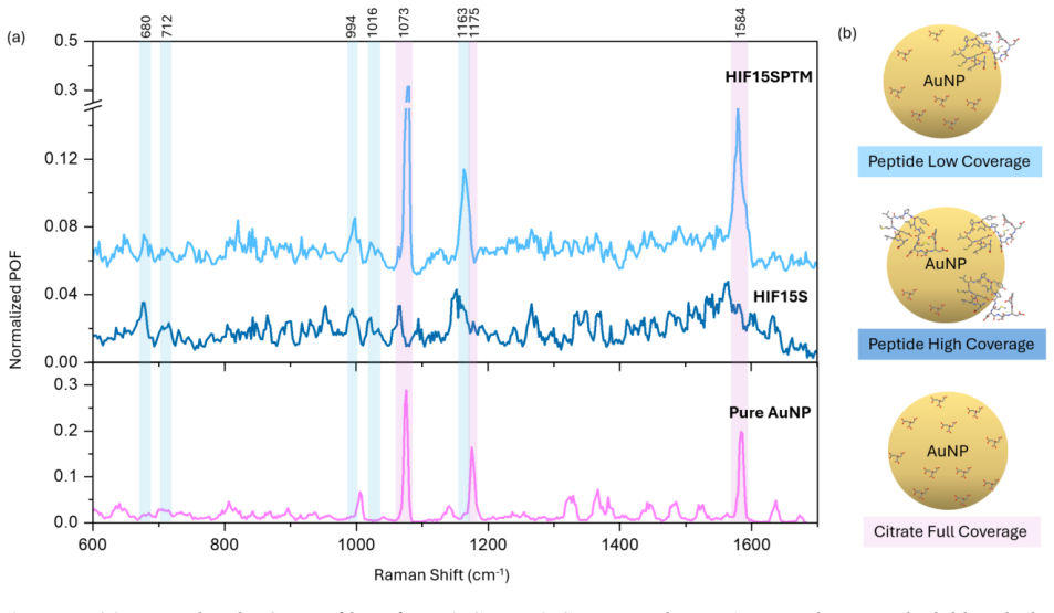
Post-translational modifications (PTMs) play essential roles in regulating protein structure, function, and cellular signalling. However, peptide level discrimination of hydroxylation at the single-molecule level remains difficult. Here, we report a particle-in-pore single-molecule surface-enhanced Raman spectroscopy (SERS) platform combined with peak occurrence frequency (POF) analysis and a one-dimensional convolutional neural network (1D-CNN) for discriminating hydroxylated and non-hydroxylated HIF peptide fragments. Three peptide pairs containing the Pro-564 hydroxylation site, with lengths of 7, 9, and 15 amino acids (AAs), were investigated. POF analysis revealed reproducible hydroxylation-dependent spectral changes in the 7AA and 9AA peptide pairs, which were attributed to changes in adsorption conformation and surface interactions. CNN-based classification achieved post-evaluation accuracies of 72.98%, 78.55%, and 89.74% for the 7AA, 9AA, and 15AA peptide pairs, respectively, with AUC values above 0.80 for all the pairs, indicating a reliable discrimination. Gradient-weighted feature visualization further showed that CNN-sensitive regions overlapped with recurrent POF features, supporting the chemical relevance of the learned classification patterns. Notably, for the 15AA peptide pair, the enhanced citrate-associated band suggests that hydroxylation can substantially alter peptide-gold nanoparticle adsorption behaviour. This adsorption-mediated effect may amplify hydroxylation-induced spectral differences and contribute to the improved discrimination accuracy despite the increased structural complexity. These results demonstrate that the particle-in-pore sensor, assisted by deep learning, can capture hydroxylation-induced spectral and adsorption changes in peptide fragments, providing a promising strategy for ultrasensitive analysis of weak PTM signatures in peptides.
Editorial analysis
A structured set of objections, weighed in public.
Referee Report
Summary. The manuscript presents a particle-in-pore single-molecule SERS platform integrated with peak occurrence frequency (POF) analysis and a 1D-CNN for label-free discrimination of hydroxylated versus non-hydroxylated HIF peptide fragments at the Pro-564 site. Three peptide length pairs (7AA, 9AA, 15AA) are examined; POF identifies reproducible spectral changes attributed to adsorption conformation shifts, and the CNN achieves post-evaluation accuracies of 72.98%, 78.55%, and 89.74% with AUC values >0.80. Gradient-weighted visualizations link CNN features to POF peaks, and an enhanced citrate band is noted for the 15AA pair as evidence of hydroxylation-altered AuNP adsorption.
Significance. If the spectral and classification differences prove specific to proline hydroxylation rather than uncontrolled adsorption or conformational variations, the work would offer a promising route for ultrasensitive, label-free PTM detection in peptides where conventional methods lack sensitivity. The combination of experimental SERS nanopore measurements, POF feature extraction, CNN classification with AUC>0.80, and feature visualization provides concrete empirical support and chemical interpretability that strengthens the central claim.
major comments (2)
- [Abstract] Abstract: The central claim that the platform discriminates native proline hydroxylation requires that observed spectral shifts and CNN accuracies arise specifically from the PTM. The abstract itself states that for the 15AA pair 'the enhanced citrate-associated band suggests that hydroxylation can substantially alter peptide-gold nanoparticle adsorption behaviour' and that this 'may amplify hydroxylation-induced spectral differences.' No description is given of matched controls that hold peptide concentration, ionic strength, or conformational ensemble fixed while toggling only the hydroxylation state; without such controls the CNN decision boundaries could be driven by surface-coverage or citrate-interaction differences instead of the PTM.
- [Abstract] Abstract and POF analysis section: Reproducibility of hydroxylation-dependent spectral changes is asserted, yet the manuscript provides no quantitative metrics (e.g., standard deviations on peak frequencies or intensities across independent measurements) or explicit statement of how many independent peptide preparations and nanopore runs were performed for each pair. This information is load-bearing for the claim that the 72.98–89.74% accuracies reflect hydroxylation rather than batch-to-batch variability.
minor comments (2)
- [Abstract] The abstract reports concrete accuracies and AUC thresholds but omits raw spectra, training/validation splits, cross-validation procedure, or error bars on the reported accuracies.
- Notation for the 1D-CNN architecture (layer counts, kernel sizes, training hyperparameters) is not summarized in the abstract or methods overview, making it difficult to assess reproducibility of the 72.98–89.74% figures.
Simulated Author's Rebuttal
We are grateful to the referee for the thorough review and valuable suggestions. We address the major comments point by point below, indicating where revisions will be made to improve the manuscript.
read point-by-point responses
-
Referee: [Abstract] Abstract: The central claim that the platform discriminates native proline hydroxylation requires that observed spectral shifts and CNN accuracies arise specifically from the PTM. The abstract itself states that for the 15AA pair 'the enhanced citrate-associated band suggests that hydroxylation can substantially alter peptide-gold nanoparticle adsorption behaviour' and that this 'may amplify hydroxylation-induced spectral differences.' No description is given of matched controls that hold peptide concentration, ionic strength, or conformational ensemble fixed while toggling only the hydroxylation state; without such controls the CNN decision boundaries could be driven by surface-coverage or citrate-interaction differences instead of the PTM.
Authors: We appreciate this concern regarding the specificity to the PTM. The hydroxylated and non-hydroxylated peptides differ only at the Pro-564 site, with all other experimental conditions, including concentration and buffer, kept identical for each pair. The observed differences in POF and the CNN's reliance on specific spectral regions (as shown by gradient-weighted visualizations) support that the discrimination is due to hydroxylation-induced changes in adsorption and conformation. However, to address the referee's point directly, we will revise the abstract and methods to explicitly describe the matched controls and provide additional justification that citrate effects are a consequence of the PTM rather than a confounding factor. We will also discuss potential limitations in controlling conformational ensembles at the single-molecule level. revision: yes
-
Referee: [Abstract] Abstract and POF analysis section: Reproducibility of hydroxylation-dependent spectral changes is asserted, yet the manuscript provides no quantitative metrics (e.g., standard deviations on peak frequencies or intensities across independent measurements) or explicit statement of how many independent peptide preparations and nanopore runs were performed for each pair. This information is load-bearing for the claim that the 72.98–89.74% accuracies reflect hydroxylation rather than batch-to-batch variability.
Authors: We concur that providing quantitative metrics and replicate information is crucial. The experiments were conducted with multiple independent preparations and runs to ensure reproducibility, but these details were omitted from the main text for brevity. In the revised manuscript, we will include a dedicated subsection on experimental reproducibility, reporting the number of independent preparations (n ≥ 3 for each peptide pair) and total measurements, along with standard deviations for the key POF peak positions and intensities. This will substantiate that the classification accuracies are not due to batch variability. revision: yes
Circularity Check
No circularity: empirical spectra + independent CNN training
full rationale
The paper presents an experimental workflow: measured SERS spectra from hydroxylated vs non-hydroxylated peptide samples, POF feature extraction on those spectra, and standard 1D-CNN training/evaluation yielding reported accuracies (72.98–89.74 %, AUC > 0.80). No equations, fitted parameters, or self-citations are invoked such that any output reduces to the input by construction. The classification result is obtained from held-out evaluation on independently acquired spectral data; the interpretation that differences arise from hydroxylation is an empirical claim open to controls, not a definitional or self-referential loop. The derivation chain is therefore self-contained against external benchmarks.
Axiom & Free-Parameter Ledger
axioms (2)
- domain assumption Plasmonic enhancement in the particle-in-pore geometry produces measurable SERS spectra from single peptides
- domain assumption Hydroxylation-induced changes in peptide adsorption produce reproducible shifts in peak occurrence frequencies
Lean theorems connected to this paper
-
IndisputableMonolith/Cost/FunctionalEquation.leanwashburn_uniqueness_aczel unclear?
unclearRelation between the paper passage and the cited Recognition theorem.
POF analysis revealed reproducible hydroxylation-dependent spectral changes... attributed to changes in adsorption conformation and surface interactions. CNN-based classification achieved post-evaluation accuracies of 72.98%, 78.55%, and 89.74%
What do these tags mean?
- matches
- The paper's claim is directly supported by a theorem in the formal canon.
- supports
- The theorem supports part of the paper's argument, but the paper may add assumptions or extra steps.
- extends
- The paper goes beyond the formal theorem; the theorem is a base layer rather than the whole result.
- uses
- The paper appears to rely on the theorem as machinery.
- contradicts
- The paper's claim conflicts with a theorem or certificate in the canon.
- unclear
- Pith found a possible connection, but the passage is too broad, indirect, or ambiguous to say the theorem truly supports the claim.
Reference graph
Works this paper leans on
-
[1]
Iommarini, L., et al., Non-Canonical Mechanisms Regulating Hypoxia-Inducible Factor 1 Alpha in Cancer. Frontiers in Oncology, 2017. 7
work page 2017
-
[2]
Ma, H., X.X. Han, and B. Zhao, Enhanced Raman spectroscopic analysis of protein post- translational modifications. TrAC Trends in Analytical Chemistry, 2020. 131: p. 116019
work page 2020
-
[3]
Trends in Pharmacological Sciences, 2012
Semenza, G.L., Hypoxia-inducible factors: mediators of cancer progression and targets for cancer therapy. Trends in Pharmacological Sciences, 2012. 33(4): p. 207-214
work page 2012
-
[4]
International Journal of Analytical Chemistry, 2016, 7436849, 6 pages, 2016
Ponomarenko, E., et al., The Size of the Human Proteome: The Width and Depth. International Journal of Analytical Chemistry, 2016, 7436849, 6 pages, 2016
work page 2016
-
[5]
Angewandte Chemie-International Edition, 2025
Li, Z., et al., Nanopore-Based High-Resolution Detection of Multiple Post-Translational Modifications in Protein. Angewandte Chemie-International Edition, 2025. 64(15)
work page 2025
-
[6]
Rosen, C., D. Rodriguez-Larrea, and H. Bayley, Single-molecule site-specific detection of protein phosphorylation with a nanopore. Nature Biotechnology, 2014. 32(2): p. 179-181
work page 2014
-
[7]
Ensslen, T., et al., Resolving Isomeric Posttranslational Modifications Using a Biological Nanopore as a Sensor of Molecular Shape. J Am Chem Soc, 2022. 144(35): p. 16060-16068
work page 2022
-
[8]
Zhang, L., et al., Detection of single peptide with only one amino acid modification via electronic fingerprinting using reengineered durable channel of Phi29 DNA packaging motor. Biomaterials, 2021. 276: p. 121022
work page 2021
-
[9]
Fahie, M.A. and M. Chen, Electrostatic Interactions between OmpG Nanopore and Analyte Protein Surface Can Distinguish between Glycosylated Isoforms. J Phys Chem B, 2015. 119(32): p. 10198-206
work page 2015
-
[10]
ACS Nano 2024, 18, 2, 1504–1515
Chan Cao , et al., Deep learning-assisted single-molecule detection of protein post- translational modifications with a biological nanopore. ACS Nano 2024, 18, 2, 1504–1515
work page 2024
-
[11]
Angewandte Chemie-International Edition, 2025
Chang, J., et al., Precise Identification of Native Peptides with Posttranslational Proline Hydroxylation by Nanopore. Angewandte Chemie-International Edition, 2025. 64(9)
work page 2025
-
[12]
Sundararajan, N., et al., Ultrasensitive detection and characterization of posttranslational modifications using surface-enhanced Raman spectroscopy. Analytical Chemistry, 2006. 78(11): p. 3543-3550
work page 2006
-
[13]
Advanced Functional Materials,
Iarossi, M., et al., High-Density Plasmonic Nanopores for DNA Sensing at Ultra-Low Concentrations by Plasmon-Enhanced Raman Spectroscopy. Advanced Functional Materials,
-
[14]
Khabarov, K., et al., Raman identification of single nucleotides flowing through permeable plasmonic films. Nature Communications, 2025. 16(1): p. 9113
work page 2025
-
[15]
Du, J., et al., The role of phosphorylation and dephosphorylation of shell matrix proteins in shell formation: an in vivo and in vitro study. Crystengcomm, 2018. 20(27): p. 3905-3916
work page 2018
-
[16]
Analytical Chemistry, 2024, 96, 45, 17978–17983
Geddis, A., et al., Label-Free SERS Sensors for Real-Time Monitoring of Tyrosine Phosphorylation. Analytical Chemistry, 2024, 96, 45, 17978–17983
work page 2024
-
[17]
Biosensors & Bioelectronics, 2015
Li, Y ., et al., Label-free in situ monitoring of protein tyrosine nitration in blood by surface- enhanced Raman spectroscopy. Biosensors & Bioelectronics, 2015. 69: p. 1-7
work page 2015
-
[18]
Journal of Photochemistry and Photobiology B- Biology, 2018
Sjöberg, B., et al., Methionine oxidation by hydrogen peroxide in peptides and proteins: A theoretical and Raman spectroscopy study. Journal of Photochemistry and Photobiology B- Biology, 2018. 188: p. 95-99
work page 2018
-
[19]
Guerrero, A. and R. Aroca, Surface-enhanced Raman scattering of hydroxyproline. Journal of Raman Spectroscopy, 2012. 43(4): p. 478-481
work page 2012
-
[20]
A Raman, SERS and theoretical study
Cárcamo, J., et al., Proline and hydroxyproline deposited on silver nanoparticles. A Raman, SERS and theoretical study. Journal of Raman Spectroscopy, 2012. 43(6): p. 750-755
work page 2012
-
[21]
Chemical Society Reviews, 2019
Lee, H., et al., Designing surface-enhanced Raman scattering (SERS) platforms beyond hotspot engineering: emerging opportunities in analyte manipulations and hybrid materials. Chemical Society Reviews, 2019. 48(3): p. 731-756
work page 2019
-
[22]
Angewandte Chemie International Edition, 2020
Huang, J.-A., et al., Multiplexed Discrimination of Single Amino Acid Residues in Polypeptides in a Single SERS Hot Spot. Angewandte Chemie International Edition, 2020. 59(28): p. 11423- 11431
work page 2020
-
[23]
Huang, J.-A., et al., SERS discrimination of single DNA bases in single oligonucleotides by electro-plasmonic trapping. Nature Communications, 2019. 10(1): p. 5321
work page 2019
-
[24]
Zhao, Y ., et al., Label-Free Optical Analysis of Biomolecules in Solid-State Nanopores: Toward Single-Molecule Protein Sequencing. Acs Photonics, 2022. 9(3): p. 730-742
work page 2022
-
[25]
Angewandte Chemie- International Edition, 2025
Lim, S., et al., Unlocking the Full Potential of SERS: Merging Direct and Indirect Approaches for Enhanced Analysis of Multiplex Plasticizer Analogs in Matrices. Angewandte Chemie- International Edition, 2025. 64(40)
work page 2025
-
[26]
Angewandte Chemie-International Edition, 2026
Tang, W., et al., Digital Decoding of Multicomponent Protein Systems via Nanocavity-Confined Single-Molecule Raman Fingerprinting. Angewandte Chemie-International Edition, 2026. 65(6)
work page 2026
-
[27]
Khabarov, K., et al., Single-Molecule Peptide Discrimination via Flow-Through SERS and Machine Learning. Acs Photonics, 2026. 13(7): p. 1840-1845
work page 2026
-
[28]
Nano Letters, 2025, 25, 18, 7499–7506
Zhao, Y ., et al., Single-Molecule SERS Discrimination of Proline from Hydroxyproline Assisted by a Deep Learning Model. Nano Letters, 2025, 25, 18, 7499–7506
work page 2025
-
[29]
Abramson, J., et al., Accurate structure prediction of biomolecular interactions with AlphaFold
- [30]
-
[32]
Franco-Ulloa, S., et al., Dispersion state phase diagram of citrate-coated metallic nanoparticles in saline solutions. Nature Communications, 2020. 11(1)
work page 2020
-
[33]
Buglak, A. and A. Kononov, Comparative study of gold and silver interactions with amino acids and nucleobases. Rsc Advances, 2020. 10(56): p. 34149-34160
work page 2020
-
[34]
Zrimsek, A., et al., Single-Molecule Chemistry with Surface- and Tip-Enhanced Raman Spectroscopy. Chemical Reviews, 2017. 117(11): p. 7583-7613
work page 2017
-
[35]
Shin, H., et al., Frequency-Domain Proof of the Existence of Atomic-Scale SERS Hot-Spots. Nano Letters, 2018. 18(1): p. 262-271
work page 2018
-
[36]
Li, W., et al., High-Throughput Single-Molecule Surface-Enhanced Raman Spectroscopic Profiling of Single-Amino Acid Substitutions in Peptides by a Gold Plasmonic Nanopore. Acs Nano, 2024. 18(29): p. 19200-19207
work page 2024
-
[37]
Graff, M. and J. Bukowska, Surface-enhanced Raman scattering (SERS) spectroscopy of enantiomeric and racemic methionine on a silver electrode-evidence for chiral discrimination in interactions between adsorbed molecules. Chemical Physics Letters, 2011. 509(1-3): p. 58- 61
work page 2011
-
[38]
Spectrochimica Acta Part a-Molecular and Biomolecular Spectroscopy, 2011
Zhu, G., et al., Raman spectra of amino acids and their aqueous solutions. Spectrochimica Acta Part a-Molecular and Biomolecular Spectroscopy, 2011. 78(3): p. 1187-1195
work page 2011
-
[39]
Spectrochimica Acta Part a-Molecular and Biomolecular Spectroscopy, 2024, 308, 123681
Hernández, B., et al., The relationship between the tyrosine residue 850-830 cm-1Raman doublet intensity ratio and the aromatic side chain x1 torsion angle. Spectrochimica Acta Part a-Molecular and Biomolecular Spectroscopy, 2024, 308, 123681
work page 2024
-
[40]
Wolpert, M. and P . Hellwig, Infrared spectra and molar absorption coefficients of the 20 alpha amino acids in aqueous solutions in the spectral range from 1800 to 500 cm-1. Spectrochimica Acta Part a-Molecular and Biomolecular Spectroscopy, 2006. 64(4): p. 987-1001
work page 2006
-
[41]
Journal of Molecular Liquids, 2025
Smirnov, M., et al., Interaction of gold and platinum nanoparticles with L-tyrosine in aqueous solution: Conformational and dynamic changes. Journal of Molecular Liquids, 2025. 420: p. 126822
work page 2025
-
[42]
Stewart, S. and P . Fredericks, Surface-enhanced Raman spectroscopy of amino acids adsorbed on an electrochemically prepared silver surface. Spectrochimica Acta Part a- Molecular and Biomolecular Spectroscopy, 1999. 55(7-8): p. 1641-1660
work page 1999
-
[43]
Hernández, B., et al., Vibrational Analysis of Amino Acids and Short Peptides in Hydrated Media. IV . Amino Acids with Hydrophobic Side Chains: L-Alanine, L-Valine, and L-Isoleucine. Journal of Physical Chemistry B, 2009. 113(10): p. 3169-3178
work page 2009
-
[44]
Stewart, S. and P . Fredericks, Surface-enhanced Raman spectroscopy of peptides and proteins adsorbed on an electrochemically prepared silver surface. Spectrochimica Acta Part a-Molecular and Biomolecular Spectroscopy, 1999. 55(7-8): p. 1615-1640
work page 1999
- [45]
-
[46]
Journal of Physical Chemistry, 1983
SIIMAN, O., et al., SURFACE-ENHANCED RAMAN-SCATTERING BY CITRATE ON COLLOIDAL SILVER. Journal of Physical Chemistry, 1983. 87(6): p. 1014-1023
work page 1983
-
[47]
MUNRO, C., et al., CHARACTERIZATION OF THE SURFACE OF A CITRATE-REDUCED COLLOID OPTIMIZED FOR USE AS A SUBSTRATE FOR SURFACE-ENHANCED RESONANCE RAMAN- SCATTERING. Langmuir, 1995. 11(10): p. 3712-3720
work page 1995
-
[48]
Kurouski, D., et al., Amide I vibrational mode suppression in surface (SERS) and tip (TERS) enhanced Raman spectra of protein specimens. Analyst, 2013. 138(6): p. 1665-1673
work page 2013
-
[49]
Deckert-Gaudig, T., E. Rauls, and V . Deckert, Aromatic Amino Acid Monolayers Sandwiched between Gold and Silver: A Combined Tip-Enhanced Raman and Theoretical Approach. Journal of Physical Chemistry C, 2010. 114(16): p. 7412-7420
work page 2010
-
[50]
Hernández, B., et al., Vibrational Analysis of Amino Acids and Short Peptides in Hydrated Media. VIII. Amino Acids with Aromatic Side Chains: L-Phenylalanine, L-Tyrosine, and L- Tryptophan. Journal of Physical Chemistry B, 2010. 114(46): p. 15319-15330
work page 2010
-
[51]
Journal of Raman Spectroscopy, 2022
Li, J., et al., Machine learning using convolutional neural networks for SERS analysis of biomarkers in medical diagnostics. Journal of Raman Spectroscopy, 2022. 53(12): p. 2044- 2057
work page 2022
-
[52]
Lu, P ., et al., CNN-assisted SERS enables ultra-sensitive and simultaneous detection of Scr and BUN for rapid kidney function assessment. Analytical Methods, 2023. 15(3): p. 322-332
work page 2023
-
[53]
Shi, G., et al., 1D Gradient-Weighted Class Activation Mapping, Visualizing Decision Process of Convolutional Neural Network-Based Models in Spectroscopy Analysis. Analytical Chemistry,
- [54]
-
[55]
Analytical and Bioanalytical Chemistry, 2023
Xiong, C., et al., Rapid and precise detection of cancers via label-free SERS and deep learning. Analytical and Bioanalytical Chemistry, 2023. 415(17): p. 3449-3462
work page 2023
- [56]
-
[57]
8(11): p. e80635. Supporting Information Label-free SERS Discrimination of Native Proline Hydroxylation at Single-molecule peptide by Deep Learning-assisted plasmonic nanopore Yingqi Zhao1,3 Kuo Zhan 1,3, Pei-Lin Xin1,3, Yuge Liang1,3, Enock Adjei Agyekum4, Matti Putkonen5, Shuai Li4, Francesco De Angelis6 and Jianan Huang 1,2,3,* 1 Research Unit of Healt...
work page 2024
discussion (0)
Sign in with ORCID, Apple, or X to comment. Anyone can read and Pith papers without signing in.