ROI Extraction in Thermographic Breast Images Using Genetic Algorithms
Pith reviewed 2026-05-25 02:29 UTC · model grok-4.3
The pith
Genetic algorithms with cardioid fitness functions extract breast regions from thermographic images automatically.
A machine-rendered reading of the paper's core claim, the machinery that carries it, and where it could break.
Core claim
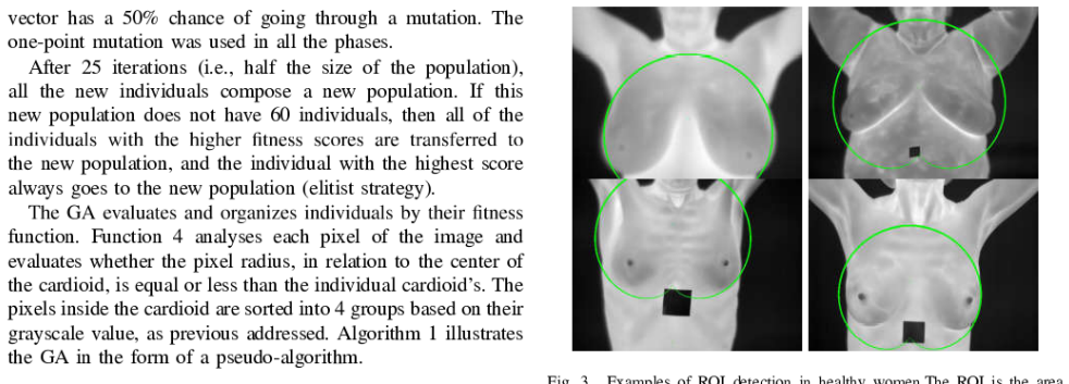
A genetic algorithm that uses pixel color information and a cardioid-based fitness function can delineate the breast region of interest in thermographic breast images, achieving correct separation in 52 out of 58 images without any manual seed-point input.
What carries the argument
Genetic algorithm search driven by a cardioid-shaped fitness function that scores candidate regions according to color consistency and boundary shape.
If this is right
- ROI extraction improves the accuracy of subsequent cancer detection steps.
- The method assists with standardization of image acquisition protocols.
- No manual seed points are required, making the pipeline fully automatic.
- This is presented as the first use of genetic algorithms and cardioids for this task.
Where Pith is reading between the lines
- The approach could reduce inter-operator variability that arises when clinicians choose seed points by hand.
- Similar shape-constrained genetic search might apply to other curved anatomical structures in thermal or infrared imaging.
- Parameter tuning of the genetic algorithm or the addition of texture features could raise the success rate above 52/58.
- The extracted ROIs could serve as input masks for training supervised classifiers on larger thermogram datasets.
Load-bearing premise
Breast regions in thermographic images can be effectively modeled and separated using a cardioid-based fitness function combined with color information alone.
What would settle it
Running the same algorithm on an independent collection of 100 thermographic breast images and measuring the fraction that produce accurate breast outlines without manual correction.
Figures
read the original abstract
This work proposes the use of Genetic Algorithms (GA) to identify the area of the breast from the background in thermographic breast images. The proposed method uses color information, a fitness function based on cardioids, and GA. This is the first work in the literature to propose a Region of Interest (ROI) extraction based on GA and cariods. ROI extraction can improve the accuracy of cancer detection and assist with the standardization of acquisition protocols. The method is able to successfully separate the breast region in 52 out of 58 images, while being fully automatic, and not requiring manual selection of seed points.
Editorial analysis
A structured set of objections, weighed in public.
Referee Report
Summary. The paper proposes a genetic algorithm (GA) method that combines color information with a cardioid-based fitness function to automatically extract the breast region of interest (ROI) from thermographic images. It claims this is the first such GA-and-cardioid approach in the literature and reports successful separation on 52 of 58 images without manual seed-point selection, with the goal of improving cancer-detection accuracy and standardizing acquisition protocols.
Significance. If properly validated, an automatic, seed-point-free ROI method could reduce operator dependence in breast thermography and support more reproducible downstream analysis. The approach is novel in its specific combination of GA search with a parametric cardioid model, but its practical significance cannot yet be assessed from the reported evidence.
major comments (2)
- [Abstract] Abstract: the headline claim of 'successful separation' in 52/58 images is unsupported by any quantitative validation metrics (Dice/Jaccard overlap, Hausdorff distance, or pixel-wise agreement against manual ground-truth delineations on the same 58 cases). Without these, it is impossible to determine whether the 52 successes meet a reproducible accuracy standard or are only visually plausible.
- [Abstract] Abstract: no description is given of how 'success' is defined, the image acquisition parameters or patient cohort for the 58 cases, the GA parameter settings, or any baseline comparison (e.g., thresholding, active contours, or other published thermographic ROI methods). These omissions make the central empirical claim impossible to evaluate or reproduce.
minor comments (1)
- [Abstract] Abstract contains the apparent typo 'cariods' (should be 'cardioids').
Simulated Author's Rebuttal
We thank the referee for the constructive comments on our manuscript. We address each major comment below and will revise the manuscript to improve the rigor and reproducibility of the reported results.
read point-by-point responses
-
Referee: [Abstract] Abstract: the headline claim of 'successful separation' in 52/58 images is unsupported by any quantitative validation metrics (Dice/Jaccard overlap, Hausdorff distance, or pixel-wise agreement against manual ground-truth delineations on the same 58 cases). Without these, it is impossible to determine whether the 52 successes meet a reproducible accuracy standard or are only visually plausible.
Authors: We agree that the manuscript currently relies on a count of visually successful cases without reporting quantitative overlap metrics. The 52/58 figure reflects cases where the extracted ROI appeared appropriate upon visual inspection. In the revised version we will compute and report Dice, Jaccard, and Hausdorff metrics against manual ground-truth delineations for the full set of 58 images. revision: yes
-
Referee: [Abstract] Abstract: no description is given of how 'success' is defined, the image acquisition parameters or patient cohort for the 58 cases, the GA parameter settings, or any baseline comparison (e.g., thresholding, active contours, or other published thermographic ROI methods). These omissions make the central empirical claim impossible to evaluate or reproduce.
Authors: We acknowledge that the abstract and current text omit these details. Success was defined by visual agreement with expected breast anatomy; the 58 images come from a clinical thermography dataset acquired under standard protocols. In revision we will expand the methods section with the definition of success, acquisition parameters, patient cohort description, GA settings (population size, generations, operators), and comparisons against baseline methods such as simple thresholding and active contours. revision: yes
Circularity Check
No circularity: purely empirical GA application with no derivation chain
full rationale
The paper presents an application of genetic algorithms to segment thermographic breast images using a cardioid-based fitness function and color cues. No equations, first-principles derivations, or mathematical predictions are present in the abstract or described method. The central claim (successful separation in 52/58 images) is an empirical outcome evaluated by visual inspection rather than any reduction of a fitted parameter or self-referential definition back to inputs. No self-citation load-bearing steps, uniqueness theorems, or ansatz smuggling occur. The work is self-contained as a practical implementation without any claimed derivation that could be circular.
Axiom & Free-Parameter Ledger
axioms (1)
- domain assumption Breast regions in thermographic images can be modeled using cardioid shapes for fitness evaluation
Reference graph
Works this paper leans on
-
[1]
Nimmi Arora, Diana Martins, Danielle Ruggerio, Eleni Tousimis, Alexan- der . Swistel, Michael P. Osbome, and Rache M. Simmons. Effectiveness of a noninvasive digital infrared thermal imaging system in the detection of breast cancer. The American Journal of Surgery, 196(4):523 — 526, 2008 Deborah A. Kennedy, Tanya Lee, and Dugald Seely. A comparative re- v...
work page 2008
-
[2]
Digital Image Processing and Pattern Recognition Techniques for the Detection of Cancer. U. Rajendra Acharya, E. Y. K. Ng, Jen-Hong Tan, and S. Vinitha Sree. Thermography based breast cancer detection using texture features and support vector machine. Journal of Medical Systems, 36(3): 15031510, 2012 World Health Organization. Breast cancer,
work page 2012
-
[3]
[Online; accessed 08- November-2019]. E L. Silva, D. C. M. Saade, G. O. Sequeiros, A. C. Silva, A. C. Paiva, R. S. Bravo, and A. Conci. A New Database for Breast Research with Infrared Image. Journal of Medical Imaging and Health Informatics, 4(1)92-100,
work page 2019
-
[4]
E. O. Rodrigues, A. Conci, TB. Borchart, A.C. Paiva, A.C. Silva, and T. MacHenry. Comparing results of thermographic images based diagnosis for breast diseases. IWSSIP 2014 Proceedings, pages 39-42, 2014, E. O. Rodrigues, P. Liatsis, L. Satoru, and A. Conci. Fractal triangular search: a metaheurisic for image content search. IET Image Processing, 12:1475-1484,
work page 2014
-
[5]
Richard A. Caruana and J. David Schaffer. Representation and hidden bias: Gray vs. binary coding for genetic algorithms. In John Laird, editor, Machine Learning Proceedings 1988, pages 153 —
work page 1988
-
[6]
Springer London. É. O. Rodriguesi. Combining Minkowski and Cheyshev: New distance proposal and survey of distance mefrics using k-nearest neighbours classifier. Pattern Recognition Letters, 110:66-71, 2018
work page 2018
discussion (0)
Sign in with ORCID, Apple, or X to comment. Anyone can read and Pith papers without signing in.