Towards 3D heart mesh generation using contactless radar imaging and physics-informed neural network
Pith reviewed 2026-06-29 23:11 UTC · model grok-4.3
The pith
SAR2Mesh generates accurate 3D cardiac meshes from mmWave radar SAR images by deforming an anatomical template under a physics-informed loss.
A machine-rendered reading of the paper's core claim, the machinery that carries it, and where it could break.
Core claim
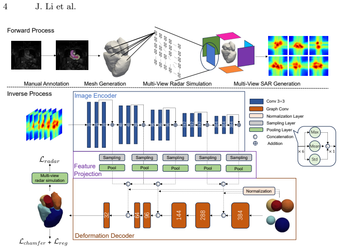
SAR2Mesh reformulates 3D cardiac mesh generation as a coarse-to-fine deformation process starting from a topological template. It uses a geometry-aware feature projection module to extract multi-view features via 3D-to-2D sampling and a physics-informed radar loss to enforce consistency between the predicted geometry and raw radar echoes. This produces anatomically correct and physically consistent meshes on the new Cardiac Mesh-SAR dataset and outperforms existing image-based baselines.
What carries the argument
Coarse-to-fine mesh deformation initialized from a topological template, driven by a geometry-aware feature projection module and a physics-informed radar loss.
If this is right
- The resulting meshes preserve anatomical connectivity through the deformation process.
- Reconstructions remain consistent with the original radar echo data.
- The method works despite speckle noise and ambiguous boundaries in SAR images.
- A large paired dataset enables training and evaluation of such models.
Where Pith is reading between the lines
- This approach could support continuous at-home heart monitoring if integrated with wearable radar devices.
- The physics loss might generalize to other radar-based imaging tasks like lung or liver reconstruction.
- Future work could test whether the meshes improve downstream tasks such as ejection fraction calculation.
Load-bearing premise
Initializing from a topological template and using progressive deformation with a physics loss will yield correct heart shapes even when SAR images have severe noise and unclear edges.
What would settle it
A direct comparison showing that the predicted meshes deviate significantly from MRI-derived ground truth shapes or fail to match the radar signals in withheld test cases would disprove the claim.
Figures
read the original abstract
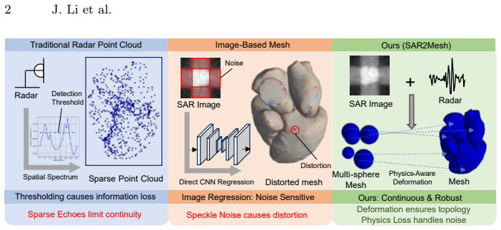
Cardiac function evaluation necessitates continuous, non-invasive monitoring, a capability limited in MRI. Millimeter-wave (mmWave) radar and its Synthetic Aperture Radar (SAR) mode offer a privacy-preserving and portable point-of-care clinical applications. However, reconstructing high-fidelity 3D cardiac geometry from SAR remains an open challenge. Traditional radar methods generate sparse point clouds that lack continuous surface topology. Meanwhile, direct application of optical reconstruction networks performs poorly due to the severe speckle noise and ambiguous boundaries inherent in SAR images. To bridge this gap, we propose SAR2Mesh, a novel framework that reformulates the task as a coarse-to-fine mesh deformation process. By initializing with a topological template, our approach explicitly preserves anatomical connectivity through progressive mesh deformation.We introduce a geometry-aware feature projection module to extract multi-view features via 3D-to-2D sampling, and a physics-informed radar loss to enforce consistency between the predicted geometry and raw radar echoes. Furthermore, we present Cardiac Mesh-SAR, the first large-scale paired SAR-mesh dataset. Extensive experiments demonstrate that SAR2Mesh significantly outperforms existing image-based baselines, achieving accurate and physically consistent cardiac reconstructions.
Editorial analysis
A structured set of objections, weighed in public.
Referee Report
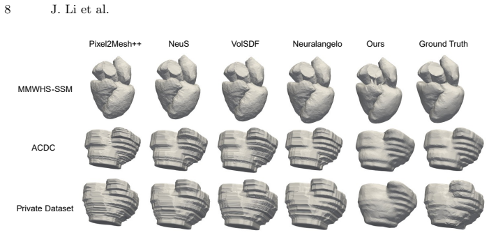
Summary. The paper introduces SAR2Mesh, a framework that reformulates 3D cardiac mesh reconstruction from mmWave SAR images as a coarse-to-fine mesh deformation process. It initializes from a topological template, applies progressive deformation with a geometry-aware feature projection module for multi-view 3D-to-2D sampling, and uses a physics-informed radar loss to enforce consistency with raw radar echoes. The authors also release the Cardiac Mesh-SAR paired dataset and claim that SAR2Mesh significantly outperforms image-based baselines in producing accurate and physically consistent reconstructions.
Significance. If the performance and consistency claims hold with quantitative validation, the work would address a clear gap in portable, privacy-preserving cardiac imaging by moving beyond sparse point clouds to topologically correct meshes. The dataset release would be a concrete enabling contribution for the community.
major comments (2)
- [Abstract] Abstract: the central claim that the physics-informed radar loss produces 'physically consistent' reconstructions cannot be evaluated because no loss formulation, weighting terms, or radar forward model is provided; without these it is impossible to determine whether the loss is load-bearing or reduces to a data-fitting term by construction.
- [Abstract] Abstract (method paragraph): the assertion that template initialization plus progressive deformation will overcome severe speckle noise and ambiguous boundaries is presented without any supporting derivation, noise model, or preliminary result; this is the load-bearing assumption for the entire pipeline yet remains untested in the given text.
minor comments (1)
- [Abstract] The abstract states 'extensive experiments' but supplies no metrics, baselines, error bars, or ablation results, making the performance claim impossible to assess.
Simulated Author's Rebuttal
We thank the referee for their constructive comments. We address each major comment point by point below, proposing revisions to the abstract where the concerns are valid.
read point-by-point responses
-
Referee: [Abstract] Abstract: the central claim that the physics-informed radar loss produces 'physically consistent' reconstructions cannot be evaluated because no loss formulation, weighting terms, or radar forward model is provided; without these it is impossible to determine whether the loss is load-bearing or reduces to a data-fitting term by construction.
Authors: The referee is correct that the abstract does not contain the explicit loss formulation, weighting terms, or radar forward model. These details appear in Section 3.3 of the manuscript. We will revise the abstract to include a concise statement of the loss components and forward model so that the physical-consistency claim can be evaluated directly from the abstract. revision: yes
-
Referee: [Abstract] Abstract (method paragraph): the assertion that template initialization plus progressive deformation will overcome severe speckle noise and ambiguous boundaries is presented without any supporting derivation, noise model, or preliminary result; this is the load-bearing assumption for the entire pipeline yet remains untested in the given text.
Authors: The referee correctly observes that the abstract states the assumption without supporting material. The noise model, derivation of the progressive deformation strategy, and preliminary results appear in Sections 2 and 4.1. We will revise the abstract to add a brief reference to this supporting analysis, making the assumption testable from the abstract alone. revision: yes
Circularity Check
No significant circularity detected
full rationale
The abstract outlines a pipeline of template initialization, progressive mesh deformation, geometry-aware projection, and a physics-informed radar loss, plus a new paired dataset. No equations, derivations, or self-citations are presented that reduce any claimed prediction or result to a fitted input or prior self-result by construction. The central claim of outperformance is presented as empirically testable on external data and does not rely on load-bearing self-citations or self-definitional steps. This is the normal case of a self-contained empirical method description.
Axiom & Free-Parameter Ledger
Reference graph
Works this paper leans on
-
[1]
Right ventricle segmentation via registrationandmulti-inputmodalitiesincardiacmagneticresonanceimagingfrom multi-disease, multi-view and multi-center,
X. Sun, L.-H. Cheng, and R. J. van der Geest, “Right ventricle segmentation via registrationandmulti-inputmodalitiesincardiacmagneticresonanceimagingfrom multi-disease, multi-view and multi-center,” inInternational Workshop on Statisti- cal Atlases and Computational Models of the Heart. Springer, 2021, pp. 241–249
2021
-
[2]
Random style transfer based domain generalization networks integrating shape and spatial information,
L. Li, V. A. Zimmer, W. Ding, F. Wu, L. Huang, J. A. Schnabel, and X. Zhuang, “Random style transfer based domain generalization networks integrating shape and spatial information,” inInternational Workshop on Statistical Atlases and Computational Models of the Heart. Springer, 2020, pp. 208–218
2020
-
[3]
Modelling cardiac motion via spatio- temporal graph convolutional networks to boost the diagnosis of heart conditions,
P. Lu, W. Bai, D. Rueckert, and J. A. Noble, “Modelling cardiac motion via spatio- temporal graph convolutional networks to boost the diagnosis of heart conditions,” inInternational Workshop on Statistical Atlases and Computational Models of the Heart. Springer, 2020, pp. 56–65
2020
-
[4]
Studying robustness of semantic segmentation under domain shift in cardiac mri,
P. M. Full, F. Isensee, P. F. Jäger, and K. Maier-Hein, “Studying robustness of semantic segmentation under domain shift in cardiac mri,” inInternational Work- shop on Statistical Atlases and Computational Models of the Heart. Springer, 2020, pp. 238–249
2020
-
[5]
A generalizable deep-learning approach for cardiac magnetic resonance image segmentation using image augmentation and attention u-net,
F. Kong and S. C. Shadden, “A generalizable deep-learning approach for cardiac magnetic resonance image segmentation using image augmentation and attention u-net,” inInternational Workshop on Statistical Atlases and Computational Models of the Heart. Springer, 2020, pp. 287–296
2020
-
[6]
Multiple emitter location and signal parameter estimation,
R. Schmidt, “Multiple emitter location and signal parameter estimation,”IEEE transactions on antennas and propagation, vol. 34, no. 3, pp. 276–280, 1986
1986
-
[7]
Esprit-estimation of signal parameters via rotational in- variance techniques,
R. Roy and T. Kailath, “Esprit-estimation of signal parameters via rotational in- variance techniques,”IEEE Transactions on acoustics, speech, and signal process- ing, vol. 37, no. 7, pp. 984–995, 2002
2002
-
[8]
Pixel2mesh++: Multi-view 3d mesh gener- ation via deformation,
C. Wen, Y. Zhang, Z. Li, and Y. Fu, “Pixel2mesh++: Multi-view 3d mesh gener- ation via deformation,” inProceedings of the IEEE/CVF international conference on computer vision, 2019, pp. 1042–1051. 10 J. Li et al
2019
-
[9]
Nerf: Representing scenes as neural radiance fields for view synthesis,
B. Mildenhall, P. P. Srinivasan, M. Tancik, J. T. Barron, R. Ramamoorthi, and R. Ng, “Nerf: Representing scenes as neural radiance fields for view synthesis,” Communications of the ACM, vol. 65, no. 1, pp. 99–106, 2021
2021
-
[10]
Instant neural graphics primi- tives with a multiresolution hash encoding,
T. Müller, A. Evans, C. Schied, and A. Keller, “Instant neural graphics primi- tives with a multiresolution hash encoding,”ACM transactions on graphics (TOG), vol. 41, no. 4, pp. 1–15, 2022
2022
-
[11]
Volume rendering of neural implicit surfaces,
L. Yariv, J. Gu, Y. Kasten, and Y. Lipman, “Volume rendering of neural implicit surfaces,”Advances in neural information processing systems, vol. 34, pp. 4805– 4815, 2021
2021
-
[12]
NeuS: Learning Neural Implicit Surfaces by Volume Rendering for Multi-view Reconstruction
P. Wang, L. Liu, Y. Liu, C. Theobalt, T. Komura, and W. Wang, “Neus: Learning neural implicit surfaces by volume rendering for multi-view reconstruction,”arXiv preprint arXiv:2106.10689, 2021
work page internal anchor Pith review Pith/arXiv arXiv 2021
-
[13]
Neuralangelo: High-fidelity neural surface reconstruction,
Z. Li, T. Müller, A. Evans, R. H. Taylor, M. Unberath, M.-Y. Liu, and C.-H. Lin, “Neuralangelo: High-fidelity neural surface reconstruction,” inProceedings of the IEEE/CVF conference on computer vision and pattern recognition, 2023, pp. 8456–8465
2023
-
[14]
A coherent through-wall mimo phased array imaging radar based on time-duplexed switching,
Q. Chen, K. Chetty, P. Brennan, L. B. Lok, M. Ritchie, and K. Woodbridge, “A coherent through-wall mimo phased array imaging radar based on time-duplexed switching,” inRadar Sensor Technology XXI, vol. 10188. SPIE, 2017, pp. 74–84
2017
-
[15]
Open- source 4d statistical shape model of the heart for x-ray projection imaging,
M. Unberath, A. Maier, D. Fleischmann, J. Hornegger, and R. Fahrig, “Open- source 4d statistical shape model of the heart for x-ray projection imaging,” in 2015 IEEE 12th International Symposium on Biomedical Imaging (ISBI). IEEE, 2015, pp. 739–742
2015
-
[16]
Multi-scale patch and multi-modality atlases for whole heart segmentation of mri,
X. Zhuang and J. Shen, “Multi-scale patch and multi-modality atlases for whole heart segmentation of mri,”Medical image analysis, vol. 31, pp. 77–87, 2016
2016
-
[17]
Deep learning techniques for automatic mri cardiac multi-structures segmentation and diagnosis: is the problem solved?
O. Bernard, A. Lalande, C. Zotti, F. Cervenansky, X. Yang, P.-A. Heng, I. Cetin, K. Lekadir, O. Camara, M. A. G. Ballesteret al., “Deep learning techniques for automatic mri cardiac multi-structures segmentation and diagnosis: is the problem solved?”IEEE transactions on medical imaging, vol. 37, no. 11, pp. 2514–2525, 2018
2018
-
[18]
Cinemyops: Segmenting myocardial pathologies from cine cardiac mr,
W. Ding, L. Li, J. Qiu, B. Lin, M. Yang, L. Huang, L. Wu, S. Wang, and X. Zhuang, “Cinemyops: Segmenting myocardial pathologies from cine cardiac mr,” IEEE Transactions on Medical Imaging, 2025
2025
-
[19]
a delaunay-based quality tetrahedral mesh generatoracm,
S. HTetGen, “a delaunay-based quality tetrahedral mesh generatoracm,”Trans. Math. Softw, vol. 201541211, pp. 1–11
discussion (0)
Sign in with ORCID, Apple, or X to comment. Anyone can read and Pith papers without signing in.