Multi-Class Brain Tumor Classification Using Advanced Deep Learning Models: A Comparative Study
Pith reviewed 2026-06-26 21:36 UTC · model grok-4.3
The pith
EfficientNetB0 reaches 95 percent accuracy on multi-class brain tumor classification from MRI and lifts meningioma recall from 20 to 89 percent.
A machine-rendered reading of the paper's core claim, the machinery that carries it, and where it could break.
Core claim
EfficientNetB0 supplies the best overall performance for multi-class brain tumor classification on MRI, recording 95 percent accuracy together with an 89 percent recall rate on meningiomas, while the same experimental conditions show that model efficiency can outweigh greater depth.
What carries the argument
Side-by-side training and evaluation of VGG16, VGG19, DenseNet121, EfficientNetB0, and a custom CNN on identical MRI data using accuracy plus per-tumor recall as the performance measures.
If this is right
- EfficientNetB0 supplies the strongest practical option among the tested models for reducing missed meningioma cases.
- Architectural efficiency can be more decisive than network depth when classifying medical images.
- Pre-trained models deliver substantially higher per-class recall than a simple custom CNN on this task.
- The reported ranking of the five architectures rests on both aggregate accuracy and clinically relevant tumor-specific recall.
Where Pith is reading between the lines
- Hospitals could adopt EfficientNetB0 as a first-line screening tool to lower the rate of undetected meningiomas.
- The depth-versus-efficiency observation may generalize to other subtle-feature medical imaging problems.
- Repeating the comparison on multi-center datasets would test whether the 95 percent figure and meningioma recall hold outside the original collection.
Load-bearing premise
The same training setup applied to all models on a single 10,000-image dataset produces performance differences that will hold for other clinical MRI collections.
What would settle it
Retraining the five models on an independent collection of MRI scans and observing that any architecture other than EfficientNetB0 exceeds 95 percent accuracy or that its meningioma recall falls below 80 percent.
Figures
read the original abstract
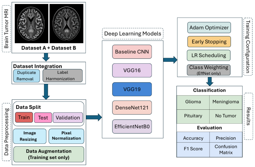
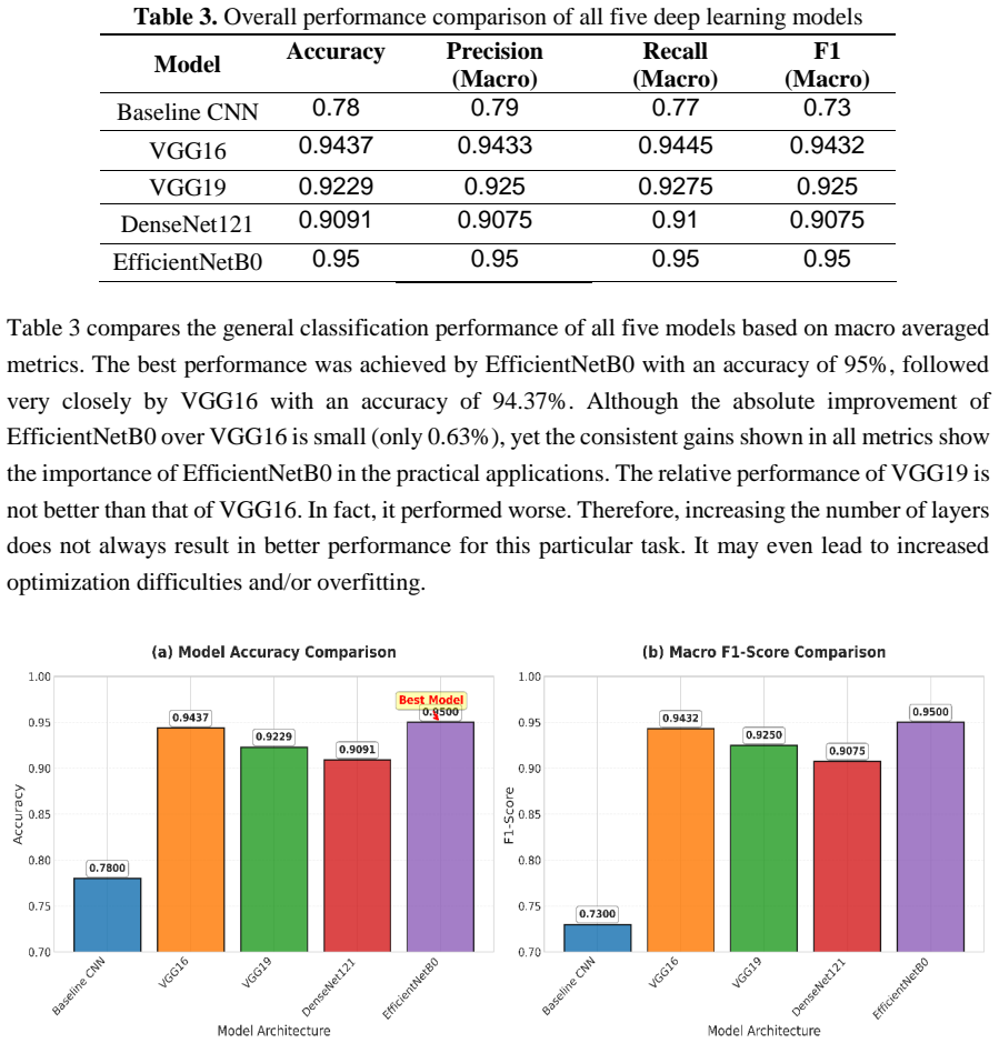
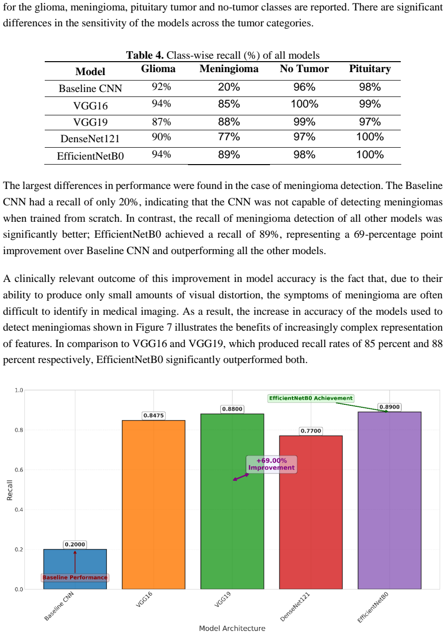
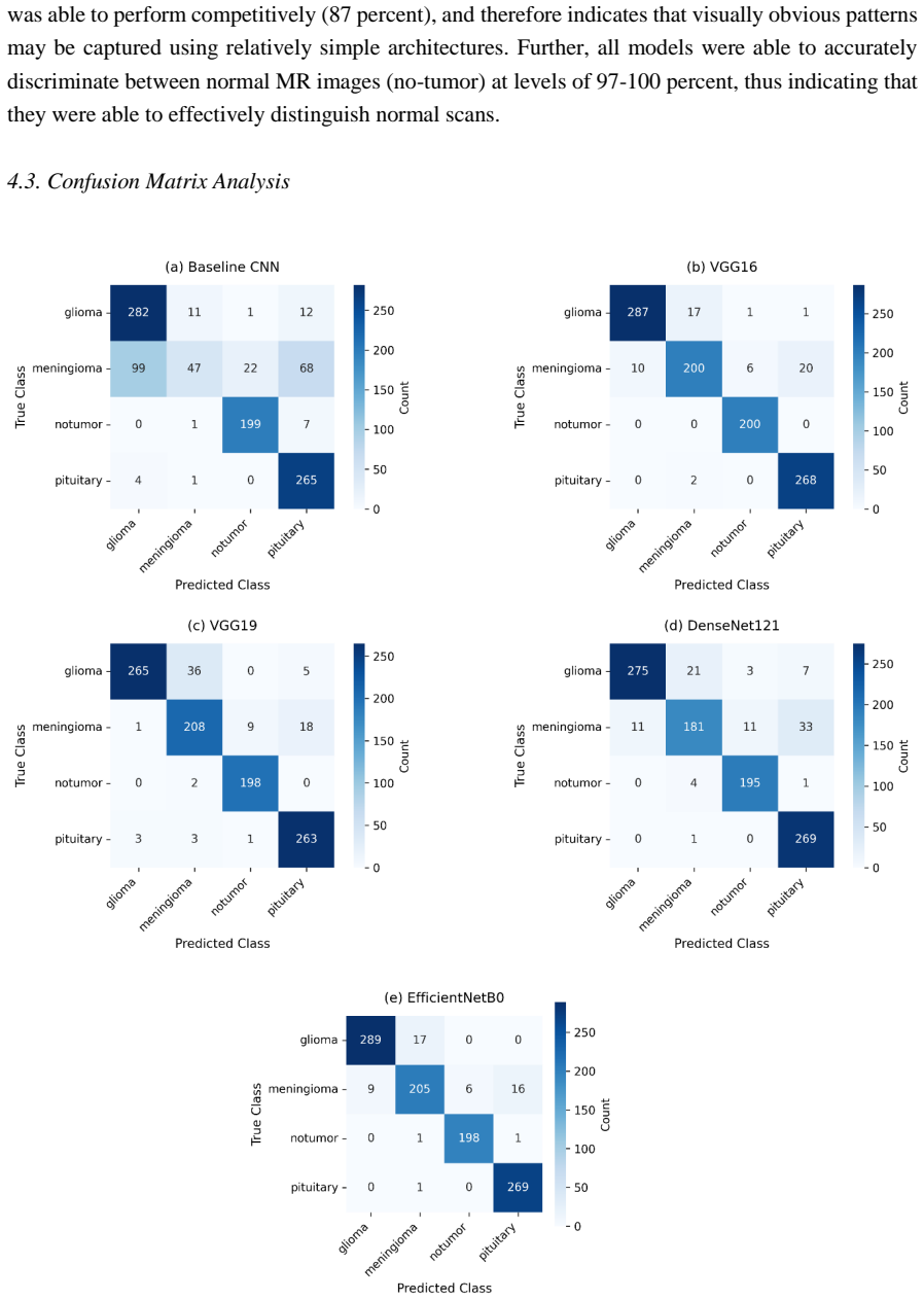
Despite recent advancements in deep learning, accurately classifying brain tumors from MRI images continues to pose challenges. In this research, we present a comprehensive evaluation of five different convolutional neural networks (CNN) architectures, including a customized baseline model and four pre-trained models - for use in classifying multi-class brain tumors using a clinically-sourced dataset of approximately 10,000 MRI images. We have utilized five different architectures; VGG16, VGG19, DenseNet121, and EfficientNetB0, which were all tested and trained within an identical experimental framework. Performance was measured by both overall accuracy and tumor-wise recall as a means to measure the clinically-relevant performance of each architecture. We found that EfficientNetB0 had the best overall classification accuracy at 95%, when compared to the other architectures tested; specifically VGG16 (94.37%), VGG19 (92.29%), DenseNet121 (90.91%) and the customized CNN (78.00%). An especially important finding of our research was the considerable improvement in detecting meningiomas; specifically, while simple CNNs could detect meningiomas with a recall rate of approximately 20%, EfficientNetB0 was able to detect meningiomas with a recall rate of 89%. Meningiomas are often difficult to detect because they can appear very subtly on MRI images. Additionally, an interesting finding was that the deeper VGG19 performed worse than the shallower VGG16. This indicates that in many cases the architectural efficiency of a CNN model may be more important than its depth when working with medical images. Overall, EfficientNetB0 appears to provide the optimal trade-off between classification accuracy, number of parameters used in the model and clinically meaningful performance.
Editorial analysis
A structured set of objections, weighed in public.
Referee Report
Summary. The manuscript evaluates five CNN architectures (a custom baseline CNN plus VGG16, VGG19, DenseNet121, and EfficientNetB0) for multi-class brain tumor classification on a clinically sourced dataset of approximately 10,000 MRI images. All models are stated to have been trained and tested under an identical experimental framework. The central empirical claims are that EfficientNetB0 attains the highest overall accuracy (95 %) and markedly higher meningioma recall (89 %) than the custom CNN (~20 %), with secondary observations that VGG16 outperforms the deeper VGG19 and that EfficientNetB0 offers the best accuracy–parameter trade-off.
Significance. If the reported performance differences can be reproduced under documented, identical conditions and on a properly stratified dataset, the results would supply a concrete, clinically relevant ranking of standard architectures for meningioma detection, where subtle appearance makes recall especially important. The finding that architectural efficiency can outweigh depth on medical images is also potentially useful for resource-constrained clinical deployment.
major comments (3)
- [Abstract] Abstract and Methods: the abstract reports accuracy and recall figures (EfficientNetB0 95 %, meningioma recall 89 %) but supplies no information on train/validation/test splits, cross-validation procedure, or class-imbalance handling. Without these details the central performance claims cannot be verified or compared across architectures.
- [Abstract] Abstract: the claim that all five architectures were evaluated 'within an identical experimental framework' is load-bearing for attributing the observed differences (e.g., 95 % vs. 78 %) to model choice rather than to differing preprocessing, data augmentation, learning-rate schedules, or early-stopping criteria; no supporting protocol is provided.
- [Abstract] Abstract: meningioma recall is highlighted as clinically important, yet no per-class precision, F1, or confusion-matrix data are supplied, nor is any statistical significance test reported for the 89 % vs. ~20 % difference, leaving the strength of the improvement unquantified.
minor comments (2)
- [Abstract] The exact dataset size, class distribution, and source (hospital, scanner types) are given only approximately; these details are needed for reproducibility and to assess generalizability.
- [Abstract] No mention is made of whether the pre-trained models were fine-tuned from ImageNet weights or trained from scratch, nor of the optimizer, batch size, or number of epochs used.
Simulated Author's Rebuttal
We thank the referee for the constructive comments highlighting the need for greater methodological transparency in the abstract. We agree that these details strengthen the manuscript and will revise the abstract and, where appropriate, the methods section to incorporate the requested information on splits, protocols, and metrics.
read point-by-point responses
-
Referee: [Abstract] Abstract and Methods: the abstract reports accuracy and recall figures (EfficientNetB0 95 %, meningioma recall 89 %) but supplies no information on train/validation/test splits, cross-validation procedure, or class-imbalance handling. Without these details the central performance claims cannot be verified or compared across architectures.
Authors: We agree these details belong in the abstract. The dataset was partitioned via stratified sampling into 70 % training, 15 % validation and 15 % test sets to preserve class proportions; no cross-validation was performed owing to computational cost. We will add a concise statement of the split and stratification procedure to the abstract. revision: yes
-
Referee: [Abstract] Abstract: the claim that all five architectures were evaluated 'within an identical experimental framework' is load-bearing for attributing the observed differences (e.g., 95 % vs. 78 %) to model choice rather than to differing preprocessing, data augmentation, learning-rate schedules, or early-stopping criteria; no supporting protocol is provided.
Authors: All models shared identical preprocessing (resize to 224×224, ImageNet mean/std normalization), the same augmentation pipeline (random rotation ±15°, horizontal flip, zoom 0.8–1.2), Adam optimizer (lr = 0.001, batch size 32), and early-stopping patience of 10 epochs on validation loss. We will insert a brief protocol summary into the abstract. revision: yes
-
Referee: [Abstract] Abstract: meningioma recall is highlighted as clinically important, yet no per-class precision, F1, or confusion-matrix data are supplied, nor is any statistical significance test reported for the 89 % vs. ~20 % difference, leaving the strength of the improvement unquantified.
Authors: We will add per-class precision and F1 scores for all models to the abstract. The full manuscript already contains the confusion matrix; we will ensure it is clearly referenced. No statistical significance test was performed originally; we can report the raw difference magnitude or add a post-hoc test in revision if the editor deems it essential. revision: partial
Circularity Check
No significant circularity
full rationale
The paper reports empirical classification accuracies and recall rates from training five CNN architectures (VGG16, VGG19, DenseNet121, EfficientNetB0, and a custom CNN) on a fixed ~10k-image MRI dataset under an asserted identical experimental framework. No equations, derivations, fitted parameters, or predictions are presented that could reduce to inputs by construction. All load-bearing claims rest on measured performance differences, with no self-citation chains, ansatzes, or uniqueness theorems invoked. This is a standard empirical comparison study with no circularity.
Axiom & Free-Parameter Ledger
axioms (2)
- domain assumption The MRI images are independent and identically distributed within each tumor class.
- domain assumption The training procedure (optimizer, learning rate schedule, data augmentation) is identical and fair across all five models.
Reference graph
Works this paper leans on
-
[1]
Dorfner, F.J., Patel, J.B., Kalpathy -Cramer, J. et al. A review of deep learning for brain tumor analysis in MRI. npj Precis. Onc. 9, 2 (2025). https://doi.org/10.1038/s41698-024-00789-2
-
[2]
Price M, Ballard C, Benedetti J, et al. CBTRUS Statistical Report: Primary Brain and Other Central Nervous System Tumors Diagnosed in the Uni ted States in 2017 -2021. Neuro Oncol. 2024;26(Supplement_6):vi1-vi85. doi:10.1093/neuonc/noae145
-
[3]
Assessing inter-rater reliability of MRI features in glioma: a multi -radiologist agre ement study
Mohebbi A, Mohammadzadeh S, Zare AH, Moradi Z, Ardakani AA, Mohammadi A. Assessing inter-rater reliability of MRI features in glioma: a multi -radiologist agre ement study. BMC Medical Imaging. 2025 Dec;25(1):480
2025
-
[4]
Stathopoulos I, Serio L, Karavasilis E, et al. Evaluating Brain Tumor Detection with Deep Learning Convolutional Neural Networks Across Multiple MRI Modalities. J Imaging . 2024;10(12):296. Published 2024 Nov 21. doi:10.3390/jimaging10120296
-
[5]
Iris van der Loo, Teresa M. Tareco Bucho, James A. Hanley, Regina G.H. Beets-Tan, Alex L.T. Imholz, Stefano Trebeschi, Measurement variability of radiologists when measuring brain tumors, European Journal of Radiology , Volume 183, 2025, 111874, ISSN 0720-048X, https://doi.org/10.1016/j.ejrad.2024.111874
-
[6]
Shiri I, Hajianfar G, Sohrabi A, et al. Repeatability of radiomic features in magnetic resonance imaging of glioblastoma: Test -retest and image registration analyses. Med Phys . 2020;47(9):4265-4280. doi:10.1002/mp.14368
-
[7]
Laustsen AF, Dineen R, Ilginiene J, et al. Interobserver variabil ity in assessing preoperative imaging biomarkers for cerebellar mutism syndrome: a multiobserver pilot study. Pediatric Radiology 2025;55:1915–1926. https://doi.org/10.1007/s00247-025-06326-y
-
[8]
Salama A, Daradkeh YI, Javeed D, Ur Rehman A, Hamam H
Raza A, Ayub H, Khan JA, Ahmad I, S. Salama A, Daradkeh YI, Javeed D, Ur Rehman A, Hamam H. A hybrid deep learning -based approach for brain tumor classification. Electronics. 2022 Apr 5;11(7):1146
2022
-
[9]
Enhanced brain tumor classification using graph convolutional neural network architecture
Ravinder M, Saluja G, Allabun S, Alqahtani MS, Abbas M, Othman M, Soufiene BO. Enhanced brain tumor classification using graph convolutional neural network architecture. Scientific Reports. 2023 Sep 11;13(1):14938
2023
-
[10]
Brain tumor classification from MRI using image enhancement and convolutional neural network techniques
Rasheed Z, Ma YK, Ullah I, Ghadi YY, Khan MZ, Khan MA, Abdusalomov A, Alqahtani F, Shehata AM. Brain tumor classification from MRI using image enhancement and convolutional neural network techniques. Brain Sciences. 2023 Sep 14;13(9):1320
2023
-
[11]
Convolutional Neural Network in Medical Image Analysis: A Review
Kshatri, S.S., Singh, D. Convolutional Neural Network in Medical Image Analysis: A Review. Arch Computat Methods Eng 30, 2793 –2810 (2023). https://doi.org/10.1007/s11831-023-09898-w
-
[12]
Development of hybrid models based on deep learning and optimized machine learning algorithms for brain tumor Multi -Classification
Celik M, Inik O. Development of hybrid models based on deep learning and optimized machine learning algorithms for brain tumor Multi -Classification. Expert Systems with Applications. 2024 Mar 15;238:122159
2024
-
[13]
T1 -weighted MRI-based brain tumor classification using hybrid deep learning models
Ilani MA, Shi D, Banad YM. T1 -weighted MRI-based brain tumor classification using hybrid deep learning models. Scientific Reports. 2025 Feb 27;15(1):7010
2025
-
[14]
Saeedi S, Rezayi S, Keshavarz H, R. Niakan Kalhori S. MRI-based brain tumor detection using convolutional deep learning methods and chosen machine learning techniques. BMC Medical Informatics and Decision Making. 2023 Jan 23;23(1):16. https://doi.org/10.1186/s12911-023-02114-6
-
[15]
Imagenet: A large-scale hierarchical image database
Deng J, Dong W, Socher R, Li LJ, Li K, Fei -Fei L. Imagenet: A large-scale hierarchical image database. In2009 IEEE conference on computer vision and pattern recognition 2009 Jun 20 (pp. 248-255). Ieee
2009
-
[16]
Brain tumor classification for MR images using transfer learning and fine -tuning
Swati ZN, Zhao Q, Kabir M, Ali F, Ali Z, Ahmed S, Lu J. Brain tumor classification for MR images using transfer learning and fine -tuning. Computerized Medical Imaging and Graphics. 2019 Jul 1;75:34-46
2019
-
[17]
Disci R, Gurcan F, Soylu A. Advanced brain tumor classi fication in MR images using transfer learning and pre -trained deep CNN models. Cancers. 2025 Jan 2;17(1):121. https://doi.org/10.3390/cancers17010121
-
[18]
Transfer learning for medical image classification: a literature review
Kim HE, Cosa-Linan A, Santhanam N, Jannesari M, Maros ME, Ganslandt T. Transfer learning for medical image classification: a literature review. BMC medical imaging. 2022 Apr 13;22(1):69. https://doi.org/10.1186/s12880-022-00793-7
-
[19]
Brain tumor classification using VGG 16, ResNet50, and inception V3 transfer learning models
Pillai R, Sharma A, Sharma N, Gupta R. Brain tumor classification using VGG 16, ResNet50, and inception V3 transfer learning models. In2023 2nd International Conference for Innovation in Technology (INOCON) 2023 Mar 3 (pp. 1-5). IEEE
2023
-
[20]
Abnormal brain tumors classification using resnet50 and its comprehensive evaluation
Younis A, Li Q, Afzal Z, Adamu MJ, Kawuwa HB, Hussain F, Hussain H. Abnormal brain tumors classification using resnet50 and its comprehensive evaluation. IEEE Access. 2024 May 21;12:78843-53
2024
-
[21]
Brainnet: precision brain tumor classification with optimized efficientnet architecture
Islam MM, Talukder MA, Uddin MA, Akhter A, Khalid M. Brainnet: precision brain tumor classification with optimized efficientnet architecture. International Journal of Intelligent Systems. 2024;2024(1):3583612
2024
-
[22]
Comparative study on architecture of deep neural networks for segmentation of brain tumor using magnetic resonance images
Preetha R, Priyadarsini MJ, Nisha JS. Comparative study on architecture of deep neural networks for segmentation of brain tumor using magnetic resonance images. IEEE Access. 2023 Dec 7;11:138549-67
2023
-
[23]
Multiple Brain Tumor Classification with Dense CNN Architecture Using Brain MRI Images
Özkaraca, O.; Bağrıaçık, O.İ.; Gürüler, H.; Khan, .; Hussain, J.; Khan, J.; Laila, U.e. Multiple Brain Tumor Classification with Dense CNN Architecture Using Brain MRI Images. Life 2023, 13, 349. https://doi.org/10.3390/life13020349
-
[24]
Irmak E. Multi -classification of brain tumor MRI images using deep convolutional neural network with fully optimized framework. Iranian Journal of Science and Technology, Transactions of Electrical Engineering. 2021 Sep;45(3):1015 -36. https://doi.org/10.1007/s40998-021-00426-9
-
[25]
Brain tumour detection using machine and deep learning: a systematic review
Rasool N, Bhat JI. Brain tumour detection using machine and deep learning: a systematic review. Multimedia tools and applications. 2025 Apr;84(13):11551-604
2025
-
[26]
Convolutional neural networks in medical image understanding: a survey
Sarvamangala DR, Kulkarni RV. Convolutional neural networks in medical image understanding: a survey. Evolutionary intelligence. 2022 Mar;15(1):1 -22. https://doi.org/10.1007/s12065-020-00540-3
-
[27]
Susanto, A.; Sari, C.A.; Rahmalan, H.; Doheir, M. a. S. Support Vector Machine Based Discrete Wavelet Transform for Magnetic Resonance Imaging Brain Tumor Classification. TELKOMNIKA (Telecom munication Computing Electronics and Control) 2023, 21, 592, doi:10.12928/telkomnika.v21i3.24928
-
[28]
Early Brain Tumor Detection Using Random Forest Classification
Upadhyay, A.; Palival, U.; Jaiswal, S. Early Brain Tumor Detection Using Random Forest Classification. In Advances in intelligent systems and computing; 2020; pp. 258–264
2020
-
[29]
-N.; Guillevin, C.; Lallouette, A.; Thomas, C.; Rittano, G.; Wager, M.; Guillevin, R.; Vallée, A
Vallée, R.; Vallée, J. -N.; Guillevin, C.; Lallouette, A.; Thomas, C.; Rittano, G.; Wager, M.; Guillevin, R.; Vallée, A. Machine Learning Decision Tree Models for Multiclass Classification of Common Malignant Brain Tumors Using Perfusion and Spectroscopy MRI Data. Frontiers in Oncology 2023, 13, doi:10.3389/fonc.2023.1089998
-
[30]
Brain tumor detection from MRI images using Naive classifier
Divyamary D, Gopika S, Pradeeba S, Bhuvaneswari M. Brain tumor detection from MRI images using Naive classifier. In2020 6th international conference on advanced computing and communication systems (ICACCS) 2020 Mar 6 (pp. 620-622). IEEE
2020
-
[31]
Jiang, S.; Gu, Y.; Kumar, E. Magnetic Resonance Imaging (MRI) Brain Tumor Image Classification Based on Five Machine Learning Algorithms. Cloud Computing and Data Science 2023, 122–133, doi:10.37256/ccds.4220232740
-
[32]
and Mengko, T.R., 2018, May
Abiwinanda, N., Hanif, M., Hesaputra, S.T., Handayani, A. and Mengko, T.R., 2018, May. Brain tumor classification using convolutional neural network. In World Congress on Medical Physics and Biomedical Engineering 2018: June 3 –8, 2018, Prague, Czech Republic (Vol. 1) (pp. 183-189). Singapore: Springer Nature Singapore
2018
-
[33]
Cinar, N.; Ka a, M.; Ka a, B. A Novel Convolutional Neural Network‐based Approach for Brain Tumor Classification Using Magnetic Resonance Images. International J ournal of Imaging Systems and Technology 2022, 33, 895–908, doi:10.1002/ima.22839
-
[34]
Multi -grade brain tumor classification using deep CNN with extensive data augmentation
Sajjad M, Khan S, Muhammad K, Wu W, Ullah A, Baik SW. Multi -grade brain tumor classification using deep CNN with extensive data augmentation. Journal of computational science. 2019 Jan 1;30:174-82
2019
-
[35]
Capsule networks for brain tumor classification based on MRI images and coarse tumor boundaries
Afshar P, Plataniotis KN, Mohammadi A. Capsule networks for brain tumor classification based on MRI images and coarse tumor boundaries. InICASSP 2019 -2019 IEEE international conference on acoustics, speech and signal processing (ICASSP) 2019 May 12 (pp. 1368-1372). IEEE
2019
-
[36]
Deep learning for brain tumor classification
Paul JS, Plassard AJ, Landman BA, Fabbri D. Deep learning for brain tumor classification. InMedical Imaging 2017: Biomedical Applications in Molecular, Structural, and Functional Imaging 2017 Mar 13 (Vol. 10137, pp. 253-268). SPIE
2017
-
[37]
Brain Tumor Classification in MRI Image Using Convolutional Neural Network
Khan, H.A.; Jue, W.; Mushtaq, M.; Mushtaq, M.U. Brain Tumor Classification in MRI Image Using Convolutional Neural Network. Mathematical Biosciences & Engineering 2020, 17, 6203–6216, doi:10.3934/mbe.2020328
-
[38]
Deep transfer learning approaches in performance analysis of brain tumor classification using MRI images
Srinivas C, KS NP, Zakariah M, Alothaibi YA, Shaukat K, Partibane B, Awal H. Deep transfer learning approaches in performance analysis of brain tumor classification using MRI images. Journal of Healthcare Engineering. 2022;2022(1):3264367
2022
-
[39]
Rasheed, M., Jaffar, M.A., Akram, A. et al. Improved brain tumor classification through DenseNet121 based transfer learning. Discov Onc 16, 1645 (2025). https://doi.org/10.1007/s12672-025-03501-3
-
[40]
Rastogi, D.; Johri, P.; Donelli, M.; Kumar, L.; Bindewari, S.; Raghav, A.; Khatri, S.K. Brain Tumor Detection and Prediction in MRI Images Utilizing a Fine-Tuned Transfer Learning Model Integrated Within Deep Learning Frameworks. Life 2025, 15, 327. https://doi.org/10.3390/life15030327
-
[41]
A hybrid transfer learning framework for brain tumor diagnosis
Tonni SI, Sheakh MA, Tahosin MS, Hasan MZ, Shuva TF, Bhuiyan T, Almoyad MA, Orka NA, Rahman MT, Khan RT, Kaiser MS. A hybrid transfer learning framework for brain tumor diagnosis. Advanced Intelligent Systems. 2025 Mar;7(3):2400495
2025
-
[42]
A transfer learning-based approach for brain tumor classification
Bibi N, Wahid F, Ma Y, Ali S, Abbasi IA, Alkhayyat A. A transfer learning-based approach for brain tumor classification. IEEE Access. 2024 Jul 9;12:111218-38
2024
-
[43]
Brain tumor identification using data augmentation and transfer learning approach
Kumar KK, Dinesh PM, Rayavel P, Vijayaraja L, Dhanasekar R, Kesavan R, Raju K, Khan AA, Wechtaisong C, Haq MA, Alzamil ZS. Brain tumor identification using data augmentation and transfer learning approach. Computer Systems Science & Engineering. 2023 Aug 1;46(2)
2023
-
[44]
Brain Tumor Classification Using Dense Efficient-Net
Nayak, D.R.; Padhy, N.; Mall ick, P.K.; Zymbler, M.; Kumar, S. Brain Tumor Classification Using Dense Efficient-Net. Axioms 2022, 11, 34. https://doi.org/10.3390/axioms11010034
-
[45]
Comparative analysis for accurate multi -classification of brain tumor based on significant deep learning models
Elhadidy MS, Elgohr AT, El -Geneedy M, Akram S, Kasem HM. Comparative analysis for accurate multi -classification of brain tumor based on significant deep learning models. Computers in Biology and Medicine. 2025 Apr 1;188:109872
2025
-
[46]
Cheng, Jun (2017). brain tumor dataset. figshare. Dataset. https://doi.org/10.6084/m9.figshare.1512427.v5
-
[47]
Very deep convolutional networks for large-scale image recognition
Simonyan K, Zisserman A. Very deep convolutional networks for large-scale image recognition. arXiv preprint arXiv:1409.1556. 2014 Sep 4
Pith/arXiv arXiv 2014
-
[48]
Densely connected convo lutional networks
Huang G, Liu Z, Van Der Maaten L, Weinberger KQ. Densely connected convo lutional networks. InProceedings of the IEEE conference on computer vision and pattern recognition 2017 (pp. 4700-4708)
2017
-
[49]
Efficientnet: Rethinking model scaling for convolutional neural networks
Tan M, Le Q. Efficientnet: Rethinking model scaling for convolutional neural networks. InInternational conference on machine learning 2019 May 24 (pp. 6105-6114). PMLR
2019
-
[50]
Adam: A method for stochastic optimization
Kingma DP. Adam: A method for stochastic optimization. arXiv preprint arXiv:1412.6980. 2014
Pith/arXiv arXiv 2014
-
[51]
Information Processing and Management45(4), 427–437 (2009)
Sokolova, M.; Lapalme, G. A Systematic Analysis of Performance Measures for Classification Tasks. Information Processing & Management 2009, 45, 427 –437, doi:10.1016/j.ipm.2009.03.002
-
[52]
Identification and prediction of brain tumor using VGG-16 empowered with explainable artificial intelligence
Ahmed F, Asif M, Saleem M, Mushtaq UF, Imran M. Identification and prediction of brain tumor using VGG-16 empowered with explainable artificial intelligence. International Journal of Computational and Innovative Sciences. 2023 Jun 30;2(2):24-33
2023
-
[53]
Brain Tumor Classification Based on Fine-Tuned Models and the Ensemble Method
Noreen N, Palaniappan S, Qayyum A, Ahmad I, Alassafi MO. Brain Tumor Classification Based on Fine-Tuned Models and the Ensemble Method. Computers, Materials & Continua. 2021 Jun 1;67(3)
2021
-
[54]
Multi -class classification of brain tumor types from MR images using EfficientNets
Zulfiqar F, Bajwa UI, Mehmood Y. Multi -class classification of brain tumor types from MR images using EfficientNets. Biomedical Signal Processing and Control. 2023 Jul 1;84:104777
2023
-
[55]
Refined automatic brain tumor classification using hybrid convolutional neural networks for MRI scans
AlTahhan FE, Khouqeer GA, Saadi S, Elgarayhi A, Sallah M. Refined automatic brain tumor classification using hybrid convolutional neural networks for MRI scans. Diagnostics. 2023 Feb 23;13(5):864
2023
-
[56]
Brain tumor detection and classification using an optimized convolutional neural network
Aamir M, Namoun A, Munir S, Aljohani N, Alanazi MH, Alsahafi Y, Alotibi F. Brain tumor detection and classification using an optimized convolutional neural network. Diagnostics. 2024 Aug 7;14(16):1714
2024
discussion (0)
Sign in with ORCID, Apple, or X to comment. Anyone can read and Pith papers without signing in.