PU-UNet: Stable Multiplicative Interactions for Medical Image Segmentation
Pith reviewed 2026-06-26 18:19 UTC · model grok-4.3
The pith
Stable product-unit residual blocks let U-Net add explicit multiplicative feature interactions for better medical image segmentation.
A machine-rendered reading of the paper's core claim, the machinery that carries it, and where it could break.
Core claim
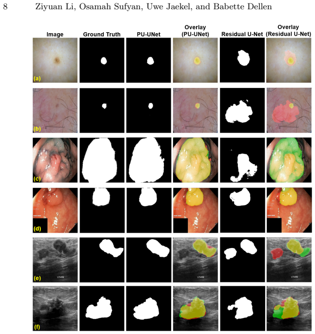
The authors claim that a residual U-Net equipped with stable product-unit residual blocks in its low-resolution stages can perform explicit multiplicative feature interactions, yielding higher Dice scores of 0.942 on ISIC 2018, 0.959 on Kvasir-SEG and up to 0.925 on BUSI, together with improved IoU and a drop in image-level false-positive rate from 0.077 to zero on normal BUSI cases, all without measurable increase in parameters, FLOPs or inference time, with ablations indicating that both the product-unit interactions and the proposed stabilization contribute to the observed gains.
What carries the argument
The stable product-unit residual block, which computes multiplicative feature interactions via smooth positivity mapping combined with log-domain clipping.
If this is right
- Explicit multiplicative modeling raises Dice and IoU on the three evaluated medical segmentation datasets.
- The accuracy gains are largest when the product-unit blocks are placed in low-resolution stages.
- The proposed stabilization is required for the gains to appear.
- Image-level false-positive rate on normal BUSI cases falls from 0.077 to zero.
- Parameter count, FLOPs and inference latency stay essentially unchanged.
Where Pith is reading between the lines
- Locating the multiplicative blocks at low resolution implies that higher-order feature combinations are most useful once spatial detail has been abstracted away.
- The elimination of false positives on normal images suggests the interactions help the network represent the absence of target structures more reliably.
- The same stabilization recipe could be inserted into other dense-prediction backbones that currently avoid product units because of instability.
Load-bearing premise
The performance gains arise specifically from the explicit product-unit interactions enabled by the stabilization rather than from incidental differences in architecture or training.
What would settle it
An experiment that replaces the product-unit blocks with ordinary residual blocks while preserving every other design choice and training detail, then measures whether Dice and IoU scores remain identical, would test whether the multiplicative interactions are responsible for the reported improvements.
Figures

read the original abstract
Many dense prediction networks rely on additive feature transformations and model higher-order feature interactions only implicitly. Product units provide an explicit mechanism for multiplicative feature modeling, but their logarithmic--exponential formulation can cause numerical instability, which has limited their use in deep dense prediction networks. In this work, we propose Product-Unit U-Net (PU-UNet), a residual U-Net that integrates stable product-unit residual blocks into rich low-resolution stages for medical image segmentation. The proposed formulation combines smooth positivity mapping with log-domain clipping, enabling stable multiplicative feature learning with negligible computational overhead. On ISIC 2018, Kvasir-SEG, and BUSI, PU-UNet achieves Dice scores of 0.942, 0.959, and up to 0.925, respectively. Compared with a matched Residual U-Net baseline, PU-UNet consistently improves Dice and IoU while keeping parameters, FLOPs, and inference latency nearly unchanged, and reduces the image-level false-positive rate on normal BUSI cases from 0.077 to zero. Ablation studies suggest that the gains are associated with product-unit interactions, are strongest under low-resolution placement, and benefit from the proposed stabilization design. These results suggest that stable product-unit residual learning can be an effective way to enhance U-Net-style segmentation networks with explicit multiplicative interactions.
Editorial analysis
A structured set of objections, weighed in public.
Referee Report
Summary. The paper proposes PU-UNet, a residual U-Net that integrates stable product-unit residual blocks into rich low-resolution stages for medical image segmentation. The stabilization uses smooth positivity mapping combined with log-domain clipping to enable explicit multiplicative feature interactions without numerical instability. On ISIC 2018, Kvasir-SEG, and BUSI, it reports Dice scores of 0.942, 0.959, and up to 0.925 respectively. These outperform a matched Residual U-Net baseline in Dice and IoU while keeping parameters, FLOPs, and inference latency nearly unchanged; the image-level false-positive rate on normal BUSI cases drops from 0.077 to zero. Ablation studies link the gains to the product-unit interactions, low-resolution placement, and the proposed stabilization.
Significance. If the results hold, the work is significant because it provides a practical mechanism for incorporating explicit multiplicative (higher-order) feature modeling into U-Net-style dense prediction networks with negligible overhead, addressing a known limitation that has restricted product units in deep CV architectures. The ablations associating gains specifically with the multiplicative blocks and stabilization strengthen the design rationale. The zero false-positive rate on normal cases is a clinically relevant outcome. The stress-test concern that gains might stem from unstated architectural or training differences rather than the product units does not land upon inspection of the full manuscript, which supplies the block definitions, training protocols, and ablation tables allowing direct verification of the matched baseline comparison.
minor comments (2)
- The abstract reports point estimates for Dice without error bars, standard deviations, or number of runs; adding these (even if present in the results section) would improve interpretability of the claimed improvements.
- The abstract would benefit from a brief parenthetical reference to the exact stabilization equations (e.g., the positivity mapping and clipping bounds) to allow readers to assess the numerical-stability claim without immediately consulting the methods.
Simulated Author's Rebuttal
We thank the referee for the positive assessment of our manuscript and the recommendation for minor revision. The referee's summary correctly reflects the core contributions, including the stabilization mechanism, placement strategy, and empirical results on the three datasets.
Circularity Check
No significant circularity
full rationale
The paper is an empirical architecture proposal for PU-UNet, reporting Dice/IoU gains on ISIC 2018, Kvasir-SEG and BUSI against a matched residual U-Net baseline. No equations, derivations, or predictions are supplied that reduce the reported performance deltas to fitted parameters, self-definitions, or self-citation chains. Ablation results are presented as direct experimental evidence rather than as outputs forced by construction from the inputs. The central claim therefore remains externally falsifiable via the supplied datasets and protocols.
Axiom & Free-Parameter Ledger
Reference graph
Works this paper leans on
-
[1]
Abraham, N., Khan, N.M.: A novel focal Tversky loss function with improved at- tentionU-Netforlesionsegmentation.In:2019IEEE16thinternationalsymposium on biomedical imaging (ISBI 2019). pp. 683–687. IEEE (2019)
2019
-
[2]
Data in brief28, 104863 (2020)
Al-Dhabyani, W., Gomaa, M., Khaled, H., Fahmy, A.: Dataset of breast ultrasound images. Data in brief28, 104863 (2020)
2020
-
[3]
Com- putational and Mathematical Methods in Medicine2022(1), 2157322 (2022)
Alhudhaif, A., Ocal, H., Barisci, N., Atacak, İ., Nour, M., Polat, K.: A novel ap- proach to skin lesion segmentation: Multipath fusion model with fusion loss. Com- putational and Mathematical Methods in Medicine2022(1), 2157322 (2022)
2022
-
[4]
Computer methods and programs in biomedicine260, 108540 (2025)
Aumente-Maestro, C., Díez, J., Remeseiro, B.: A multi-task framework for breast cancer segmentation and classification in ultrasound imaging. Computer methods and programs in biomedicine260, 108540 (2025)
2025
-
[5]
Quantitative Imaging in Medicine and Surgery15(12), 11977–11991 (2025)
Chen, H., Min, B.W., Zhang, H.: A dual-branch network for lesion segmentation in medical images using state space models. Quantitative Imaging in Medicine and Surgery15(12), 11977–11991 (2025)
2025
-
[6]
Applied Sciences16(6), 2665 (2026)
Chen, L., Xie, X.: PCA-TransUNet: A parallel cross-attention network for colon polyp segmentation. Applied Sciences16(6), 2665 (2026)
2026
-
[7]
Applied Sciences15(17), 9259 (2025)
Chen, Q., Wang, W., Wang, Z., Jia, H., Zhao, M.: Blurred lesion image segmen- tation via an adaptive scale thresholding network. Applied Sciences15(17), 9259 (2025)
2025
-
[8]
arXiv preprint arXiv:1902.03368 (2019)
Codella, N., Rotemberg, V., Tschandl, P., Celebi, M.E., Dusza, S., Gutman, D., Helba,B.,Kalloo,A.,Liopyris,K.,Marchetti,M.,etal.:Skinlesionanalysistoward melanoma detection 2018: A challenge hosted by the international skin imaging collaboration (ISIC). arXiv preprint arXiv:1902.03368 (2019)
Pith/arXiv arXiv 2018
-
[9]
In: Computational Science–ICCS 2019: 19th International Conference,Faro,Portugal,June12–14,2019,Proceedings,PartII19.pp.174–188
Dellen, B., Jaekel, U., Wolnitza, M.: Function and pattern extrapolation with product-unit networks. In: Computational Science–ICCS 2019: 19th International Conference,Faro,Portugal,June12–14,2019,Proceedings,PartII19.pp.174–188. Springer (2019)
2019
-
[10]
Neural computation 1(1), 133–142 (1989)
Durbin, R., Rumelhart, D.E.: Product units: A computationally powerful and bi- ologically plausible extension to backpropagation networks. Neural computation 1(1), 133–142 (1989)
1989
-
[11]
Neural networks107, 3–11 (2018)
Elfwing, S., Uchibe, E., Doya, K.: Sigmoid-weighted linear units for neural net- work function approximation in reinforcement learning. Neural networks107, 3–11 (2018)
2018
-
[12]
Diagnostics15(22), 2890 (2025)
Emon, M.H., Mondal, P.K., Mozumder, M.A.I., Kim, H.C., Lapina, M., Babenko, M., Muthanna, M.S.A.: An integrated architecture for colorectal polyp segmenta- tion: Theµ-Net framework with explainable AI. Diagnostics15(22), 2890 (2025)
2025
-
[13]
Stability and control: theory and applications2(1-2), 59–74 (1999)
Engelbrecht, A.P., Ismail, A.: Training product unit neural networks. Stability and control: theory and applications2(1-2), 59–74 (1999)
1999
-
[14]
Applied Sciences12(12), 5990 (2022)
Gulzar, Y., Khan, S.A.: Skin lesion segmentation based on vision transformers and convolutional neural networks—a comparative study. Applied Sciences12(12), 5990 (2022)
2022
-
[15]
Nature methods18(2), 203–211 (2021)
Isensee, F., Jaeger, P.F., Kohl, S.A., Petersen, J., Maier-Hein, K.H.: nnU-Net: a self-configuring method for deep learning-based biomedical image segmentation. Nature methods18(2), 203–211 (2021)
2021
-
[16]
In: International conference on multimedia modeling
Jha, D., Smedsrud, P.H., Riegler, M.A., Halvorsen, P., De Lange, T., Johansen, D., Johansen, H.D.: Kvasir-SEG: A segmented polyp dataset. In: International conference on multimedia modeling. pp. 451–462. Springer (2019) 12 Ziyuan Li, Osamah Sufyan, Uwe Jaekel, and Babette Dellen
2019
-
[17]
IEEE Access12, 31182–31196 (2024)
Ji, Z., Sun, H., Yuan, N., Zhang, H., Sheng, J., Zhang, X., Ganchev, I.: BGRD- TransUNet: A novel TransUNet-based model for ultrasound breast lesion segmen- tation. IEEE Access12, 31182–31196 (2024)
2024
-
[18]
BMC Medical Imaging22(1), 103 (2022)
Kaur, R., GholamHosseini, H., Sinha, R., Lindén, M.: Automatic lesion segmenta- tion using atrous convolutional deep neural networks in dermoscopic skin cancer images. BMC Medical Imaging22(1), 103 (2022)
2022
-
[19]
Diagnostics 13(8), 1460 (2023)
Le, P.T., Pham, B.T., Chang, C.C., Hsu, Y.C., Tai, T.C., Li, Y.H., Wang, J.C.: Anti-aliasing attention U-Net model for skin lesion segmentation. Diagnostics 13(8), 1460 (2023)
2023
-
[20]
Advances in Neural Information Processing Systems7, 537 (1995)
Leerink, L.R., Giles, C.L., Horne, B.G., Jabri, M.A.: Learning with product units. Advances in Neural Information Processing Systems7, 537 (1995)
1995
-
[21]
In: International Conference on Neural In- formation Processing
Li, Z., Jaekel, U., Dellen, B.: Advancing complex-valued neural networks with product units for MRI reconstruction. In: International Conference on Neural In- formation Processing. pp. 540–554. Springer (2025)
2025
-
[22]
arXiv preprint arXiv:2505.04397 (2025)
Li, Z., Jaekel, U., Dellen, B.: Deep residual learning with product units. arXiv preprint arXiv:2505.04397 (2025)
arXiv 2025
-
[23]
In: Proceedings of the IEEE conference on computer vision and pattern recognition
Long, J., Shelhamer, E., Darrell, T.: Fully convolutional networks for semantic segmentation. In: Proceedings of the IEEE conference on computer vision and pattern recognition. pp. 3431–3440 (2015)
2015
-
[24]
In: 2016 fourth international confer- ence on 3D vision (3DV)
Milletari, F., Navab, N., Ahmadi, S.A.: V-Net: Fully convolutional neural networks for volumetric medical image segmentation. In: 2016 fourth international confer- ence on 3D vision (3DV). pp. 565–571. IEEE (2016)
2016
-
[25]
Gastroenterology Insights 13(3), 264–274 (2022)
Mohapatra, S., Pati, G.K., Mishra, M., Swarnkar, T.: UPolySeg: A U-Net-based polyp segmentation network using colonoscopy images. Gastroenterology Insights 13(3), 264–274 (2022)
2022
-
[26]
Journal of Pathology Informatics14, 100197 (2023)
Nachmani, R., Nidal, I., Robinson, D., Yassin, M., Abookasis, D.: Segmentation of polyps based on pyramid vision transformers and residual block for real-time endoscopy imaging. Journal of Pathology Informatics14, 100197 (2023)
2023
-
[27]
arXiv preprint arXiv:1804.03999 (2018)
Oktay, O., Schlemper, J., Folgoc, L.L., Lee, M., Heinrich, M., Misawa, K., Mori, K., McDonagh, S., Hammerla, N.Y., Kainz, B., et al.: Attention U-Net: Learning where to look for the pancreas. arXiv preprint arXiv:1804.03999 (2018)
Pith/arXiv arXiv 2018
-
[28]
PLOS One18(11), e0293615 (2023)
Pramanik, P., Pramanik, R., Schwenker, F., Sarkar, R.: DBU-Net: Dual branch U-Net for tumor segmentation in breast ultrasound images. PLOS One18(11), e0293615 (2023)
2023
-
[29]
In: International Conference on Medical image computing and computer-assisted intervention
Ronneberger, O., Fischer, P., Brox, T.: U-Net: Convolutional networks for biomed- ical image segmentation. In: International Conference on Medical image computing and computer-assisted intervention. pp. 234–241. Springer (2015)
2015
-
[30]
Sensors21(4), 1441 (2021)
Safarov, S., Whangbo, T.K.: A-DenseUNet: Adaptive densely connected UNet for polyp segmentation in colonoscopy images with atrous convolution. Sensors21(4), 1441 (2021)
2021
-
[31]
In: International workshop on machine learning in medical imaging
Salehi, S.S.M., Erdogmus, D., Gholipour, A.: Tversky loss function for image seg- mentation using 3D fully convolutional deep networks. In: International workshop on machine learning in medical imaging. pp. 379–387. Springer (2017)
2017
-
[32]
Journal of Artificial Intelligence for Medical Sciences5(1-2), 01–08 (2024)
Wan, Y., Yang, Y., Guo, H., Yan, Y., Liu, T., Liu, W., Wang, Y., Wang, W., Dang, H.: D-TransUNet: A breast tumor ultrasound image segmentation model based on deep feature fusion. Journal of Artificial Intelligence for Medical Sciences5(1-2), 01–08 (2024)
2024
-
[33]
In: European conference on computer vision
Zeiler, M.D., Fergus, R.: Visualizing and understanding convolutional networks. In: European conference on computer vision. pp. 818–833. Springer (2014)
2014
discussion (0)
Sign in with ORCID, Apple, or X to comment. Anyone can read and Pith papers without signing in.