REVIEW 3 major objections 6 minor 62 references
EPRA U-Net: An Efficient Pyramid Residual Attention Framework for Accurate Infarct Segmentation in Diffusion-Weighted MRI
T0 review · 3 major / 6 minor · reviewed 2026-07-12 · grok-4.5
Pith's one-line read A task-tuned U-Net cuts missed stroke lesions on DWI by up to 29% versus strong baselines.
desk verdict Solid single-center engineering paper with thorough stats and a real efficiency/recall trade-off; Abstract overstates overall superiority once nnU-Net is in the picture. read the letter →
The pith
A machine-rendered reading of the paper's core claim, the machinery that carries it, and where it could break.
The reading
What carries the argument
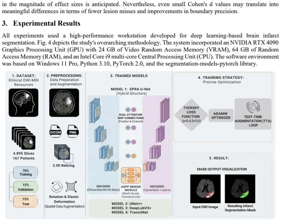
EPRA U-Net: a hybrid encoder–decoder that uses EfficientNet-B0 for compact hierarchical features, R2 residual-recurrent blocks for spatial continuity, ASPP dilated multi-scale context, dual (position + channel) attention for lesion focus, and Tversky loss with β>α to penalize missed infarcts more than false alarms.
What would settle it
An external multi-center DWI test set of comparable size on which EPRA U-Net no longer reduces false-negative lesion counts relative to the same three baselines under identical patient-level splitting and Tversky training.
Extended reading notes
Core claim
EPRA U-Net, by combining an EfficientNet-B0 encoder, residual-recurrent (R2) blocks, ASPP multi-scale context, dual attention, and Tversky loss (α=0.4, β=0.6), attains superior infarct segmentation on DWI—pixel Dice 0.8984, per-sample Dice 0.9469, IoU 0.8155, recall 0.8887, lesion F1 0.9378, HD95 11.62 px—and reduces missed lesions by 16%, 25%, and 29% versus UNet++, DeepLabV3+, and TransUNet on a 167-patient held-out test set.
Load-bearing premise
That scores measured on one hospital’s retrospectively collected DWI scans of 167 patients will hold for other scanners, protocols, and patient populations.
Editorial analysis
A structured set of objections, weighed in public.
Referee Report
Summary. The manuscript proposes EPRA U-Net, a hybrid encoder–decoder for acute ischemic infarct segmentation on DWI that combines an EfficientNet-B0 backbone, Residual-Recurrent (R2) blocks (t=2), ASPP multi-scale context, dual (position+channel) attention on skip pathways, and Tversky loss (α=0.4, β=0.6) to favor recall. On a single-center, patient-level split of 167 patients / 4,895 slices (14.7% positive), the model is compared with UNet++, DeepLabV3+, TransUNet under a matched training protocol and with 2D nnU-Net under its default self-configuring protocol. Headline results are pixel-aggregated Dice 0.8984, per-sample Dice 0.9469, IoU 0.8155, Recall 0.8887, Lesion F1 0.9378, HD95 11.62 px (median 2.00), with 16–29% fewer missed lesions versus the three matched baselines; ablation (Table 10) and multi-layered statistics (Wilcoxon, t-test, McNemar, bootstrap CIs, Cohen’s d) support the internal comparisons. The authors emphasize clinical prioritization of false-negative reduction and report a substantial training/inference cost advantage over nnU-Net.
Significance. If the reported gains hold under broader validation, the work is a solid, practically useful contribution to DWI infarct segmentation: it packages known components (EfficientNet, R2, ASPP, dual attention, Tversky) into a parameter-efficient pipeline (7.44M params, ~132 min training, ~34 ms/slice) that demonstrably reduces missed lesions relative to common matched-protocol baselines, with thorough lesion-level error analysis, ablations, and statistical testing. Strengths include patient-level splitting, clinically motivated loss design, multi-metric reporting (including FN lesion counts and HD95 median/mean), and explicit efficiency comparison to nnU-Net. The main scientific value is therefore a carefully engineered, efficiency-aware clinical tool rather than a fundamental architectural breakthrough; that is still of interest to medical imaging venues if claims are scoped accurately and external validity is strengthened.
major comments (3)
- Abstract and §3 / Table 3 frame EPRA as attaining “superior performance” (Dice 0.8984, HD95 11.62, fewer missed lesions vs UNet++/DeepLabV3+/TransUNet). Under its intended default protocol, 2D nnU-Net reaches Dice 0.8997, IoU 0.8177, mean HD95 7.98 (median 2.05 vs EPRA 2.00), Lesion F1 0.9376—essentially tied or better on overlap and boundary metrics—while EPRA leads mainly on Recall (0.8887 vs 0.8798) and lesion-level FP count (32 vs 69). The paper correctly notes the protocol/cost difference and excludes nnU-Net from paired tests, but the Abstract’s unqualified superiority language is not supported once the strongest practical baseline is included. Please reframe the claim as a favorable recall/efficiency trade-off against matched-protocol baselines (and competitive with nnU-Net at far lower cost), and align Abstract/Conclusion wording with Tables 3–4.
- Training protocol is not fully matched: EPRA is allowed 60 epochs while baselines use a 50-epoch ceiling (early stopping patience=15 applied uniformly; §2.4.2, Fig. 5). EPRA’s best validation Dice is reported at epoch 52. Although early stopping is cited, the extra budget and the fact that the selected checkpoint occurs after the baseline ceiling leave open a systematic training advantage. Either retrain all models under an identical epoch budget / identical early-stopping rule with the same patience clock, or provide a controlled ablation showing that the extra 10 epochs do not drive the reported gains.
- All quantitative claims rest on a single-institution retrospective cohort of 167 patients with no external test set (§2.1, §4.2). Patient-level splitting is correctly used, but scanner/protocol/population shift is untested. Given that the central clinical claim is reduced missed lesions and more reliable treatment-eligibility estimation, at least one external multi-center or multi-scanner hold-out (or a clear, quantified domain-shift experiment) is needed before the superiority/robustness language is appropriate for a journal audience. If external data cannot be added in revision, the claim scope must be narrowed throughout Abstract, Results, and Conclusion.
minor comments (6)
- Table 1 lists “Number of Epochs: Varies by model” without stating the actual ceilings; make the 50 vs 60 distinction explicit in the table for reproducibility.
- Data-availability URL contains a typo (“htps://” missing a “t”); correct to a working link and confirm that code/weights sufficient to reproduce Table 3 are released.
- Inter-annotator agreement is not quantified (§4.2 acknowledges this). Even a small double-read kappa or Dice on a subset would strengthen confidence in the ground truth used for all metrics.
- Fig. 5 / training curves: clarify whether the CosineAnnealing T_max equals the per-model epoch ceiling and whether any model was allowed to continue past early-stopping for plotting only.
- Notation: ASPP dilation rates (1, 6, 12, 18) and R2 unrolling t=2 are stated but not justified by a short sensitivity check; a one-row ablation or reference to the original formulations would help.
- Minor language/typos: “Atrous Spatial” double spaces; “Trans UNet” vs “TransUNet” inconsistency; “Yozgat Bozok” double spaces; Abstract omits nnU-Net while body includes it—align for consistency.
Circularity Check
No circularity: empirical supervised segmentation with held-out evaluation; architecture and loss choices are design decisions, not self-defining predictions.
full rationale
EPRA U-Net is a composite CNN (EfficientNet-B0 encoder + R2 blocks + ASPP + dual attention) trained with a fixed Tversky loss (α=0.4, β=0.6) on a patient-level split of an in-house DWI dataset. Performance claims (Dice, IoU, Recall, Lesion F1, HD95, missed-lesion reductions) are measured on held-out patients against baselines under a matched protocol; nnU-Net is reported separately under its own protocol. No quantity is fitted from the test set and then re-presented as a prediction; no uniqueness theorem or load-bearing premise is imported solely via overlapping-author citation; R2 unrolling t=2 and ASPP rates follow cited original formulations without redefining the evaluation metrics. Self-citations (e.g., authors’ prior COVID/R-CNN work) are ordinary background and do not close any definitional loop. The paper is therefore self-contained against its external benchmarks; circularity score is 0.
Assumptions & free parameters
free parameters (4)
- Tversky α, β =
α=0.4, β=0.6
- R2 recurrent unrolling steps t =
t=2
- ASPP dilation rates =
1,6,12,18
- Learning-rate / epoch budget =
1e-4 / 60 vs 50 epochs
assumptions (5)
- domain assumption Patient-level stratified split prevents data leakage and yields unbiased estimates of clinical performance.
- domain assumption 2.5D three-slice stacks supply sufficient inter-slice context for acute infarct segmentation without full 3D modeling.
- domain assumption Tversky loss with β>α correctly encodes the clinical cost asymmetry of false negatives over false positives for stroke.
- domain assumption Single-radiologist two-session polygon annotations are sufficiently reliable ground truth.
- standard math Standard U-Net skip-connection and encoder-decoder inductive biases remain appropriate for DWI infarcts.
invented entities (1)
-
EPRA U-Net architecture
Cite this review
Pith. "Pith review of EPRA U-Net: An Efficient Pyramid Residual Attention Framework for Accurate Infarct Segmentation in Diffusion-Weighted MRI." pith.science (2026). https://pith.science/paper/AAVTGPWB
@misc{pith2026260703568,
  author       = {Pith},
  title        = {Pith review of: EPRA U-Net: An Efficient Pyramid Residual Attention Framework for Accurate Infarct Segmentation in Diffusion-Weighted MRI},
  year         = {2026},
  howpublished = {\url{https://pith.science/paper/AAVTGPWB}},
  note         = {Machine review of arXiv:2607.03568}
}
read the original abstract
Objective: Accurate identification of acute ischemic infarcts on diffusion-weighted magnetic resonance imaging (DWI) is a critical prerequisite for reliable lesion quantification and effective clinical decision support in the management of cerebrovascular events. Methods: This study presents EPRA U-Net (Efficient Pyramid Residual Attention U-Net), a task-specific integrated architecture for efficient and accurate infarct segmentation of DWI images. In the proposed architecture, an EfficientNet-based encoder was used as a hierarchical feature extractor with a minimized parameterization. In addition, a Residual-Recurrent (R2) block (recurrent unrolling step t = 2, following the original formulation) and Atrous Spatial Pyramid Pooling (ASPP) were integrated to enhance the performance of spatial dependency modeling. Additionally, a dual attention mechanism was incorporated to highlight lesion-related activations while concurrently enabling the suppression of extraneous background responses. To prioritize lesion detection consistent with clinical imperative, a Tversky loss function was adopted, emphasizing the sensitivity of detection over its specificity during the optimization process. Results: Experimental evaluations were conducted utilizing an in-house dataset comprising 167 patients with 4,895 DWI slices; subsequently, the performance of the proposed EPRA U-Net was assessed in comparison with state-of-the-art models, specifically UNet++, DeepLabV3+, and TransUNet. The experimental results suggest that EPRA U-Net attained superior performance, evidenced by a pixel-aggregated Dice of 0.8984, a per-sample Dice of 0.9469, an IoU of 0.8155, a Recall of 0.8887, a Lesion F1 of 0.9378, and an HD95 of 11.62 px. Furthermore, a clear reduction in the rate of missed lesions, specifically by 16%, 25%, and 29%, was observed when compared with UNet++, DeepLabV3+, and TransUNet, respectively.
Figures


Figures from the paper (10 more)


Reference graph
Works this paper leans on
-
[1]
Current concepts on magnetic resonance imaging (MRI) perfusion- diffusion assessment in acute ischaemic stroke: a review & an update for the clinicians.,
E. Roldán-Valadéz and M. López-Mejía, “Current concepts on magnetic resonance imaging (MRI) perfusion- diffusion assessment in acute ischaemic stroke: a review & an update for the clinicians.,” Indian J Med Res. , vol. 140, no. 6, pp. 717–28, Dec. 2014, PMID: 25758570; PMCID: PMC4365345
2014
-
[2]
World Stroke Organization: Global Stroke Fact Sheet 2025,
V. L. Feigin et al., “World Stroke Organization: Global Stroke Fact Sheet 2025,” International Journal of Stroke, vol. 20, no. 2, pp. 132–144, Dec. 2024, doi: 10.1177/17474930241308142
-
[3]
TeleStroke: real -time stroke detection with federated learning and YOLOv8 on edge devices,
A. Elhanashi, P. Dini, S. Saponara, and Q. Zheng, “TeleStroke: real -time stroke detection with federated learning and YOLOv8 on edge devices,” Journal of Real -Time Image Processing, vol. 21, no. 4, Jun. 2024, doi: 10.1007/s11554-024-01500-1
-
[4]
N. Nagaraja, “Diffusion weighted imaging in acute ischemic stroke: A review of its interpretation pitfalls and advanced diffusion imaging application,” Journal of the Neurological Sciences , vol. 425, pp. 117435 –117435, Apr. 2021, doi: 10.1016/j.jns.2021.117435
-
[5]
V. T. Heeralal, S. E. Chadee, B. Ilyaev, R. Ilyaev, and S. Ilyayeva, “Artificial Intelligence in Stroke Care: A Narrative Review of Diagnostic, Predictive, and Workflow Applications,” Cureus , vol. 17, no. 9, Sep. 2025, doi: 10.7759/cureus.93430
-
[6]
U -Net: Convolutional Networks for Biomedical Image Segmentation,
O. Ronneberger, P. Fischer, and T. Brox, “U -Net: Convolutional Networks for Biomedical Image Segmentation,” in Lecture notes in computer science, Springer Science+Business Media, 2015, pp. 234–241. doi: 10.1007/978-3-319-24574-4_28
-
[7]
UNet++: A Nested U-Net Architecture for Medical Image Segmentation,
Z. Zhou, M. M. R. Siddiquee, N. Tajbakhsh, and J. Liang, “UNet++: A Nested U-Net Architecture for Medical Image Segmentation,” Lecture notes in computer science , vol. 11045, pp. 3 –11, Jan. 2018, doi: 10.1007/978 -3- 030-00889-5_1
doi:10.1007/978 2018
-
[8]
Attention U-Net: Learning Where to Look for the Pancreas,
O. Oktay et al., “Attention U-Net: Learning Where to Look for the Pancreas,” arXiv (Cornell University), Apr. 2018, doi: 10.48550/arxiv.1804.03999
Show all 62 references
-
[9]
Recurrent Residual Convolutional Neural Network based on U -Net (R2U-Net) for Medical Image Segmentation,
M. Z. Alom, Md. M. Hasan, C. Yakopcic, T. M. Taha, and V. K. Asari, “Recurrent Residual Convolutional Neural Network based on U -Net (R2U-Net) for Medical Image Segmentation,” arXiv (Cornell University), Feb. 2018, doi: 10.48550/arxiv.1802.06955
- [10]
- [11]
-
[12]
Medical Image Segmentation Review: The Success of U-Net,
R. Azad et al., “Medical Image Segmentation Review: The Success of U-Net,” IEEE Transactions on Pattern Analysis and Machine Intelligence, vol. 46, no. 12. IEEE Computer Society, pp. 10076 –10095, Aug. 21, 2024. doi: 10.1109/tpami.2024.3435571
2024 doi
-
[13]
3D U -Net: Learning Dense Volumetric Segmentation from Sparse Annotation,
Ö. Çiçek, A. Abdulkadir, S. S. Lienkamp, T. Brox, and O. Ronneberger, “3D U -Net: Learning Dense Volumetric Segmentation from Sparse Annotation,” in Lecture notes in computer science, Springer Science+Business Media, 2016, pp. 424–432. doi: 10.1007/978-3-319-46723-8_49
2016 doi
-
[14]
V-Net: Fully Convolutional Neural Networks for Volumetric Medical Image Segmentation,
F. Milletarì, N. Navab, and S. Ahmadi, “V-Net: Fully Convolutional Neural Networks for Volumetric Medical Image Segmentation,” pp. 565–571, Oct. 2016, doi: 10.1109/3dv.2016.79
2016 doi
-
[15]
Efficient multi -scale 3D CNN with fully connected CRF for accurate brain lesion segmentation,
K. Kamnitsas et al., “Efficient multi -scale 3D CNN with fully connected CRF for accurate brain lesion segmentation,” Medical Image Analysis, vol. 36, pp. 61–78, Oct. 2016, doi: 10.1016/j.media.2016.10.004
2016 doi
-
[16]
nnU-Net: a self -configuring method for deep learning -based biomedical image segmentation,
F. Isensee, P. F. Jaeger, S. A. A. Kohl, J. Petersen, and K. H. Maier-Hein, “nnU-Net: a self -configuring method for deep learning -based biomedical image segmentation,” Nature Methods, vol. 18, no. 2, pp. 203 –211, Dec. 2020, doi: 10.1038/s41592-020-01008-z
2020 doi
-
[17]
Encoder -Decoder with Atrous Separable Convolution for Semantic Image Segmentation,
L. -C. Chen, Y. Zhu, G. Papandreou, F. Schroff, and H. Adam, “Encoder -Decoder with Atrous Separable Convolution for Semantic Image Segmentation,” in Lecture notes in computer science, Springer Science+Business Media, 2018, pp. 833–851. doi: 10.1007/978-3-030-01234-2_49
2018 doi
-
[18]
Pyramid Scene Parsing Network,
H. Zhao, J. Shi, X. Qi, X. Wang, and J. Jia, “Pyramid Scene Parsing Network,” pp. 6230 –6239, Jul. 2017, doi: 10.1109/cvpr.2017.660
2017 doi
-
[19]
Attention is All you Need,
A. Vaswani et al., “Attention is All you Need,” in Adv. Neural Inf. Process. Syst., 2017
2017
-
[20]
CBAM: Convolutional Block Attention Module,
S. Woo, J. Park, J. Lee, and I. S. Kweon, “CBAM: Convolutional Block Attention Module,” in Lecture notes in computer science, Springer Science+Business Media, 2018, pp. 3 –19. doi: 10.1007/978-3-030-01234-2_1
2018 doi
-
[21]
Dual Attention Network for Scene Segmentation,
J. Fu et al., “Dual Attention Network for Scene Segmentation,” pp. 3141 –3149, Jun. 2019, doi : 10.1109/cvpr.2019.00326
2019 doi
-
[22]
Generalised Dice Overlap as a Deep Learning Loss Function for Highly Unbalanced Segmentations,
C. H. Sudre, W. Li, T. Vercauteren, S. Ourselin, and M. J. Cardoso, “Generalised Dice Overlap as a Deep Learning Loss Function for Highly Unbalanced Segmentations,” Lecture notes in computer science, vol. 2017, pp. 240–248, Jan. 2017, doi: 10.1007/978-3-319-67558-9_28
2017 doi
-
[23]
Tackling the class imbalance problem of deep learning-based head and neck organ segmentation,
E. Tappeiner, M. Welk, and R. Schubert, “Tackling the class imbalance problem of deep learning-based head and neck organ segmentation,” International Journal of Computer Assisted Radiology and Surgery, vol . 17, no. 11, pp. 2103–2111, May 2022, doi: 10.1007/s11548-022-02649-5
2022 doi
-
[24]
Tversky Loss Function for Image Segmentation Using 3D Fully Convolutional Deep Networks,
S. S. M. Salehi, D. Erdoğmuş, and A. Gholipour, “Tversky Loss Function for Image Segmentation Using 3D Fully Convolutional Deep Networks,” Lecture notes in computer science, pp. 379 –387, Jan. 2017, doi: 10.1007/978-3-319-67389-9_44
2017 doi
-
[25]
A comprehensive survey of loss functions and metrics in deep learning,
J. Terven, D. Córdova‐Esparza, J.-A. Romero-González, A. Ramírez-Pedraza, and E. A. Chávez‐Urbiola, “A comprehensive survey of loss functions and metrics in deep learning,” Artificial Intelligence Review, vol. 58, no. 7, Apr. 2025, doi: 10.1007/s10462-025-11198-7
2025 doi
-
[26]
A Novel Focal Tversky Loss Function With Improved Attention U-Net for Lesion Segmentation,
N. Abraham and N. Khan, “A Novel Focal Tversky Loss Function With Improved Attention U-Net for Lesion Segmentation,” pp. 683–687, Apr. 2019, doi: 10.1109/isbi.2019.8759329
2019 doi
-
[27]
The Multimodal Brain Tumor Image Segmentation Benchmark (BRATS),
B. Menze et al., “The Multimodal Brain Tumor Image Segmentation Benchmark (BRATS),” IEEE Transactions on Medical Imaging, vol. 34, no. 10, pp. 1993–2024, Dec. 2014, doi: 10.1109/tmi.2014.2377694
1993 doi
- [28]
-
[29]
Random effects during training: Implications for deep learning -based medical image segmentation,
J. Åkesson, J. Töger, and E. Heiberg, “Random effects during training: Implications for deep learning -based medical image segmentation,” Computers in Biology and Medicine, vol. 180, pp. 108944 –108944, Aug. 2024, doi: 10.1016/j.compbiomed.2024.108944
2024 doi
-
[30]
Robust chest CT image segmentation of COVID -19 lung infection based on limited data,
D. Müller, I. Soto‐Rey, and F. Krämer, “Robust chest CT image segmentation of COVID -19 lung infection based on limited data,” Informatics in Medicine Unlocked, vol. 25, pp. 100681 –100681, Jan. 2021, doi: 10.1016/j.imu.2021.100681
2021 doi
-
[31]
Detection, Diagnosis and Treatment of Acute Ischemic Stroke: Current and Future Perspectives,
S. Patil, R. Rossi, D. Jabrah, and K. Doyle, “Detection, Diagnosis and Treatment of Acute Ischemic Stroke: Current and Future Perspectives,” Frontiers in Medical Technology, vol. 4. Frontiers Media, Jun. 24, 2022. doi: 10.3389/fmedt.2022.748949
2022 doi
-
[32]
Make Sense: Free to use online annotation tool,
P. Skalski, "Make Sense: Free to use online annotation tool," GitHub, 2019. [Online]. Available: https://github.com/SkalskiP/make-sense
2019
-
[33]
Effect of data leakage in brain MRI classification using 2D convolutional neural networks,
E. Yağış et al. , “Effect of data leakage in brain MRI classification using 2D convolutional neural networks,” Scientific Reports, vol. 11, no. 1, Nov. 2021, doi: 10.1038/s41598-021-01681-w
2021 doi
- [34]
- [35]
-
[36]
Deep Residual Learning for Image Recognition,
K. He, X. Zhang, S. Ren, and J. Sun, “Deep Residual Learning for Image Recognition,” pp. 770 –778, Jun. 2016, doi: 10.1109/cvpr.2016.90
2016 doi
-
[37]
DeepLab: Semantic Image Segmentation with Deep Convolutional Nets, Atrous Convolution, and Fully Connected CRFs,
L.-C. Chen, G. Papandreou, I. Kokkinos, K. Murphy, and A. Yuille, “DeepLab: Semantic Image Segmentation with Deep Convolutional Nets, Atrous Convolution, and Fully Connected CRFs,” IEEE Transactions on Pattern Analysis and Machine Intelligence, vol. 40, no. 4, pp. 834–848, Apr...
2017 doi
-
[38]
Densely Connected Recurrent Residual (Dense R2UNet) Convolutional Neural Network for Segmentation of Lung CT Images,
K. Dutta, “Densely Connected Recurrent Residual (Dense R2UNet) Convolutional Neural Network for Segmentation of Lung CT Images,” arXiv (Cornell University) , Feb. 2021, doi: 10.48550/arxiv.2102.00663
-
[39]
A semi -automatic segmentation method for meningioma developed using a variational approach model,
L. Burrows, J. Patel, A. I. Islim, M. D. Jenkinson, S. J. Mills, and K. Chen, “A semi -automatic segmentation method for meningioma developed using a variational approach model,” The Neuroradiology Journal, vol. 37, no. 2, pp. 199–205, Dec. 2023, doi: 10.1177/19714009231224442
2023 doi
-
[40]
Image Segmentation Evaluation With the Dice Index: Methodological Issues,
M. L. Seghier, “Image Segmentation Evaluation With the Dice Index: Methodological Issues,” International Journal of Imaging Systems and Technology, vol. 34, no. 6, Oct. 2024, doi: 10.1002/ima.23203
2024 doi
-
[41]
Towards a guideline for evaluation metrics in medical image segmentation,
D. Müller, I. Soto‐Rey, and F. Krämer, “Towards a guideline for evaluation metrics in medical image segmentation,” BMC Research Notes, vol. 15, no. 1. BioMed Central, Jun. 20, 2022. doi: 10.1186/s13104 -022- 06096-y
2022 doi
- [42]
-
[43]
Detection and classification of COVID-19 by using faster R-CNN and mask R-CNN on CT images,
M. E. Şahin, H. Ulutaş, E. Yüce, and M. F. Erkoç, “Detection and classification of COVID-19 by using faster R-CNN and mask R-CNN on CT images,” Neural Computing and Applications, vol. 35, no. 18, pp. 13597–13611, Mar. 2023, doi: 10.1007/s00521-023-08450-y
2023 doi
-
[44]
U -Net and Its Variants for Medical Image Segmentation: A Review of Theory and Applications,
N. Siddique, S. Paheding, C. Elkin, and V. Devabhaktuni, “U -Net and Its Variants for Medical Image Segmentation: A Review of Theory and Applications,” IEEE Access, vol. 9, pp . 82031–82057, Jan. 2021, doi: 10.1109/access.2021.3086020
2021 doi
-
[45]
Deep learning -based approach for detecting COVID -19 in chest X -rays,
M. E. Şahin, “Deep learning -based approach for detecting COVID -19 in chest X -rays,” Biomedical Signal Processing and Control, vol. 78, pp. 103977–103977, Jul. 2022, doi: 10.1016/j.bspc.2022.103977
2022 doi
-
[46]
Calibrating the Dice Loss to Handle Neural Network Overconfidence for Biomedical Image Segmentation,
M. Yeung, L. Rundo, N. Yang, E. Sala, C. Schönlieb, and G. Yang, “Calibrating the Dice Loss to Handle Neural Network Overconfidence for Biomedical Image Segmentation,” Journal of Digital Imaging, vol. 36, no. 2, pp. 739–752, Dec. 2022, doi: 10.1007/s10278-022-00735-3
2022 doi
-
[47]
Retinoblastoma Detection via Image Processing and Interpretable Artificial Intelligence Techniques,
S. Duraivenkatesh, A. Narayan, V. Srikanth, and A. F. Made, “Retinoblastoma Detection via Image Processing and Interpretable Artificial Intelligence Techniques,” medRxiv (Cold Spring Harbor Laboratory), May 2023, doi: 10.1101/2023.05.02.23289419
2023 doi
-
[48]
From coarse to fine: a deep 3D probability volume contours framework for tumour segmentation and dose painting in PET images,
W. Zhang and S. Ray, “From coarse to fine: a deep 3D probability volume contours framework for tumour segmentation and dose painting in PET images,” Frontiers in Radiology, vol. 3, Sep. 2023, doi: 10.3389/fradi.2023.1225215
2023 doi
-
[49]
Individual Comparisons by Ranking Methods,
F. Wilcoxon, “Individual Comparisons by Ranking Methods,” Biometrics Bulletin, vol. 1, no. 6, pp. 80 –80, Dec. 1945, doi: 10.2307/3001968
1945 doi
-
[50]
Parametric versus non -parametric statistics in the analysis of randomized trials with non - normally distributed data,
A. J. Vickers, "Parametric versus non -parametric statistics in the analysis of randomized trials with non - normally distributed data," BMC Medical Research Methodology, vol. 5, no. 1, p. 35, 2005, doi: 10.1186/1471 - 2288-5-35
2005 doi
-
[51]
Note on the Sampling Error of the Difference Between Correlated Proportions or Percentages,
Q. McNemar, “Note on the Sampling Error of the Difference Between Correlated Proportions or Percentages,” Psychometrika, vol. 12, no. 2, pp. 153–157, Jun. 1947, doi: 10.1007/bf02295996
1947 doi
-
[52]
Efron and R
B. Efron and R. Tibshirani, An Introduction to the Bootstrap. 1994. doi: 10.1201/9780429246593
1994 doi
-
[53]
Cohen, Statistical Power Analysis for the Behavioral Sciences, 2nd ed
J. Cohen, Statistical Power Analysis for the Behavioral Sciences, 2nd ed. Hillsdale, NJ: Lawrence Erlbaum Associates, 1988
1988
-
[54]
Resolving power: a general approach to compare the distinguishing ability of threshold -free evaluation metrics,
C. A. Beam, “Resolving power: a general approach to compare the distinguishing ability of threshold -free evaluation metrics,” Machine Learning, vol. 114, no. 1, Jan. 2025, doi: 10.1007/s10994 -024-06723-8
2025 doi
-
[55]
Risk-based Evaluation of ML Classification Methods Used for Medical Devices,
M. Haimerl and C. Reich, “Risk-based Evaluation of ML Classification Methods Used for Medical Devices,” Research Square (Research Square), Sep. 2023, doi: 10.21203/rs.3.rs -3317894/v1
2023 doi
-
[56]
Challenges in Deep Learning-Based Small Organ Segmentation: A Benchmarking Perspective for Medical Research with Limited Datasets,
P. M. Konrad, A. -A. Popa, Y. Sabzehmeidani, L. Zhong, E. A. Liehn, and S. Ayvaz, “Challenges in Deep Learning-Based Small Organ Segmentation: A Benchmarking Perspective for Medical Research with Limited Datasets,” arXiv (Cornell University), Sep. 2025, doi: 10.48550/arxiv.2509.05892
-
[57]
Accuracy of CT perfusion ischemic core volume and location estimation: A comparison between four ischemic core estimation approaches using syngo.via,
J. W. Hoving et al., “Accuracy of CT perfusion ischemic core volume and location estimation: A comparison between four ischemic core estimation approaches using syngo.via,” PLoS ONE, vol. 17, no. 8, Aug. 2022, doi: 10.1371/journal.pone.0272276
2022 doi
-
[58]
Automated identification of thrombectomy amenable vessel occlusion on computed tomography angiography using deep learning,
J. H. Han et al., “Automated identification of thrombectomy amenable vessel occlusion on computed tomography angiography using deep learning,” Frontiers in Neurology, vol. 15, Jul. 2024, doi: 10.3389/fneur.2024.1442025
2024 doi
-
[59]
Early detection of white matter hyperintensities using SHIVA‐WMH detector,
A. Tsuchida, P. Boutinaud, V. Verrecchia, C. Tzourio, S. Debette, and M. Joliot, “Early detection of white matter hyperintensities using SHIVA‐WMH detector,” Human Brain Mapping, vol. 45, no. 1, Dec. 2023, doi: 10.1002/hbm.26548
2023 doi
-
[60]
Automatic Liver Segmentation from Multiphase CT Using Modified SegNet and ASPP Module,
P. V. Nayantara, S. Kamath, R. Kadavigere, and K. Manjunath, “Automatic Liver Segmentation from Multiphase CT Using Modified SegNet and ASPP Module,” SN Computer Science, vol. 5, no. 4, Mar. 2024, doi: 10.1007/s42979-024-02719-2
2024 doi
-
[61]
Rapid risk stratification of acute ischemic stroke patients in the emergency department: the incremental prognostic role of left atrial reservoir strain
Sonaglioni, A., Di Cara, M., Nicolosi, G. L., Eusebio, A., Bordonali, M., Santalucia, P., & Lombardo, M. "Rapid risk stratification of acute ischemic stroke patients in the emergency department: the incremental prognostic role of left atrial reservoir strain." Journal of Strok...
2021
-
[62]
Weak Edge Target Segmentation Network Based on Dual Attention Mechanism,
N. Wu, D. Jia, Z. Li, and Z. He, “Weak Edge Target Segmentation Network Based on Dual Attention Mechanism,” Applied Sciences, vol. 14, no. 19, pp. 8963–8963, Oct. 2024, doi: 10.3390/app14198963
2024 doi
Reviewed July 12, 2026 · model on record in the stance chip above.
Discussion (0). Sign in to comment.