REVIEW 3 major objections 4 minor 43 references
Clinical joint angles can be read directly from a parametric body model's per-segment rotation matrices, with no inverse-kinematics or musculoskeletal-model fitting step, at a pooled mean absolute error of 4.5 degrees over fifteen comparabl
Reviewed by Pith at T0; open to challenge. T0 means a machine referee read the full paper against a public rubric. the ladder, T0–T4 →
A swing-twist decomposition plus a per-body-model calibration table converts body-model rotation matrices into clinical joint angles at 4.50° MAE on OpenCap LabValidation.
T0 review reviewed 2026-08-01 challenge →
load-bearing objection The core idea is sound and useful, but the headline accuracy parity with OpenCap Monocular is based on a post-hoc 15-angle subset and is not yet established. the 3 major comments →
Direct Clinical Joint Angle Extraction from Parametric Body Model Rotation Matrices
The pith
A machine-rendered reading of the paper's core claim, the machinery that carries it, and where it could break.
The reading
Core claim
The paper's central claim is that the mapping from a parametric body model's internal segment orientations to standardised clinical joint angles is a coordinate transformation, not an optimisation. The procedure isolates each joint's motion by expressing the child segment in the parent frame and subtracting the rest pose; decomposes the relative rotation into a twist about the bone's long axis and a swing in the perpendicular plane; then combines the three resulting magnitudes through a per-joint linear formula θ = w1 c0 + w2 c1 + w3 c2 + δ. The three axes, three weights, per-side sign, and offset are the entire content of a small calibration table, fit once per body model offline. Validated
What carries the argument
The load-bearing mechanism is the swing-twist decomposition applied to the relative rotation between parent and child segments, followed by a linear readout. For each joint, the calibration table fixes a twist axis e1 (the bone's long axis) and two perpendicular swing axes e2 and e3; the relative rotation is split into twist magnitude c0 and two swing components c1 and c2, and the clinical angle is θ = w1 c0 + w2 c1 + w3 c2 + δ. The weights and offset absorb the difference between the body model's rest pose and clinical neutral, keeping the decomposition decoupled across a wide range of motion and removing the need for joint-type-specific code.
Load-bearing premise
The calibration assumes that the linear correction learned from nine healthy adults against a marker-based inverse-kinematics reference captures how body-model rotations translate to clinical angles for all people and all movements, including poses far outside the trained range.
What would settle it
An independent instrumented measurement—such as a goniometer or a marker-based system applied to new subjects performing motions the calibration set never exercises, like deep knee flexion, full arm extension, or pathological gait—that shows the table's angle errors clearly exceeding the reported 4.5-degree envelope would falsify the generalization claim. Likewise, if applying the same fixed table to another body model or a clinical population yields pooled mean absolute errors substantially larger than 4.5 degrees under the same reference protocol, the transferability claim fails.
If this is right
- Any pose estimator that outputs per-segment rotation matrices can feed this extraction; swapping body models only requires regenerating the calibration table, with no change to the extraction code.
- The extraction adds about one millisecond per frame and is deterministic closed-form arithmetic, so it can run on smartphones, servers, browsers, or embedded devices without biomechanics software installed.
- Since no per-recording inputs are needed, clinical angles can be produced from any monocular video regardless of camera or viewing angle, and from a live single-camera stream when the upstream estimator runs in real time.
- The output angles follow a standard clinical convention, so they can feed downstream analyses of gait or forces directly from body-model output, without returning to inverse kinematics.
- The procedure transfers across body models: a second estimator gave statistically indistinguishable accuracy, supporting the claim that the extraction is body-model-agnostic.
- Because the calibration table is fit per body model, the same procedure should extend to multi-camera, inertial, or depth-sensor inputs without changing the extraction itself.
Where Pith is reading between the lines
- Editorial inference: The calibration table is the only body-model-specific component, so the extraction is sensor-agnostic; the paper leaves multi-camera, inertial, and depth-sensor validation to future work, but the method itself should carry over unchanged.
- Editorial inference: The reported 4.5-degree envelope comes from nine healthy adults performing walking, squats, sit-to-stand, and jumps; the most direct test of clinical utility is replication on pathological movement or atypical joint ranges, where calibration coverage is thinnest.
- Editorial inference: Because the calibration is fit against a reference standard that itself comes from marker-based inverse kinematics, any systematic bias in that reference will be inherited by the table; an independent instrumented check on a well-defined joint like the knee could isolate whether residual error is sensor, model, or reference.
- Editorial inference: The large shoulder and elbow offsets in the table reflect the body model's rest pose differing from clinical neutral, suggesting calibration tables would need retraining for body models with very different rest poses, even though the extraction procedure itself is unchanged.
Editorial analysis
A structured set of objections, weighed in public.
Referee Report
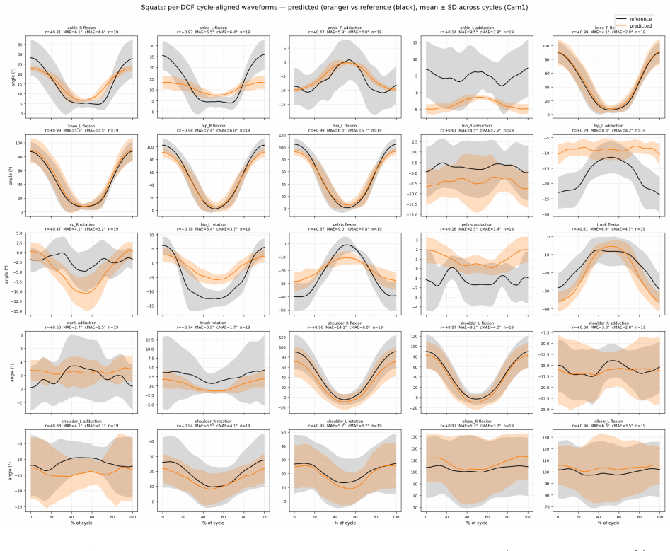
Summary. The paper presents a method for converting per-segment rotation matrices produced by parametric body models (e.g., GEM-X/SOMA, SAM 3D Body/MHR) into clinical joint angles, using a swing-twist decomposition and a per-joint calibration table. The runtime step is closed-form and requires no inverse kinematics or musculoskeletal fitting. Validation on the OpenCap LabValidation cohort (nine subjects) uses monocular smartphone video, leave-one-subject-out calibration, and compares against OpenSim inverse-kinematics reference. The headline result is a pooled MAE of 4.50° over fifteen OpenCap-comparable angles from a 45° camera view, which the paper claims is within the same accuracy range as OpenCap Monocular's published 4.8° over eighteen angles. The same procedure applied to a second body model yields 4.66°.
Significance. If the method's accuracy is confirmed, the contribution is practically useful: it removes a per-recording musculoskeletal-optimisation stage, runs in real time, and can be paired with any body-model estimator. Strengths include the transparent LOSO protocol, publication of the full calibration table in Appendix A, per-DOF results in Appendix B, and a second body-model confirmation in Appendix C. The accuracy numbers, however, are on a single cohort of nine healthy adults and the headline parity with OpenCap Monocular depends on an angle-set subset selection; those issues must be resolved before the central claim is accepted. The method is best framed as a supervised calibration of a linear mapping, not a parameter-free derivation.
major comments (3)
- [§3.1 / Abstract] The comparison with OpenCap Monocular is not like-for-like. The reported 4.50° MAE is over 15 of OpenCap's 18 joint angles, selected post hoc as having a direct counterpart, while the 4.8° reference is over all 18 angles. Without per-angle MAE from OpenCap on the same 15-angle subset, the claim that the methods are 'in the same accuracy range' is unsupported; OpenCap's subset error could be materially different. The bootstrap CI in §3.1 only characterises the 15-angle pooled mean and does not address the selection. Please either evaluate both methods on identical angle sets or explicitly weaken the headline claim.
- [§2.4 vs §3.1] The validation protocol states 'No subjects, trials, or DOFs are excluded,' but the matched comparison uses only the fifteen OpenCap-comparable angles and only non-jumping activities. Drop jumps and three OpenCap angles are excluded in the headline. This is an internal inconsistency in a claim central to the paper's credibility. Please reconcile the protocol with the analysis, e.g., by defining the matched envelope as a separate protocol with explicit inclusion/exclusion criteria.
- [§2.2 Step 3 / §2.3] The 'direct extraction' framing overstates what is derived. θ = w1 c0 + w2 c1 + w3 c2 + δ is a linear regression whose weights, offset, axes, component-to-angle assignment, and per-side signs are all fit to the OpenSim reference in Phases 1–3. The reported MAE is therefore for a calibrated mapping, not a self-contained geometric conversion. The abstract's 'no inverse-kinematics or musculoskeletal-model fitting step' should be qualified to 'at runtime'; offline calibration still relies on an IK-derived reference. This does not invalidate the method, but the current wording invites overinterpretation.
minor comments (4)
- [§2.4] The per-trial sub-frame alignment shifts the reference to minimise the residual on a subset of channels. This can only reduce reported error. Please report sensitivity to the alignment or state whether OpenCap Monocular applies the same synchronization before its MAE is computed.
- [Table A1] The r values in Table A1 are in-sample correlations from Phase 1 and should be labelled as such, to avoid confusion with the held-out r in Table B1.
- [§3.3] 'Statistically indistinguishable' should be phrased as 'not statistically significantly different'; a 95% CI spanning zero does not establish equivalence.
- [§2.4] Please provide the exact formula for centred MAE (per-trial bias removed) rather than only a verbal definition.
Circularity Check
No significant circularity: the calibration is openly disclosed and the headline accuracy is computed under leave-one-subject-out cross-validation.
full rationale
The paper's Step 3 formula, θ = w1·c0 + w2·c1 + w3·c2 + δ, is a linear model whose weights and offset are fit by least squares against the OpenSim reference (Section 2.3, Phase 3). This means the output is a calibrated regression rather than a first-principles derivation. However, the paper explicitly and repeatedly identifies this as a 'calibration table' rather than hiding the fitting step, and the reported 4.50° pooled MAE is computed under leave-one-subject-out cross-validation (Section 2.4), so the accuracy estimate is a genuine held-out evaluation of the fitted mapping, not an in-sample artifact. The swing-twist decomposition is attributed to prior work (HybrIK) and the ISB conventions are cited as context; there is no self-citation chain, no imported uniqueness theorem, and no ansatz disguised as an external result. The comparison against OpenCap Monocular uses a post-hoc 15-of-18 angle subset, which raises a legitimate statistical-fairness concern about whether the headline parity claim is apples-to-apples, but that is a comparison-design issue, not a circularity of the derivation: the paper's own equations do not reduce to the comparison. Under the required standard—that circularity be exhibited by a specific reduction or by a fitted parameter being renamed as a prediction—no such step is present. The calibration is disclosed, the validation is held out, and the central claim is an empirical one about the sufficiency of a small per-body-model calibration table.
Axiom & Free-Parameter Ledger
free parameters (5)
- Per-joint linear weights (w1,w2,w3) =
Table A3; e.g., knee flexion +0.753/+0.139/−0.991
- Per-joint offset δ =
Table A3; e.g., hip flexion +11.752°
- Swing-twist axes (e1,e2,e3) =
Table A2 per joint
- Component-to-angle assignment and per-side sign =
Table A1
- Ridge penalty strength =
not stated
axioms (5)
- standard math Swing-twist decomposition of a rotation is well-defined and closed-form.
- domain assumption Per-segment rotation matrices from GEM-X/SOMA and SAM 3D Body correspond to rigid anatomical segments in a kinematic chain.
- domain assumption OpenSim inverse kinematics on marker data with the LaiUhlrich2022 model is a valid clinical reference.
- ad hoc to paper The clinical angle is a linear function of the three swing-twist magnitudes: θ = w1 c0 + w2 c1 + w3 c2 + δ.
- domain assumption Calibration on nine healthy adults in a lab transfers to new subjects and other body models without per-subject scaling.
Cite this review
Pith. "Pith review of Direct Clinical Joint Angle Extraction from Parametric Body Model Rotation Matrices." pith.science (2026). https://pith.science/paper/AGX4IVRR
@misc{pith2026260717639,
  author       = {Pith},
  title        = {Pith review of: Direct Clinical Joint Angle Extraction from Parametric Body Model Rotation Matrices},
  year         = {2026},
  howpublished = {\url{https://pith.science/paper/AGX4IVRR}},
  note         = {Machine review of arXiv:2607.17639}
}
read the original abstract
Quantitative joint angles are rarely available in routine care because the tools are slow, costly, or confined to a laboratory. We show that clinical joint angles can be read directly from the per-segment rotation matrices a parametric body model already produces, with no inverse-kinematics or musculoskeletal-model fitting step. On the OpenCap LabValidation cohort, using the GEM-X body-model estimator on single-smartphone video, our pooled mean absolute error is 4.50 degrees over the fifteen joint angles that match the OpenCap Monocular reference set, the same accuracy range as OpenCap Monocular's 4.8 degrees on the same cohort and reference standard, from a much simpler pipeline. The step that connects a body model to clinical angles is a small calibration table rather than an optimisation, so the same procedure transfers unchanged to other body models: repeating it on SAM 3D Body, changing only the table, gives 4.66 degrees, statistically indistinguishable from GEM-X, and runs in real time from a live single-camera stream. The method needs no per-recording inputs beyond the video itself: no participant height, no camera-intrinsics database, no per-subject model scaling. This broadens where movement analysis is practical, from in-clinic and at-home recording to telerehabilitation and large-scale decentralised studies.
Figures




Reference graph
Works this paper leans on
-
[1]
R. R. Schaller. Moore’s law: past, present and future. IEEE Spectrum , 34(6):52–59,
-
[2]
M. I. Jordan and T. M. Mitchell. Machine learning: Trends, perspectives, and prospects. Science, 349(6245):255–260, 2015. doi: 10.1126/science.aaa8415
-
[3]
Krizhevsky, I
A. Krizhevsky, I. Sutskever, and G. E. Hinton. ImageNet classification with deep con- volutional neural networks. In Advances in Neural Information Processing Systems (NeurIPS), volume 25, pages 1097–1105, 2012
2012
-
[4]
Y. LeCun, Y. Bengio, and G. Hinton. Deep learning. Nature, 521(7553):436–444, 2015. doi: 10.1038/nature14539
-
[5]
Artificial intelligence index report 2025
Stanford Institute for Human-Centered Artificial Intelligence. Artificial intelligence index report 2025. Stanford University, https://hai.stanford.edu/ai-index/ 2025-ai-index-report , 2025
2025
-
[6]
A. Voulodimos, N. Doulamis, A. Doulamis, and E. Protopapadakis. Deep learning for computer vision: A brief review. Computational Intelligence and Neuroscience , 2018: 7068349, 2018. doi: 10.1155/2018/7068349
-
[7]
C. Zheng, W. Wu, C. Chen, T. Yang, S. Zhu, J. Shen, N. Kehtarnavaz, and M. Shah. Deep learning-based human pose estimation: A survey. ACM Computing Surveys , 56 (1):11:1–11:37, 2023. doi: 10.1145/3603618
doi:10.1145/3603618 2023
-
[8]
R. Baker. Gait analysis methods in rehabilitation. Journal of NeuroEngineering and Rehabilitation, 3:4, 2006. doi: 10.1186/1743-0003-3-4
-
[9]
S. D. Uhlrich, A. Falisse, L. Kidziński, et al. OpenCap: Human movement dynamics from smartphone videos. PLOS Computational Biology , 19(10):e1011462, 2023
2023
-
[10]
A. Seth, J. L. Hicks, T. K. Uchida, A. Habib, C. L. Dembia, J. J. Dunne, et al. OpenSim: Simulating musculoskeletal dynamics and neuromuscular control to study human and animal movement. PLOS Computational Biology , 14(7):e1006223, 2018
2018
-
[11]
D. Rode, A. Dunkel, R. Willi, P. Wolf, M. Xiloyannis, and R. Riener. Assessment of monocular human pose estimation models for clinical movement analysis. Scientific Reports, 15:38767, 2025. doi: 10.1038/s41598-025-22626-7
-
[12]
Loper, N
M. Loper, N. Mahmood, J. Romero, G. Pons-Moll, and M. J. Black. SMPL: A skinned multi-person linear model. ACM Transactions on Graphics , 34(6):248:1–248:16, 2015
2015
-
[13]
Pavlakos, V
G. Pavlakos, V. Choutas, N. Ghorbani, et al. Expressive body capture: 3D hands, face, and body from a single image. In CVPR, pages 10975–10985, 2019
2019
-
[14]
J. Park, J. Romero, S. Saito, et al. ATLAS: Decoupling skeletal and shape parameters for expressive parametric human modeling. arXiv:2508.15767, 2025. 12
Pith/arXiv arXiv 2025
-
[15]
A. Ferguson, A. A. A. Osman, B. Bescos, et al. MHR: Momentum human rig. arXiv:2511.15586, 2025
arXiv 2025
-
[16]
R. Brégier, G. Fiche, L. Bravo-Sánchez, et al. Human mesh modeling for Anny body. arXiv:2511.03589, 2025
Pith/arXiv arXiv 2025
- [17]
-
[18]
Kanazawa, M
A. Kanazawa, M. J. Black, D. W. Jacobs, and J. Malik. End-to-end recovery of human shape and pose. In CVPR, pages 7122–7131, 2018
2018
-
[19]
S. Shin, J. Kim, E. Halilaj, and M. J. Black. WHAM: Reconstructing world-grounded humans with accurate 3D motion. In CVPR, pages 2070–2080, 2024
2070
-
[20]
J. Li, J. Cao, H. Zhang, D. Rempe, J. Kautz, U. Iqbal, and Y. Yuan. GENMO: A generalist model for human motion. In ICCV, pages 11766–11776, 2025. URL https: //arxiv.org/abs/2505.01425. GEM-X is the SOMA-variant inference tool released as part of this framework
Pith/arXiv arXiv 2025
-
[21]
X. Yang, D. Kukreja, D. Pinkus, et al. SAM 3D Body: Robust full-body human mesh recovery. arXiv:2602.15989, 2026
arXiv 2026
- [22]
-
[23]
J. D. Peiffer, K. Shah, I. Djuraskovic, et al. Portable biomechanics laboratory: Clinically accessible movement analysis from a handheld smartphone. arXiv:2507.08268v2, 2025
arXiv 2025
-
[24]
Keller, K
M. Keller, K. Werling, S. Shin, S. Delp, S. Pujades, C. K. Liu, and M. J. Black. From skin to skeleton: Towards biomechanically accurate 3D digital humans. ACM Transactions on Graphics, 42(6):553, 2023
2023
-
[25]
Werling, N
K. Werling, N. A. Bianco, M. Raitor, J. Stingel, J. L. Hicks, S. H. Collins, S. L. Delp, and C. K. Liu. AddBiomechanics: Automating model scaling, inverse kinematics, and inverse dynamics from human motion data through sequential optimization. PLOS ONE, 18(11):e0295152, 2023
2023
-
[26]
Koleini, M
F. Koleini, M. U. Saleem, P. Wang, H. Xue, A. Helmy, and A. Fenwick. BioPose: Biomechanically-accurate 3D pose estimation from monocular videos. In W ACV, pages 6330–6339, 2025
2025
-
[27]
Y. Xia, X. Zhou, E. Vouga, Q. Huang, and G. Pavlakos. Reconstructing humans with a biomechanically accurate skeleton. In CVPR, pages 5355–5365, 2025
2025
-
[28]
E. S. Grood and W. J. Suntay. A joint coordinate system for the clinical description of three-dimensional motions: application to the knee. Journal of Biomechanical Engi- neering, 105(2):136–144, 1983. 13
1983
-
[29]
G. Wu, S. Siegler, P. Allard, et al. ISB recommendation on definitions of joint coordinate system of various joints for the reporting of human joint motion — part I: ankle, hip, and spine. Journal of Biomechanics , 35(4):543–548, 2002
2002
-
[30]
G. Wu, F. C. T. van der Helm, H. E. J. Veeger, et al. ISB recommendation on definitions of joint coordinate systems of various joints for the reporting of human joint motion — part II: shoulder, elbow, wrist and hand. Journal of Biomechanics , 38(5):981–992, 2005
2005
-
[31]
J. Li, C. Xu, Z. Chen, S. Bian, L. Yang, and C. Lu. HybrIK: A hybrid analytical-neural inverse kinematics solution for 3D human pose and shape estimation. In CVPR, pages 3383–3393, 2021
2021
-
[32]
D. A. Winter. Biomechanics and Motor Control of Human Movement . Wiley, 4 edition, 2009
2009
-
[33]
A. K. M. Lai, A. S. Arnold, and J. M. Wakeling. Why are antagonist muscles co- activated in my simulation? A musculoskeletal model for analysing human locomotor tasks. Annals of Biomedical Engineering , 45(12):2762–2774, 2017
2017
-
[34]
D. Austin. Better rigs, not bigger networks: A body model ablation for Gaussian avatars. arXiv:2604.01447, 2026
Pith/arXiv arXiv 2026
-
[35]
M. Dunn, A. Kennerley, Z. Murrell-Smith, K. Webster, K. Middleton, and J. Wheat. Ap- plication of video frame interpolation to markerless, single-camera gait analysis. Sports Engineering, 26:22, 2023. doi: 10.1007/s12283-023-00419-3
-
[36]
S. Ma, J. Zhang, Q. Cao, and D. Tao. PoseBench: Benchmarking the robustness of pose estimation models under corruptions. arXiv:2406.14367, 2024
Pith/arXiv arXiv 2024
-
[37]
T. Tan, T. Van Wouwe, K. F. Werling, et al. GaitDynamics: a generative foundation model for analyzing human walking and running. Nature Biomedical Engineering, 2026. doi: 10.1038/s41551-025-01565-8
-
[38]
R. D. Magruder, S. Gilon, A. Falisse, and S. D. Uhlrich. GaitEncoder: a foundation model of gait kinematics for diverse clinical applications and pathologies. medRxiv, 2026. doi: 10.64898/2026.07.07.26357479. preprint, not peer reviewed
- [39]
-
[40]
C. Banarjee, P. Kwon, A. Lipat, R. Xie, C. Chen, and L. Thiamwong. Fall risk and gait analysis in community-dwelling older adults using world-spaced 3D human mesh recovery. arXiv:2604.11961, 2026
Pith/arXiv arXiv 2026
- [41]
-
[42]
A. Qammaz. SAM3DBody-cpp: Real-time 3D full-body reconstruction in C++ (ONNX + ggml). GitHub, https://github.com/AmmarkoV/SAM3DBody-cpp, 2026. 14 A Full calibration tables The calibration procedure of Section 2.3 produces three sets of values per body model: the component-to-clinical-angle assignment and per-side sign (Phase 1), the three swing-twist axes...
2026
-
[1997]
doi: 10.1109/6.591665
This paper was first reviewed by deepseek-v4-flash on August 1, 2026.
discussion (0)
Sign in with ORCID, Apple, or X to comment. Anyone can read and Pith papers without signing in.