REVIEW 4 major objections 6 minor 58 references
Residual Vision Transformer (ResViT) Based Self-Supervised Learning Model for Brain Tumor Classification
T0 review · 4 major / 6 minor · reviewed 2026-08-12 · deepseek-v4-flash
Pith's one-line read This paper claims that learning to synthesize one brain MRI sequence from another, then fine-tuning the same model for classification, outperforms ImageNet pretraining on small brain tumor datasets.
desk verdict Plausible SSL pipeline whose reported gains are likely inflated by slice-level leakage between pretext pretraining and the classification test set. read the letter →
The pith
A machine-rendered reading of the paper's core claim, the machinery that carries it, and where it could break.
The reading
What carries the argument
The central machinery is ResViT, a hybrid CNN-transformer generator whose information bottleneck is made of Aggregated Residual Transformer (ART) blocks: each block runs the feature map through a downsampler, splits it into patches, applies a transformer encoder with multi-head self-attention and MLP layers, then deflates and up-samples the result and concatenates it with the residual CNN path before channel compression and a residual CNN. This lets local convolutional features and global attention features be fused at every block. The same encoder and ART blocks are reused in the fine-tuning stage with the decoder replaced by an MLP classification head, so the pretraining and downstream tasks share the same feature extractor. The pretext objective is MRI sequence synthesis (for example T1 to T2, T2 to T1, Flair to T1) trained with a pixel-wise loss, a reconstruction loss, and a PatchGAN adversarial loss; the synthesized images are then also used to double the minority classes during classification training.
What would settle it
A decisive check is to train the identical ResViT classifier under three initialization conditions—MRI-synthesis pretraining, ImageNet pretraining, and random initialization—holding the slice-selection and augmentation protocol fixed, and compare test accuracy across multiple seeds. If the MRI-pretrained model's margin over the others shrinks to noise, the pretext task is not the cause of the reported gains; additionally, rerunning the full pipeline with all available slices rather than the manually chosen tumor-maximum and center-close healthy slices would reveal whether the reported accuracies depend on that selection.
Extended reading notes
Core claim
The central discovery claimed is that generative self-supervised learning on MRI synthesis is a better pretraining strategy for brain tumor classification than supervised pretraining on ImageNet. In the pretext stage, ResViT is trained to translate between MRI sequences (T1 to T2, T2 to T1, Flair to T1, T1 to Flair) using pixel-wise, reconstruction, and PatchGAN adversarial losses, learning the distribution of MRI data without tumor-type labels. In the downstream stage, the pretrained encoder and Aggregated Residual Transformer blocks are kept and the decoder is replaced by an MLP head; the model is fine-tuned on real plus synthesized images. The paper reports that this pipeline reaches 90.56% accuracy on the augmented BraTS T1 set, 98.53% on Figshare, and 98.47% on Kaggle, and that the MRI-pretrained model outperforms ConvNeXtTiny, ResNet-101, DenseNet-121, and several ImageNet-pretrained ViT variants on every dataset and sequence tested.
Load-bearing premise
The load-bearing premise is that learning to synthesize one MRI sequence from another teaches general MRI features that transfer to tumor classification, and that the manually chosen slices—the five with the largest tumor region and the healthy slices nearest the brain center—are representative enough that their selection does not inflate the accuracy numbers.
Editorial extensions
If this is right
- MRI-specific self-supervised pretraining can replace ImageNet pretraining for brain tumor classification on small labeled sets; the proposed model surpasses every ImageNet-pretrained baseline tested on BraTS, Figshare, and Kaggle.
- The pretrained model transfers across MRI datasets: a checkpoint pretrained on BraTS sequences fine-tunes to 98.53% on Figshare and 98.47% on Kaggle, which would mean the learned MRI features are not specific to one acquisition protocol.
- The same synthesis model serves as a data augmentation tool: adding ResViT-synthesized MRIs to the BraTS training set raises accuracy from 83.44–88.89% to 88.89–90.56% depending on the MRI sequence.
- T1-weighted images are the most informative sequence for the downstream classification among T1, T2, and FLAIR, with the highest reported accuracy on BraTS.
- Combining residual CNN and vision transformer branches in one architecture outperforms using either branch alone, supporting the local-plus-global feature rationale.
Reading between the lines
- Editorial inference: the largest confound is the manual slice-selection protocol; rerunning the comparison on all slices or on automatically selected slices would test whether the reported margins come from the pretraining signal or from picking the most informative slices.
- Editorial inference: because the pretext task does not use tumor labels, the same ResViT checkpoint should transfer to other downstream tasks such as tumor segmentation or survival prediction on BraTS; a positive result would strengthen the representation-learning interpretation.
- Editorial inference: the ImageNet baselines differ in architecture from ResViT, so the cleanest controlled test of the paper's core claim would be to pretrain the identical ResViT backbone on ImageNet (or on natural images) and compare fine-tuning from that checkpoint with fine-tuning from the MRI-synthesis checkpoint.
- Editorial inference: the reported accuracy gains from synthetic MRI augmentation suggest that generation quality and classification transfer are coupled; a targeted ablation varying the number of synthetic images per class would reveal whether the benefit is from class balancing or from the diversity of synthesized examples.
Signed reviews
Editorial analysis
A structured set of objections, weighed in public.
Referee Report
Summary. The paper proposes a two-stage generative self-supervised learning (SSL) framework for brain tumor classification. In the first stage, a Residual Vision Transformer (ResViT) is pretrained for MRI sequence synthesis (e.g., T1 to T2, Flair to T1) as a pretext task on a selected subset of BraTS 2023 slices. In the second stage, a ResViT-based classifier is fine-tuned from the pretrained encoder, with synthetic MRI images used as data augmentation. The method is evaluated on BraTS (basic and augmented), Figshare, and Kaggle brain tumor MRI datasets, reporting accuracies of 90.56% on BraTS T1 (augmented), 98.53% on Figshare, and 98.47% on Kaggle, claiming superiority over ImageNet-pretrained baselines and prior work.
Significance. If the reported results are valid, the paper would provide a useful demonstration that MRI-specific generative self-supervised pretraining can outperform generic ImageNet transfer for small medical imaging datasets, and the hybrid CNN-transformer architecture is a sensible design choice. The manuscript includes comparisons across multiple datasets and MRI sequences, and the synthesis-quality metrics (PSNR, SSIM, MSE) are standard. However, the central claim rests on the integrity of the experimental protocol, especially the separation between pretext and downstream data, and on the statistical reliability of the accuracy numbers.
major comments (4)
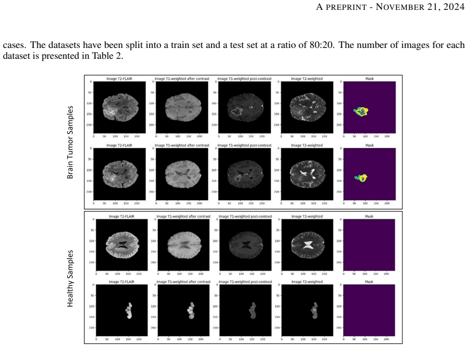
- [Section 4.1] The pretraining and classification datasets are not explicitly disjoint at the subject or slice level. The pretext set uses the top five tumor-coverage slices per BraTS case plus center-close healthy slices, while the classification set uses a single 'most relevant' slice per case from the same case pool. Since the most relevant slice is likely among the top-coverage slices, the ResViT generator is plausibly trained on the exact 2D images that later appear in the classification test set. The paper never states that classification slices were excluded from pretraining or that the train/test split was patient-level. This information leakage would directly inflate the reported gains over ImageNet-pretrained baselines in Tables 4 and 5 and undermine the central claim. Please clarify the exact overlap and rerun the main comparisons with strictly disjoint subjects and with the classification slices excluded from all pretraining data.
- [Section 4.2 and Tables 4-6] All accuracy, precision, recall, and F1 results are reported from a single run on a single split, without error bars, repeated seeds, or significance tests. Given the modest test-set sizes (900 BraTS slices, 613 Figshare, 1311 Kaggle), the differences between the proposed model and the best baselines may not be statistically meaningful. The authors should report mean and standard deviation over multiple runs and provide a significance test (e.g., McNemar's test) for the key comparisons.
- [Table 2 versus Table 5] The test set used for the augmented BraTS classification appears inconsistent with the basic BraTS test set. Table 2 lists Glioma test as 250 for the basic dataset but 200 for the augmented dataset, while the total test is 900 in both columns (the augmented total 450+200+200=850). Since augmentation should affect only the training set, the test sets should be identical. Please correct the table and state which test set was used for each reported result.
- [Section 4.1 and Table 2] The construction of the 'no tumor' class in the pretraining stage is not clearly described. The text says healthy slices were selected from the same 2,251 glioma and meningioma cases, but those cases are tumor cases; it is unclear whether the no-tumor class in the pretraining set consists of healthy slices from tumor patients or from separate healthy subjects. This ambiguity affects the interpretation of the pretext task and the classification setup, and it should be clarified.
minor comments (6)
- [Abstract and Section 3.3] The abstract refers to 'DenseNet12' while the text and tables use 'DenseNet121'; please correct the typo.
- [Section 4.2] There is a typo in 'pre-trained ResV˙IT model'; it should read 'ResViT'.
- [Section 4.1 and Table 2] The naming 'Brats' and 'BraTS' is used inconsistently throughout the paper; please standardize.
- [Section 4.4 and Table 7] The comparison in Table 7 lists only a subset of prior Figshare results and omits several high-performing recent methods; please clarify the selection criteria for the comparison.
- [Section 4.3 and Table 3] The synthesis-quality differences between ResViT and the next-best method are small (e.g., PSNR 25.663 vs. 25.105 for T1-to-T2); please indicate whether these differences are statistically significant and report confidence intervals.
- [Figure 1] Figure 1 is very dense and the two stages are not visually separated clearly; adding explicit labels and larger fonts would improve readability.
Circularity Check
No circularity: the SSL pipeline is empirical and self-contained; the only in-scope concern is a possible slice-level overlap between pretraining and classification, which is a data-hygiene risk, not a circular derivation.
full rationale
The paper is an empirical deep-learning study, not a derivation chain. The pretext task (MRI synthesis with ResViT) and the downstream task (tumor classification via fine-tuning) are measured on held-out test splits: Section 4.1 states that datasets are split 80:20 into train and test, and Tables 4-6 report accuracy, precision, recall, and F1 on those test sets. No equation in the paper defines the classification accuracy as a function of the synthesis loss, and no fitted parameter is renamed as a prediction. The architecture is taken from Dalmaz et al. [38], and the ImageNet-pretrained transformer backbones are explicitly stated in Section 3.1, so the comparison against ImageNet pretraining is a genuine empirical benchmark rather than a self-citation chain. The only in-scope concern is in Section 4.1, where the top-five tumor-coverage slices per case are used for pretraining and a single 'most relevant' slice per case is used for classification, without an explicit statement that pretraining and classification use disjoint subjects or disjoint slices. If the same 2D slices appear in both stages, the pretrained generator may have reconstructed those exact images, which could inflate reported BraTS accuracies. This is a data-leakage/validity risk and a legitimate weakness of the experimental protocol, but it is not circular reasoning: the classification results are empirical measurements on a test split, not a logical or algebraic consequence of the pretraining objective. No self-referential definition, no imported uniqueness theorem, and no ansatz smuggled in via citation was found, so the circularity score is 0.
Assumptions & free parameters
free parameters (3)
- Synthetic augmentation factor =
2x for glioma and meningioma
- Slice selection rule =
top-5 slices (tumor coverage) or center-close healthy slices
- Loss weights lambda =
not reported
assumptions (2)
- domain assumption Self-supervised synthesis pretext improves downstream classification
- domain assumption Synthetic images are valid augmentations
Cite this review
Pith. "Pith review of Residual Vision Transformer (ResViT) Based Self-Supervised Learning Model for Brain Tumor Classification." pith.science (2026). https://pith.science/paper/B3244JF2
@misc{pith2026241112874,
  author       = {Pith},
  title        = {Pith review of: Residual Vision Transformer (ResViT) Based Self-Supervised Learning Model for Brain Tumor Classification},
  year         = {2026},
  howpublished = {\url{https://pith.science/paper/B3244JF2}},
  note         = {Machine review of arXiv:2411.12874}
}
read the original abstract
Deep learning has proven very promising for interpreting MRI in brain tumor diagnosis. However, deep learning models suffer from a scarcity of brain MRI datasets for effective training. Self-supervised learning (SSL) models provide data-efficient and remarkable solutions to limited dataset problems. Therefore, this paper introduces a generative SSL model for brain tumor classification in two stages. The first stage is designed to pre-train a Residual Vision Transformer (ResViT) model for MRI synthesis as a pretext task. The second stage includes fine-tuning a ResViT-based classifier model as a downstream task. Accordingly, we aim to leverage local features via CNN and global features via ViT, employing a hybrid CNN-transformer architecture for ResViT in pretext and downstream tasks. Moreover, synthetic MRI images are utilized to balance the training set. The proposed model performs on public BraTs 2023, Figshare, and Kaggle datasets. Furthermore, we compare the proposed model with various deep learning models, including A-UNet, ResNet-9, pix2pix, pGAN for MRI synthesis, and ConvNeXtTiny, ResNet101, DenseNet12, Residual CNN, ViT for classification. According to the results, the proposed model pretraining on the MRI dataset is superior compared to the pretraining on the ImageNet dataset. Overall, the proposed model attains the highest accuracy, achieving 90.56% on the BraTs dataset with T1 sequence, 98.53% on the Figshare, and 98.47% on the Kaggle brain tumor datasets. As a result, the proposed model demonstrates a robust, effective, and successful approach to handling insufficient dataset challenges in MRI analysis by incorporating SSL, fine-tuning, data augmentation, and combining CNN and ViT.
Figures


Reference graph
Works this paper leans on
-
[1]
S. A. Abdelaziz Ismael, A. Mohammed, and H. Hefny, “An enhanced deep learning approach for brain cancer MRI images classification using residual networks,” Artif Intell Med, vol. 102, Jan. 2020, doi: 10.1016/j.artmed.2019.101779
arXiv 2020
-
[2]
Content-based brain tumor retrieval for MR images using transfer learning,
Z. N. K. Swati et al., “Content-based brain tumor retrieval for MR images using transfer learning,” IEEE Access, vol. 7, pp. 17809–17822, 2019
work page 2019
-
[3]
R. Microwave et al., “A Lightweight Deep Learning Based Microwave Brain Image Network Model for Brain Tumor Classification Using,” 2023
work page 2023
-
[4]
G. S. Tandel, A. Tiwari, O. G. Kakde, N. Gupta, L. Saba, and J. S. Suri, “Role of Ensemble Deep Learning for Brain Tumor Classification in Multiple Magnetic Resonance Imaging Sequence Data,” Diagnostics, 2023
work page 2023
-
[5]
Brain Tumor Detection and Classification Using Intelligence Techniques: An Overview,
S. Solanki and U. P. Singh, “Brain Tumor Detection and Classification Using Intelligence Techniques: An Overview,” vol. 11, no. January, 2023
work page 2023
-
[6]
P. Rani, V . Ashish, and K. Bhandari, “Role of Deep Learning in Classification of Brain MRI Images for Prediction of Disorders: A Survey of Emerging Trends,”Archives of Computational Methods in Engineering, no. 0123456789, 2023, doi: 10.1007/s11831-023-09967-0
-
[7]
Deep CNN for Brain Tumor Classification,
W. Ayadi, W. Elhamzi, I. Charfi, and M. Atri, “Deep CNN for Brain Tumor Classification,”Neural Process Lett, vol. 53, no. 1, pp. 671–700, 2021, doi: 10.1007/s11063-020-10398-2
-
[8]
S. C. Huang, A. Pareek, M. Jensen, M. P. Lungren, S. Yeung, and A. S. Chaudhari, “Self-supervised learning for medical image classification: a systematic review and implementation guidelines,” npj Digital Medicine, vol. 6, no. 1. Nature Research, Dec. 01, 2023. doi: 10.1038/s41746-023-00811-0
Show all 58 references
-
[9]
An MRI-based deep learning approach for efficient classification of brain tumors,
E. U. Haq, H. Jianjun, K. Li, H. U. Haq, and T. Zhang, “An MRI-based deep learning approach for efficient classification of brain tumors,” Journal of Ambient Intelligence and Humanized Computing, pp. 1–22, 2021
2021
-
[10]
Brain Tumor/Mass Classification Framework Using Magnetic-Resonance-Imaging-Based Isolated and Developed Transfer Deep-Learning Model,
M. F. Alanazi et al., “Brain Tumor/Mass Classification Framework Using Magnetic-Resonance-Imaging-Based Isolated and Developed Transfer Deep-Learning Model,” Sensors, vol. 22, no. 1, 2022, doi: 10.3390/s22010372. 13 A PREPRINT - NOVEMBER 21, 2024
2022 doi
-
[11]
Brain Tumor Classification Using Fine-Tuned GoogLeNet Features and Machine Learning Algorithms: IoMT Enabled CAD System,
A. Sekhar, S. Biswas, R. Hazra, A. K. Sunaniya, A. Mukherjee, and L. Yang, “Brain Tumor Classification Using Fine-Tuned GoogLeNet Features and Machine Learning Algorithms: IoMT Enabled CAD System,” IEEE journal of biomedical and health informatics, vol. 26, no. 3, pp. 983–991,...
2022
-
[12]
Deep learning,
Y . LeCun, Y . Bengio, and G. Hinton, “Deep learning,”Nature, vol. 521, no. 7553, pp. 436–444, 2015
2015
-
[13]
Convolutional neural networks in medical image understanding: a survey.,
D. R. Sarvamangala and R. V Kulkarni, “Convolutional neural networks in medical image understanding: a survey.,”Evolutionary intelligence, vol. 15, no. 1, pp. 1–22, 2022, doi: 10.1007/s12065-020-00540-3
2022 doi
-
[14]
Brain tumor classification using deep CNN features via transfer learning,
S. Deepak and P. M. Ameer, “Brain tumor classification using deep CNN features via transfer learning,”Computers in Biology and Medicine, vol. 111, Aug. 2019, doi: 10.1016/j.compbiomed.2019.103345
2019
-
[15]
Deep neural network with generative adversarial networks pre-training for brain tumor classification based on MR images,
N. Ghassemi, A. Shoeibi, and M. Rouhani, “Deep neural network with generative adversarial networks pre-training for brain tumor classification based on MR images,” Biomedical Signal Processing Control, vol. 57, p. 101678, 2020, doi: 10.1016/j.bspc.2019.101678
2020
-
[16]
Classification of brain tumors from mri images using a convolutional neural network,
M. M. Badža and M. C. Barjaktarovi´c, “Classification of brain tumors from mri images using a convolutional neural network,” Applied Sciences (Switzerland), vol. 10, no. 6, Mar. 2020, doi: 10.3390/app10061999
2020 doi
-
[17]
Enhanced brain tumor classification using an optimized multi-layered convolutional neural network architecture,
M. Alshayeji, J. Al-Buloushi, A. Ashkanani, and S. Abed, “Enhanced brain tumor classification using an optimized multi-layered convolutional neural network architecture,” Multimedia Tools and Applications, vol. 80, no. 19, pp. 28897–28917, Aug. 2021, doi: 10.1007/s11042-021-10927-8
2021 doi
-
[18]
Three-class classification of brain magnetic resonance images using average-pooling convolutional neural network,
J. Kakarla, B. V . Isunuri, K. S. Doppalapudi, and K. S. R. Bylapudi, “Three-class classification of brain magnetic resonance images using average-pooling convolutional neural network,” International Journal of Imaging Systems and Technology, vol. 31, no. 3, pp. 1731–1740, Sep...
2021 doi
-
[19]
Multi-class brain tumor classification using residual network and global average pooling,
R. L. Kumar, J. Kakarla, B. V . Isunuri, and M. Singh, “Multi-class brain tumor classification using residual network and global average pooling,” Multimedia Tools and Applications, vol. 80, no. 9, pp. 13429–13438, Apr. 2021, doi: 10.1007/s11042-020-10335-4
2021 doi
-
[20]
BTC-fCNN: Fast Convolution Neural Network for Multi-class Brain Tumor Classification,
B.S. Abd El-Wahab, M. E. Nasr, S. Khamis, and A. S. Ashour, “BTC-fCNN: Fast Convolution Neural Network for Multi-class Brain Tumor Classification,” Health Information Science and Systems, 2023, doi: 10.1007/s13755- 022-00203-w
2023 doi
-
[21]
MedViT: a robust vision transformer for generalized medical image classification,
O. N. Manzari, H. Ahmadabadi, H. Kashiani, S. B. Shokouhi, and A. Ayatollahi, “MedViT: a robust vision transformer for generalized medical image classification,” Computers in Biology and Medicine, vol. 157, p. 106791, 2023
2023
-
[22]
A survey on vision transformer,
K. Han et al., “A survey on vision transformer,” IEEE Trans Pattern Anal Mach Intell, vol. 45, no. 1, pp. 87–110, 2022
2022
-
[23]
Pyramid vision transformer: A versatile backbone for dense prediction without convolutions,
W. Wanget al., “Pyramid vision transformer: A versatile backbone for dense prediction without convolutions,” in Proceedings of the IEEE/CVF international conference on computer vision, 2021, pp. 568–578
2021
-
[24]
An image is worth 16x16 words: Transformers for image recognition at scale,
A. Dosovitskiy et al., “ An image is worth 16x16 words: Transformers for image recognition at scale,” arXiv preprint arXiv:2010.11929, 2020
2010 arXiv
-
[25]
Transmed: Transformers advance multi-modal medical image classification,
Y . Dai, Y . Gao, and F. Liu, “Transmed: Transformers advance multi-modal medical image classification,”Diag- nostics, vol. 11, no. 8, Aug. 2021, doi: 10.3390/diagnostics11081384
2021 doi
-
[26]
Combining the Transformer and Convolution for Effective Brain Tumor Classification Using MRI Images,
M. Aloraini, A. Khan, S. Aladhadh, S. Habib, M. F. Alsharekh, and M. Islam, “Combining the Transformer and Convolution for Effective Brain Tumor Classification Using MRI Images,”Applied Sciences, 2023
2023
-
[27]
Attention transformer mechanism and fusion-based deep learning architecture for MRI brain tumor classification system,
S. Tabatabaei, K. Rezaee, and M. Zhu, “Attention transformer mechanism and fusion-based deep learning architecture for MRI brain tumor classification system,” Biomedical Signal Processing Control, vol. 86, Sep. 2023, doi: 10.1016/j.bspc.2023.105119
2023
-
[28]
LCDEiT: A Linear Complexity Data-Efficient Image Transformer for MRI Brain Tumor Classification,
G. J. Ferdous, K. A. Sathi, M. A. Hossain, M. M. Hoque, and M. Ali Akber Dewan, “LCDEiT: A Linear Complexity Data-Efficient Image Transformer for MRI Brain Tumor Classification,”IEEE Access, vol. 11, pp. 20337–20350, 2023, doi: 10.1109/ACCESS.2023.3244228
2023
-
[29]
DiRA: Discriminative, Restorative, and Adversarial Learning for Self-supervised Medical Image Analysis,
F. Haghighi, M. Reza, H. Taher, M. B. Gotway, and J. Liang, “DiRA: Discriminative, Restorative, and Adversarial Learning for Self-supervised Medical Image Analysis,” in Proceedings of the IEEE/CVF Conference on Computer Vision and Pattern Recognition, 2022, pp. 20824-20834
2022
-
[30]
3D Self-Supervised Methods for Medical Imaging,
A. Taleb et al., “3D Self-Supervised Methods for Medical Imaging,” Advances in neural information processing systems, vol. 33, pp. 18158-18172, 2020
2020
-
[31]
Models Genesis,
Z. Zhou, V . Sodha, J. Pang, M. B. Gotway, and J. Liang, “Models Genesis,”Medical Image Analysis, vol. 67, Jan. 2021, doi: 10.1016/j.media.2020.101840. 14 A PREPRINT - NOVEMBER 21, 2024
2021
-
[32]
Self-supervised driven consistency training for annotation efficient histopathology image analysis,
C. L. Srinidhi, S. W. Kim, F. Der Chen, and A. L. Martel, “Self-supervised driven consistency training for annotation efficient histopathology image analysis,” Medical Image Analysis, vol. 75, Jan. 2022, doi: 10.1016/j.media.2021.102256
2022
-
[33]
Transformer-based unsupervised contrastive learning for histopathological image classification,
X. Wang et al., “Transformer-based unsupervised contrastive learning for histopathological image classification,” Medical Image Analysis, vol. 81, Oct. 2022, doi: 10.1016/j.media.2022.102559
2022
-
[34]
SAM: Self-Supervised Learning of Pixel-Wise Anatomical Embeddings in Radiologi- cal Images,
K. Yan et al., “ SAM: Self-Supervised Learning of Pixel-Wise Anatomical Embeddings in Radiologi- cal Images,” IEEE Transactions on Medical Imaging , vol. 41, no. 10, pp. 2658–2669, Oct. 2022., doi: 10.1109/TMI.2022.3169003
2022
-
[35]
Attention De-sparsification Matters: Inducing diversity in digital pathology representation learning,
S. Kapse et al., “ Attention De-sparsification Matters: Inducing diversity in digital pathology representation learning,” Medical Image Analysis, vol. 93, pp. 103070, 2024
2024
-
[36]
A General-Purpose Self-Supervised Model for Computational Pathology,
R. J. Chen et al., “A General-Purpose Self-Supervised Model for Computational Pathology,” Aug. 2023, [Online]. Available: http://arxiv.org/abs/2308.15474
2023 arXiv
-
[37]
Virchow: A Million-Slide Digital Pathology Foundation Model,
E. V orontsovet al., “ Virchow: A Million-Slide Digital Pathology Foundation Model,” Sep. 2023, [Online]. Available: http://arxiv.org/abs/2309.07778
2023 arXiv
-
[38]
ResViT: Residual Vision Transformers for Multimodal Medical Im- age Synthesis,
O. Dalmaz, M. Yurt, and T. Cukur, “ResViT: Residual Vision Transformers for Multimodal Medical Im- age Synthesis,” IEEE Transactions on Medical Imaging , vol. 41, no. 10, pp. 2598–2614, 2022, doi: 10.1109/TMI.2022.3167808
2022
-
[39]
Image-to-image translation with conditional adversarial networks,
P. Isola, J.-Y . Zhu, T. Zhou, and A. A. Efros, “Image-to-image translation with conditional adversarial networks,” in Proceedings of the IEEE conference on computer vision and pattern recognition , 2017, pp. 1125–1134
2017
-
[40]
Image synthesis in multi-contrast MRI with conditional generative adversarial networks,
S. U. H. Dar, M. Yurt, L. Karacan, A. Erdem, E. Erdem, and T. Çukur, “Image synthesis in multi-contrast MRI with conditional generative adversarial networks,”IEEE Transactions on Medical Imaging, vol. 38, no. 10, pp. 2375–2388, Oct. 2019
2019
-
[41]
Attention is all you need,
A. Vaswani et al., “Attention is all you need,” Adv Neural Inf Process Syst , vol. 30, 2017
2017
-
[42]
Haykin, Neural networks: a comprehensive foundation
S. Haykin, Neural networks: a comprehensive foundation . Prentice Hall PTR, 1998
1998
-
[43]
Attention u-net: Learning where to look for the pancreas,
O. Oktay et al., “Attention u-net: Learning where to look for the pancreas,” arXiv preprint arXiv:1804.03999, 2018
2018 arXiv
-
[44]
Deep residual learning for image recognition,
K. He, X. Zhang, S. Ren, and J. Sun, “Deep residual learning for image recognition,” in Proceedings of the IEEE conference on computer vision and pattern recognition , 2016, pp. 770–778
2016
-
[45]
A ConvNet for the 2020s,
Z. Liu, H. Mao, C.-Y . Wu, C. Feichtenhofer, T. Darrell, and S. Xie, “A ConvNet for the 2020s,”arXiv preprint arXiv:2201.03545, 2022
2022 arXiv
-
[46]
Identity mappings in deep residual networks,
K. He, X. Zhang, S. Ren, and J. Sun, “Identity mappings in deep residual networks,” in Computer Vision–ECCV 2016: 14th European Conference, Amsterdam, The Netherlands, October 11–14, 2016, Proceedings, Part IV 14 , Springer, 2016, pp. 630–645
2016
-
[47]
Densely connected convolutional networks,
G. Huang, Z. Liu, L. Van Der Maaten, and K. Q. Weinberger, “Densely connected convolutional networks,” in Proceedings of the IEEE conference on computer vision and pattern recognition , 2017, pp. 4700–4708
2017
-
[48]
Imagenet: A large-scale hierarchical image database,
J. Deng, W. Dong, R. Socher, L.-J. Li, K. Li, and L. Fei-Fei, “Imagenet: A large-scale hierarchical image database,” in 2009 IEEE conference on computer vision and pattern recognition, Jun. 2009
2009
-
[49]
Brain Tumor MRI Dataset,
M. Nickparvar, “Brain Tumor MRI Dataset,” Kaggle, 2021. [Online]. Available: https://doi.org/10.34740/KAGGLE/DSV/2645886
2021
-
[50]
brain tumor dataset
J. Cheng, “brain tumor dataset”. figshare, 2017, [Online]. Available: https://doi.org/10.6084/m9.figshare.1512427.v5 Accessed on: Apr. 2, 2017
2017 doi
-
[51]
The cancer imaging archive (TCIA): maintaining and operating a public information repository,
K. Clark, B. Vendt, K. Smith, J. Freymann, J. Kirby, P. Koppel, and L.Tarbox, “ The cancer imaging archive (TCIA): maintaining and operating a public information repository,” Journal of digital imaging, vol. 26, no. 6, pp. 1045–1057, Jul. 2013
2013
-
[52]
The rsna-asnr-miccai brats 2021 benchmark on brain tumor segmentation and radiogenomic classification,
U. Baid et al., “ The rsna-asnr-miccai brats 2021 benchmark on brain tumor segmentation and radiogenomic classification,” arXiv preprint arXiv:2107.02314, 2021
2021 arXiv
-
[53]
The multimodal brain tumor image segmentation benchmark (BRATS),
B. H. Menze et al., “The multimodal brain tumor image segmentation benchmark (BRATS),”IEEE Transactions on Medical Imaging 2014, vol. 34, no. 10, pp. 1993–2024,
2014
-
[54]
Advancing the cancer genome atlas glioma MRI collections with expert segmentation labels and radiomic features,
S. Bakas et al., “Advancing the cancer genome atlas glioma MRI collections with expert segmentation labels and radiomic features,” Sci Data, vol. 4, no. 1, pp. 1–13, 2017. 15 A PREPRINT - NOVEMBER 21, 2024
2017
-
[55]
Segmentation Labels and Radiomic Features for the Pre-operative Scans of the TCGA-GBM collection
S. Bakas, H. Akbari, A. Sotiras, M. Bilello, M. Rozycki, J. Kirby, et al., “Segmentation Labels and Radiomic Features for the Pre-operative Scans of the TCGA-GBM collection", The Cancer Imaging Archive, 2017. DOI: 10.7937/K9/TCIA.2017.KLXWJJ1Q
2017 doi
-
[56]
Segmentation Labels and Radiomic Features for the Pre-operative Scans of the TCGA-LGG collection
S. Bakas, H. Akbari, A. Sotiras, M. Bilello, M. Rozycki, J. Kirby, et al., “ Segmentation Labels and Radiomic Features for the Pre-operative Scans of the TCGA-LGG collection", The Cancer Imaging Archive, 2017. DOI: 10.7937/K9/TCIA.2017.GJQ7R0EF
2017 doi
-
[57]
The ASNR-MICCAI Brain Tumor Segmentation (BraTS) Challenge 2023: Intracranial Meningioma,
D. LaBella et al., “ The ASNR-MICCAI Brain Tumor Segmentation (BraTS) Challenge 2023: Intracranial Meningioma,” arXiv preprint arXiv:2305.07642, 2023
2023 arXiv
-
[58]
Image quality assessment: from error visibility to structural similarity,
Z. Wang, A. C. Bovik, H.R. Sheikh, E. P. Simoncelli EP. “Image quality assessment: from error visibility to structural similarity,” IEEE transactions on image processing, vol. 13, no. 4, pp. 600–612, Apr. 2004. 16
2004
Reviewed August 12, 2026 · model on record in the stance chip above.
Discussion (0). Continue with ORCID to comment.