Automated Erythrocyte Detection and Tracking for Retinal Blood Flow Quantification in Erythrocyte-Mediated Angiography
Pith reviewed 2026-06-28 17:33 UTC · model grok-4.3
The pith
EMTrack automates detection and tracking of individual erythrocytes in EMA images to enable retinal blood flow quantification.
A machine-rendered reading of the paper's core claim, the machinery that carries it, and where it could break.
Core claim
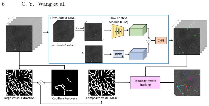
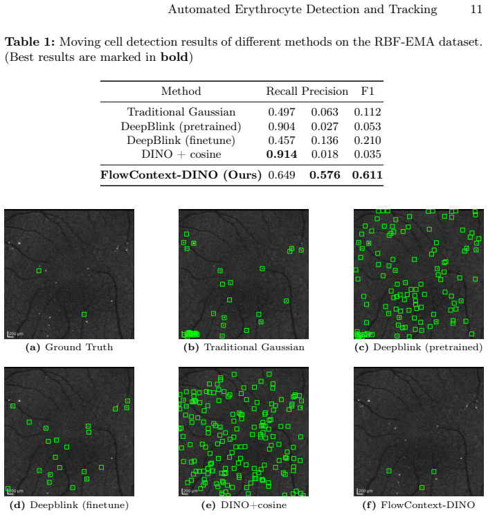
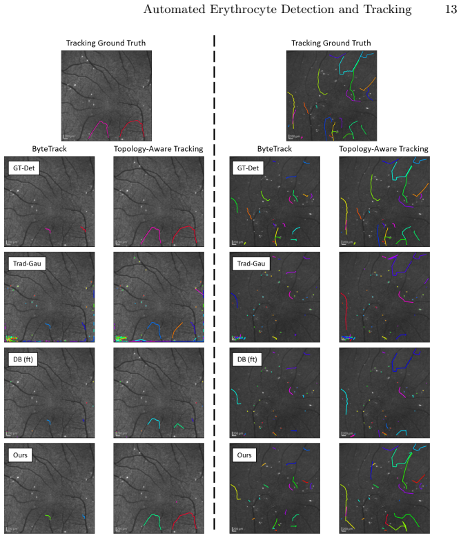
EMTrack outperforms baseline methods both quantitatively and qualitatively on erythrocyte detection and tracking tasks in the RBF-EMA dataset by employing a flow-context module that distinguishes moving from paused cells and a topology-aware tracking strategy that handles large inter-frame displacements and substantial motion variations, thereby supporting automated quantification of retinal blood flow from EMA sequences.
What carries the argument
The flow-context module for detection combined with the topology-aware tracking strategy, which together address paused cells and variable motion in EMA video sequences.
If this is right
- EMTrack exceeds baseline methods on both quantitative and qualitative measures for erythrocyte detection and tracking within the RBF-EMA dataset.
- RBF quantification produced by the framework demonstrates strong potential for automated retinal blood flow measurement.
- The approach fills the prior gap in automated erythrocyte analysis required for capillary-level RBF studies using EMA.
- The released RBF-EMA dataset supplies a new benchmark for detection and tracking methods in this imaging modality.
Where Pith is reading between the lines
- Routine automated RBF measurement could support larger clinical studies testing blood flow as a biomarker in specific eye diseases.
- The same modules for handling paused cells and large displacements may transfer to cell-tracking tasks in other dynamic vascular imaging modalities.
- Wider adoption of EMA plus automation could shift retinal blood flow assessment from research-only to more routine diagnostic use.
- Future work could test whether the quantified flow values correlate with disease progression in longitudinal patient data.
Load-bearing premise
The RBF-EMA dataset supplies accurate annotations that reflect real clinical variability in EMA imaging and the chosen baselines are appropriate comparators.
What would settle it
An independent EMA sequence collection recorded under different clinical or imaging conditions where EMTrack does not exceed the same baselines on detection or tracking metrics.
Figures
read the original abstract
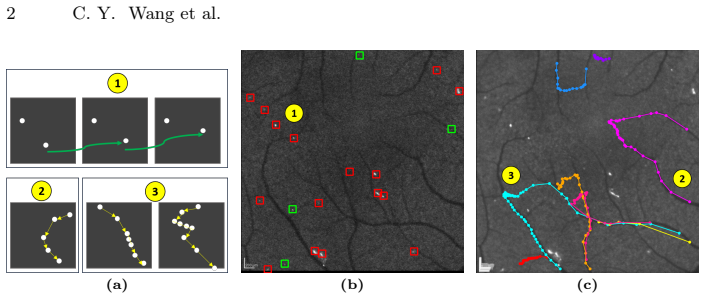
Capillary-level retinal blood flow (RBF) has strong potential as a biomarker for various ocular diseases. However, modalities for measuring capillary-level RBF remain limited. Erythrocyte-mediated angiography (EMA), an emerging imaging technique, enables capillary-level RBF measurement by visualizing individual erythrocytes, yet automated erythrocyte detection and tracking, which are essential for quantifying blood flow, remain largely unexplored. To address this gap, we propose EMTrack, a novel framework featuring a flow-context module for erythrocyte detection that distinguishes moving from paused cells and a topology-aware tracking strategy that enables tracking under large inter-frame displacements and substantial motion variations. In addition, we establish RBF-EMA, a new EMA dataset with comprehensive erythrocyte detection and tracking annotations. Experimental results demonstrate that our method outperforms baseline methods both quantitatively and qualitatively on detection and tracking tasks in the RBF-EMA dataset. Moreover, RBF quantification results highlight the strong potential of our framework for automated retinal blood flow measurement.
Editorial analysis
A structured set of objections, weighed in public.
Referee Report
Summary. The paper proposes EMTrack, a framework for erythrocyte detection and tracking in EMA images featuring a flow-context module (to distinguish moving vs. paused cells) and topology-aware tracking (for large displacements). It introduces the new RBF-EMA dataset with detection/tracking annotations and claims quantitative and qualitative outperformance over baselines on detection/tracking tasks, plus potential for automated RBF quantification.
Significance. If the results hold with validated ground truth, the work could enable scalable capillary-level RBF measurement as a biomarker for ocular diseases; the new dataset would be a useful resource for the community if annotation quality is documented.
major comments (2)
- [Dataset section (likely §4)] Dataset section (likely §4): RBF-EMA is the sole basis for the quantitative superiority claims, yet no inter-annotator agreement, annotation protocol, number of annotators, exclusion criteria, or external clinical validation is reported. This directly affects whether measured gains over baselines reflect true method improvement or annotation artifacts.
- [Abstract and §5 (Experiments)] Abstract and §5 (Experiments): The central claim of outperformance is stated without any numeric metrics, error bars, dataset split sizes, or baseline implementation details in the provided abstract; if the full experimental section similarly omits these, the empirical support for the framework's advantage is insufficient.
minor comments (1)
- [Method description] Clarify any notation for the flow-context module and topology constraints so that the method can be reproduced from the text alone.
Simulated Author's Rebuttal
We thank the referee for the constructive comments, which highlight important aspects of dataset documentation and experimental reporting. We address each major comment below and will revise the manuscript to improve clarity and completeness.
read point-by-point responses
-
Referee: Dataset section (likely §4): RBF-EMA is the sole basis for the quantitative superiority claims, yet no inter-annotator agreement, annotation protocol, number of annotators, exclusion criteria, or external clinical validation is reported. This directly affects whether measured gains over baselines reflect true method improvement or annotation artifacts.
Authors: We agree these details are essential for assessing annotation quality. In the revised manuscript, we will expand the dataset section to describe the annotation protocol, number of annotators, inter-annotator agreement (e.g., pairwise IoU and agreement rates), exclusion criteria, and any external clinical review steps performed. This addition will strengthen confidence that performance gains reflect methodological improvements rather than annotation artifacts. revision: yes
-
Referee: Abstract and §5 (Experiments): The central claim of outperformance is stated without any numeric metrics, error bars, dataset split sizes, or baseline implementation details in the provided abstract; if the full experimental section similarly omits these, the empirical support for the framework's advantage is insufficient.
Authors: The full experimental section (§5) already reports quantitative metrics (with error bars/standard deviations), dataset split sizes, and baseline implementation details. However, we concur that the abstract would be strengthened by including representative numeric results. We will revise the abstract to incorporate key metrics such as detection precision/recall and tracking accuracy scores to better support the outperformance claims. revision: yes
Circularity Check
No circularity: empirical evaluation on newly introduced dataset
full rationale
The paper introduces EMTrack (flow-context detection + topology-aware tracking) and the RBF-EMA dataset, then reports quantitative/qualitative outperformance versus baselines on detection and tracking tasks. No derivation chain exists that reduces a claimed result to its own inputs by construction; there are no fitted parameters renamed as predictions, no self-definitional equations, and no load-bearing self-citations. The evaluation is a standard empirical comparison whose validity rests on dataset quality and baseline choice rather than any internal definitional loop.
Axiom & Free-Parameter Ledger
Reference graph
Works this paper leans on
-
[1]
In: European Conference on Computer Vision
Almazán, J., Ko, B., Gu, G., Larlus, D., Kalantidis, Y.: Granularity-aware adapta- tion for image retrieval over multiple tasks. In: European Conference on Computer Vision. pp. 389–406. Springer (2022)
2022
-
[2]
Investigative ophthalmology & vi- sual science54(6), 4394–4402 (2013)
Arichika, S., Uji, A., Hangai, M., Ooto, S., Yoshimura, N.: Noninvasive and di- rect monitoring of erythrocyte aggregates in human retinal microvasculature using adaptive optics scanning laser ophthalmoscopy. Investigative ophthalmology & vi- sual science54(6), 4394–4402 (2013)
2013
-
[3]
Investigative ophthalmology & visual science48(5), 2285–2289 (2007)
Berisha, F., Feke, G.T., Trempe, C.L., McMeel, J.W., Schepens, C.L.: Retinal abnormalities in early alzheimer’s disease. Investigative ophthalmology & visual science48(5), 2285–2289 (2007)
2007
-
[4]
biorxivorg (2024)
Bragantini, J., Theodoro, I., Zhao, X., Huijben, T., Hirata-Miyasaki, E., VijayKu- mar, S., Balasubramanian, A., Lao, T., Agrawal, R., Xiao, S., et al.: Ultrack: pushing the limits of cell tracking across biological scales. biorxivorg (2024)
2024
-
[5]
In: Proceedings of the IEEE/CVF international conference on computer vision
Caron, M., Touvron, H., Misra, I., Jégou, H., Mairal, J., Bojanowski, P., Joulin, A.: Emerging properties in self-supervised vision transformers. In: Proceedings of the IEEE/CVF international conference on computer vision. pp. 9650–9660 (2021)
2021
-
[6]
Investigative Ophthalmology & Visual Science65(11), 33–33 (2024)
Chen, V.Y., Pottenburgh, J.A., Chen, S.E., Kim, S., Mayo, L., Damani, A., Cruz, M., Park, A., Im, L., Magder, L., et al.: Plexus-specific retinal capillary blood flow analysis using erythrocyte mediated angiography and optical coherence tomog- raphy angiography. Investigative Ophthalmology & Visual Science65(11), 33–33 (2024)
2024
-
[7]
Cheng, W., Li, Z., Ren, J., Jeong, H., Du, C., Pan, Y., Ling, H.: Blood flow speed estimationwithopticalcoherencetomographyangiographyimages.In:Proceedings of the Computer Vision and Pattern Recognition Conference. pp. 10466–10475 (2025)
2025
-
[8]
Nature methods11(3), 281–289 (2014)
Chenouard, N., Smal, I., De Chaumont, F., Maška, M., Sbalzarini, I.F., Gong, Y., Cardinale, J., Carthel, C., Coraluppi, S., Winter, M., et al.: Objective comparison of particle tracking methods. Nature methods11(3), 281–289 (2014)
2014
-
[9]
American journal of ophthalmology128(1), 75–80 (1999)
Ciulla, T.A., et al.: Color doppler imaging discloses reduced ocular blood flow velocities in nonexudative age-related macular degeneration. American journal of ophthalmology128(1), 75–80 (1999)
1999
-
[10]
Dijkstra, E.W.: A note on two problems in connexion with graphs. Numer. Math. 1, 269–271 (Dec 1959)
1959
-
[11]
Nu- cleic Acids Research49(13), 7292–7297 (2021)
Eichenberger, B.T., Zhan, Y., Rempfler, M., Giorgetti, L., Chao, J.A.: deepblink: threshold-independent detection and localization of diffraction-limited spots. Nu- cleic Acids Research49(13), 7292–7297 (2021)
2021
-
[12]
Ershov, D., Phan, M.S., Pylvänäinen, J.W., Rigaud, S.U., Le Blanc, L., Charles- Orszag, A., Conway, J.R., Laine, R.F., Roy, N.H., Bonazzi, D., et al.: Trackmate 7: integratingstate-of-the-artsegmentationalgorithmsintotrackingpipelines.Nature methods19(7), 829–832 (2022)
2022
-
[13]
Alzheimer’s & Dementia: Diagnosis, Assessment & Disease Monitoring1(2), 144–151 (2015) 16 C
Feke, G.T., Hyman, B.T., Stern, R.A., Pasquale, L.R.: Retinal blood flow in mild cognitive impairment and alzheimer’s disease. Alzheimer’s & Dementia: Diagnosis, Assessment & Disease Monitoring1(2), 144–151 (2015) 16 C. Y. Wang et al
2015
-
[14]
Investigative ophthalmol- ogy & visual science49(12), 5510–5516 (2008)
Flower, R., Peiretti, E., Magnani, M., Rossi, L., Serafini, S., Gryczynski, Z., Gryczynski, I.: Observation of erythrocyte dynamics in the retinal capillaries and choriocapillaris using icg-loaded erythrocyte ghost cells. Investigative ophthalmol- ogy & visual science49(12), 5510–5516 (2008)
2008
-
[15]
In: International conference on medical image computing and computer-assisted intervention
Frangi, A.F., Niessen, W.J., Vincken, K.L., Viergever, M.A.: Multiscale vessel en- hancement filtering. In: International conference on medical image computing and computer-assisted intervention. pp. 130–137. Springer (1998)
1998
-
[16]
In: European conference on computer vision
Gallusser, B., Weigert, M.: Trackastra: Transformer-based cell tracking for live-cell microscopy. In: European conference on computer vision. pp. 467–484. Springer (2024)
2024
-
[17]
Investigative ophthalmology & visual science36(5), 864–870 (1995)
Goebel, W., Lieb, W.E., Ho, A., Sergott, R.C., Farhoumand, R., Grehn, F.: Color doppler imaging: a new technique to assess orbital blood flow in patients with diabetic retinopathy. Investigative ophthalmology & visual science36(5), 864–870 (1995)
1995
-
[18]
In: 11th international database engineering and applications symposium (IDEAS 2007)
Guttoski, P.B., Sunye, M.S., Silva, F.: Kruskal’s algorithm for query tree opti- mization. In: 11th international database engineering and applications symposium (IDEAS 2007). pp. 296–302. IEEE (2007)
2007
-
[19]
Artificial Intelligence in Medicine144, 102664 (2023)
He, B., Lei, J., Lang, X., Li, Z., Cui, W., Zhang, Y.: Ultra-fast ultrasound blood flow velocimetry for carotid artery with deep learning. Artificial Intelligence in Medicine144, 102664 (2023)
2023
-
[20]
Biomedical Optics Express14(6), 2658–2677 (2023)
Hwang, Y., Won, J., Yaghy, A., Takahashi, H., Girgis, J.M., Lam, K., Chen, S., Moult, E.M., Ploner, S.B., Maier, A., et al.: Retinal blood flow speed quantification at the capillary level using temporal autocorrelation fitting octa. Biomedical Optics Express14(6), 2658–2677 (2023)
2023
-
[21]
Adam: A Method for Stochastic Optimization
Kingma, D.P., Ba, J.: Adam: A method for stochastic optimization. arXiv preprint arXiv:1412.6980 (2014)
work page internal anchor Pith review Pith/arXiv arXiv 2014
-
[22]
JAMA ophthalmology135(3), 244–251 (2017)
Lee, B., et al.: En face doppler optical coherence tomography measurement of total retinal blood flow in diabetic retinopathy and diabetic macular edema. JAMA ophthalmology135(3), 244–251 (2017)
2017
-
[23]
Iscience26(1) (2023)
Li, J., Wang, D., Pottenburgh, J., Bower, A.J., Asanad, S., Lai, E.W., Simon, C., Im, L., Huryn, L.A., Tao, Y., et al.: Visualization of erythrocyte stasis in the living human eye in health and disease. Iscience26(1) (2023)
2023
-
[24]
Analysis of approximate nearest neighbor searching with clustered point sets
Maneewongvatana, S., Mount, D.M.: Analysis of approximate nearest neighbor searching with clustered point sets. arXiv preprint cs/9901013 (1999)
work page internal anchor Pith review Pith/arXiv arXiv 1999
-
[25]
Nature Methods pp
Marks, M., Israel, U., Dilip, R., Li, Q., Yu, C., Laubscher, E., Iqbal, A., Pradhan, E., Ates, A., Abt, M., et al.: Cellsam: a foundation model for cell segmentation. Nature Methods pp. 1–9 (2025)
2025
-
[26]
In: Proceedings of the IEEE/CVF conference on computer vision and pattern recognition
Melas-Kyriazi, L., Rupprecht, C., Laina, I., Vedaldi, A.: Deep spectral methods: A surprisingly strong baseline for unsupervised semantic segmentation and localiza- tion. In: Proceedings of the IEEE/CVF conference on computer vision and pattern recognition. pp. 8364–8375 (2022)
2022
-
[27]
Journal of glaucoma5(5), 308–310 (1996)
Nicolela, M.T., Walman, B.E., Buckley, A.R., Drance, S.M.: Ocular hypertension and primary open-angle glaucoma: a comparative study of their retrobulbar blood flow velocity. Journal of glaucoma5(5), 308–310 (1996)
1996
-
[28]
British Medical Journal305(6855), 678–683 (1992)
Patel, V., Rassam, S., Newsom, R., Wiek, J., Kohner, E.: Retinal blood flow in diabetic retinopathy. British Medical Journal305(6855), 678–683 (1992)
1992
-
[29]
Biomedical optics express8(5), 2536–2562 (2017)
Pircher, M., Zawadzki, R.J.: Review of adaptive optics oct (ao-oct): principles and applications for retinal imaging. Biomedical optics express8(5), 2536–2562 (2017)
2017
-
[30]
Acta ophthalmologica88(6), 622–629 (2010) Automated Erythrocyte Detection and Tracking 17
Riva, C.E., Geiser, M., Petrig, B.L., Association, O.B.F.R.: Ocular blood flow assessment using continuous laser doppler flowmetry. Acta ophthalmologica88(6), 622–629 (2010) Automated Erythrocyte Detection and Tracking 17
2010
-
[31]
Investigative Ophthalmology & Visual Science59(9), 3950–3950 (2018)
Saeedi, O., Tracey, B., Renner, C., Li, J., Shah, K., Tsai, J., Chang, L., Ou, M.: Determination of absolute erythrocyte velocity and flow in the human retinal mi- crovasculature by direct visualization of icg-labelled erythrocytes. Investigative Ophthalmology & Visual Science59(9), 3950–3950 (2018)
2018
-
[32]
Methods115, 80–90 (2017)
Tinevez, J.Y., Perry, N., Schindelin, J., Hoopes, G.M., Reynolds, G.D., Laplantine, E.,Bednarek,S.Y.,Shorte,S.L.,Eliceiri,K.W.:Trackmate:Anopenandextensible platform for single-particle tracking. Methods115, 80–90 (2017)
2017
-
[33]
In: European Conference on Computer Vision
Tumanyan, N., Singer, A., Bagon, S., Dekel, T.: Dino-tracker: Taming dino for self- supervised point tracking in a single video. In: European Conference on Computer Vision. pp. 367–385. Springer (2024)
2024
-
[34]
Virtanen, P., Gommers, R., Oliphant, T.E., Haberland, M., Reddy, T., Cour- napeau, F., Peterson, E., Muller, A., Klőckner, A., Brett, M., et al.: SciPy 1.0: Fundamental Algorithms for Scientific Computing in Python (2020).https: //doi.org/10.1038/s41592-019-0686-2, https://rdcu.be
-
[35]
Cell systems6(4), 496–507 (2018)
van Vliet, S., Winkler, A.R., Spriewald, S., Stecher, B., Ackermann, M., et al.: Spatially correlated gene expression in bacterial groups: the role of lineage history, spatial gradients, and cell-cell interactions. Cell systems6(4), 496–507 (2018)
2018
-
[36]
PeerJ 2, e453 (2014)
Van der Walt, S., Schönberger, J.L., Nunez-Iglesias, J., Boulogne, F., Warner, J.D., Yager, N., Gouillart, E., Yu, T.: scikit-image: image processing in python. PeerJ 2, e453 (2014)
2014
-
[37]
Biomedical Optics Express15(5), 3457–3479 (2024)
Wang, C.Y., Sadrieh, F.K., Shen, Y.T., Chen, S.E., Kim, S., Chen, V., Raghaven- dra, A., Wang, D., Saeedi, O., Tao, Y.: Memo: dataset and methods for robust multimodal retinal image registration with large or small vessel density differences. Biomedical Optics Express15(5), 3457–3479 (2024)
2024
-
[38]
Biomedical optics express10(7), 3681–3697 (2019)
Wang, D., Haytham, A., Mayo, L., Tao, Y., Saeedi, O.: Automated retinal mi- crovascular velocimetry based on erythrocyte mediated angiography. Biomedical optics express10(7), 3681–3697 (2019)
2019
-
[39]
In: Proceedings of the IEEE/CVF international conference on computer vision
Zhang, L., Rao, A., Agrawala, M.: Adding conditional control to text-to-image diffusion models. In: Proceedings of the IEEE/CVF international conference on computer vision. pp. 3836–3847 (2023)
2023
-
[40]
In: Proceedings of the IEEE/CVF International Conference on Computer Vision
Zhang, W., Liu, H., Li, B., He, J., Qi, Z., Wang, Y., Zhao, S., Yu, X., Zeng, W., Jin, X.: Hybrid-grained feature aggregation with coarse-to-fine language guidance for self-supervised monocular depth estimation. In: Proceedings of the IEEE/CVF International Conference on Computer Vision. pp. 6678–6692 (2025)
2025
-
[41]
In: European conference on computer vision
Zhang, Y., Sun, P., Jiang, Y., Yu, D., Weng, F., Yuan, Z., Luo, P., Liu, W., Wang, X.: Bytetrack: Multi-object tracking by associating every detection box. In: European conference on computer vision. pp. 1–21. Springer (2022)
2022
-
[42]
In: International Conference on Medical Image Comput- ing and Computer-Assisted Intervention
Zhang, Y., Yang, G.: A motion transformer for single particle tracking in fluores- cence microscopy images. In: International Conference on Medical Image Comput- ing and Computer-Assisted Intervention. pp. 503–513. Springer (2023)
2023
-
[43]
Nature Methods pp
Zhou, H., Kim, S., Zhao, Z., Fan, J., Huang, W., Sui, X., Shao, L., An, H., Zhang, J.R., Wu, J., et al.: Cellect: contrastive embedding learning for large-scale efficient cell tracking. Nature Methods pp. 1–12 (2025)
2025
discussion (0)
Sign in with ORCID, Apple, or X to comment. Anyone can read and Pith papers without signing in.