REVIEW 4 major objections 6 minor 1 cited by
End-to-End Deep Learning for Interior Tomography with Low-Dose X-ray CT
T0 review · 4 major / 6 minor · reviewed 2026-08-10 · deepseek-v4-flash
Pith's one-line read The paper claims that image-domain CNNs cannot efficiently remove coupled cupping-plus-noise artifacts in low-dose interior CT, and that a dual-domain network which decouples the problem in projection space restores the low-rank condition…
desk verdict Useful dual-domain architecture with consistent empirical gains, but the theoretical rank argument is not established and the evaluation is thin; worth sending to review with clear requests for revision. read the letter →
The pith
A machine-rendered reading of the paper's core claim, the machinery that carries it, and where it could break.
The reading
What carries the argument
The central object is the Hankel structured matrix $H_d(f)$ and the rank condition from deep convolutional framelets, together with the decoupling mask pair $T$ and $1-T$ in the projection domain. The framelets identity $H_d(f) = \tilde{\Phi}\Phi^T H_d(f)\Psi\tilde{\Psi}^T$ and the fact that the Hankel rank equals the number of nonzero Fourier components of the image imply that a CNN with filter length $d$ can represent only artifacts whose rank stays below a bound; the coupled cupping-plus-noise artifact violates this because its Fourier support fills the spectrum. The proposed network restores the low-rank condition by splitting the projection data into two regions: a noise-estimation bridge inside $T$ and an extrapolation bridge outside $(1-T)$, so each subproblem has a narrow Fourier support and satisfies the framelets condition.
What would settle it
Compute the Fourier support or Hankel-structured rank of the artifact directly from a real scanner's truncated low-dose projections, and train an image-domain CNN whose convolutional filter length matches the signal length; if the image-domain network then matches the dual-domain network's quality, the filter-length bound is not the limiting factor and the proposed explanation would be wrong even if the method still works.
Extended reading notes
Core claim
The paper's central claim is that image-domain CNNs fail at coupled low-dose interior CT not because of capacity but because of low-rankness: for the coupled artifact $k = c_I + n_I$, the rank of the Hankel structured matrix $H_d(k)$ is close to the signal length, exceeding the filter length $d$ that a practical CNN can implement, so the network's kernels cannot satisfy the projection condition required by deep convolutional framelets. The remedy is that in the projection domain the two artifacts separate: the low-dose noise lives inside the truncation mask $T$, where it corrupts the measured sinogram, and the cupping artifact corresponds to the missing data outside $T$. The proposed projection-domain CNN therefore solves two decoupled low-rank subproblems — denoising the measured projections inside $T$ and extrapolating the projections outside $T$ — after which the filtered backprojection image enters a second image-domain CNN. The authors report that this projection-domain-first design outperforms the same-size image-domain CNN, a U-Net with twice the parameters, and a W-Net unrolled twice in the image domain, and that it also improves the isolated interior-tomography and low-dose problems.
Load-bearing premise
The load-bearing premise is that cupping artifacts sit in the low-frequency range while low-dose image noise sits in the high-frequency range, so their combined artifact spans nearly the whole spectrum and exceeds what a network with practical filter sizes can represent; if real truncated low-dose scans do not separate this way, the theoretical motivation for the dual-domain design loses its force.
Editorial extensions
If this is right
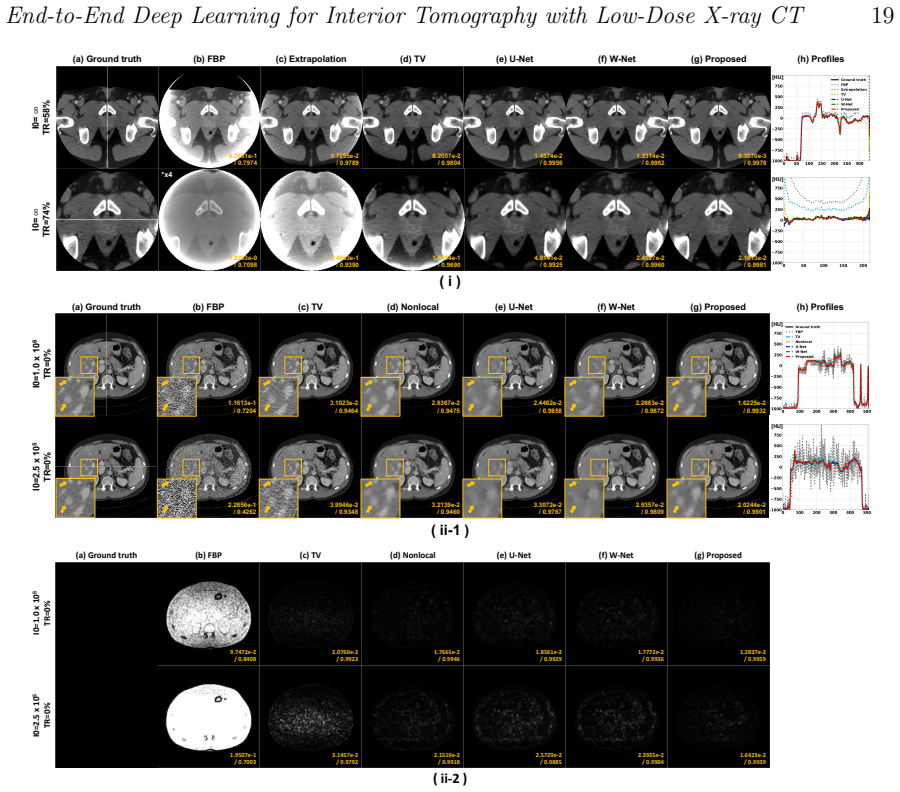
- On the ten-subject dataset with simulated fan-beam truncation and Poisson low-dose noise, the proposed network achieves the best NMSE and SSIM at every tested photon count and truncation ratio.
- A projection-domain CNN used as the first stage outperforms an image-domain CNN with the same architecture, showing the training domain matters for coupled artifacts.
- The singular-value spectra of the backbone's last feature maps are lower for the projection-domain CNN, consistent with the claim that it better satisfies the low-rank condition.
- Two-times unrolled networks beat a single larger network: the W-Net with fewer parameters outperforms a U-Net with roughly twice the parameters.
- The projection-domain-first design remains superior even when the problems are isolated, i.e., when there is no truncation or no projection noise.
Reading between the lines
- Beyond the paper, the same decoupling logic should transfer to other coupled CT distortions, such as sparse-view streaking combined with motion artifacts, whenever the two artifact types occupy disjoint regions of the projection or frequency domain.
- The authors' argument implies a testable failure threshold: an image-domain CNN should degrade sharply once the Fourier support of the coupled artifact exceeds the network's effective filter length, so plotting reconstruction quality against filter length would directly probe the proposed mechanism.
- If the mechanism holds, projection-domain auxiliary losses could regularize image-domain networks in general, since the paper treats projection data and FBP images as carrying the same information up to a linear transform.
Signed reviews
Editorial analysis
A structured set of objections, weighed in public.
Referee Report
Summary. This manuscript addresses combined interior (region-of-interest) and low-dose X-ray CT reconstruction. The authors argue, via the theory of deep convolutional framelets, that an image-domain CNN cannot efficiently remove the coupled cupping-plus-noise artifact because the combined artifact has a near-full Fourier support and hence a Hankel-matrix rank close to the signal length, exceeding the filter-length bound. They therefore propose a dual-domain network: a projection-domain CNN with two bridge modules, one estimating projection noise inside the measured (truncated) region and one extrapolating projections outside it, followed by FBP and an image-domain CNN. End-to-end training with an FBP layer is described. Experiments on simulated truncated low-dose projections from 10 AAPM Low-Dose CT Grand Challenge subjects (9 training, 1 test) compare the proposed Dual-Net against U-Net, W-Net, TV, nonlocal prior, and extrapolation methods, reporting consistent NMSE/SSIM/PSNR improvements across six truncation ratios and six photon-count levels.
Significance. If the empirical claims hold, the paper would make a useful contribution to combining interior and low-dose CT reconstruction: the dual-domain formulation is a natural and technically sound way to separate a measurement-domain noise problem from a missing-data (extrapolation) problem, and the consistent gains over U-Net and W-Net across all tested configurations are encouraging. The inclusion of an FBP layer in an end-to-end trainable pipeline, the controlled comparison of one-time and two-time unrolled architectures, and the comparison with classical methods (TV, nonlocal prior, extrapolation) are strengths. However, the significance is currently limited by the narrow experimental basis: all quantitative conclusions rest on a single test subject, with no error bars or statistical testing, and the central theoretical explanation, while interesting, is not rigorously established for the actual data.
major comments (4)
- [Sec. 4.1, Eq. (23)] The rank inequality RANKH_d(k) <= r*_max ≈ d is derived from Eq. (9), RANKH_d(f) = COUNT(F(f)≠0), but this identity is not exact for finite-length arbitrary signals; it holds only for signals composed of a few complex exponentials when the matrix pencil parameter d is sufficiently large. Low-dose CT image noise is roughly broadband, not a sum of a few exponentials, so its Hankel-matrix rank is not necessarily bounded by a small r_max, and the cupping artifact contains boundary singularities at the ROI edge that spread energy across the Fourier domain. The spectral separation in Fig. 4 is only qualitative, with no quantitative bandlimit measurement. Consequently, Eq. (23) does not follow, and the claimed theoretical reason that image-domain CNNs fail on the coupled artifact is not established. This is the load-bearing theoretical claim of the paper, so it needs either a rigorous demonstration on the actual artifact distributions or a substantial softening of the claim.
- [Sec. 6.2, Fig. 9] The experimental check of low-rankness is performed on the Hankel singular values of the last feature maps of the trained backbone networks, not on the input artifacts (the coupled image-domain artifact k or the projection-domain data p_T and its components). Thus Fig. 9 does not validate the input-side rank claim of Eq. (23); it only shows that the projection-domain CNN learns lower-rank feature representations. The paper should either measure the rank of the actual input artifact (e.g., via the Hankel matrix of q_I in the image domain and of p_T in the projection domain) or explicitly state that Fig. 9 is a feature-space observation, not a test of the theoretical premise.
- [Sec. 3.1 and Tables 2-5] All quantitative results are computed on a single test subject (486 slices) from the AAPM challenge, with point estimates only and no standard deviations, confidence intervals, or statistical significance tests. The improvement of the proposed method over W-Net is consistent in the tables, but a journal-level claim of superiority requires at least multiple test subjects or a per-slice variability analysis. The paper should also release code and, where permissible, the test dataset or a reproducible simulation pipeline, because the current evaluation is not independently verifiable.
- [Sec. 4.2 and Table 1(e)] The paper claims that 'the capacity of the neural network Q is fixed, but it is possible to apply low-rank constraints to individual regions while satisfying the theory of the deep convolutional framelets.' However, no low-rank constraint is actually enforced in the objective functions of Table 1; the two bridge heads share a single backbone and are jointly optimized with a weighted sum of two losses. The low-rank behavior is only observed post hoc in Fig. 9. The link between the loss decomposition and the low-rank condition is therefore asserted rather than demonstrated. An ablation that trains with explicit rank regularization or that compares separate backbones for the two heads would help support the claim.
minor comments (6)
- [Sec. 2.1, affiliation footer] The affiliation contains a typo: 'Bostan' should be 'Boston'.
- [Eq. (17)] The PSNR formula uses N M ||f^*||_∞ in the numerator, which is not the standard PSNR definition (which uses the maximum pixel value, not the number of pixels times the maximum). This appears to be a typo and should be corrected, since it affects the reported PSNR values in Tables 4 and 5.
- [Sec. 3.3] The training is described as 'end-to-end,' but the gradient of the second network is blocked from propagating to the first network. This is a specific design choice; clarify that the first network is trained on its own loss plus an image-domain loss term, but not on the final image loss, and discuss the implication for the term 'end-to-end.'
- [Sec. 3.2] The statement that 'the U-Net is set up to twice the size of the channels of other networks' is clear from Fig. 5 but could be stated more precisely: the U-Net's first layer has 64 channels while the others have 32, giving approximately twice the parameters.
- [Sec. 6.3] The authors note that 'the NMSE and SSIM valuse do not seem to reflect the well-corrected image quality' and then recompute metrics in body regions. This admission suggests that global image metrics may be misleading for texture-preservation; consider including the body-region metrics in the main tables or justifying the choice of global metrics.
- [Fig. 4] The Fourier spectra of image noise and cupping artifact are shown without a quantitative scale or a quantitative measure of 'dominant low/high frequency support.' A simple percentage-of-energy threshold would make the spectral-separation premise testable.
Circularity Check
No circularity: the empirical comparison is external and the self-cited framelets theory is independent mathematical support; the rank argument is a validity concern, not a circular reduction.
full rationale
No load-bearing circular step is present. The paper's derivation chain is: (i) an empirical observation that image-domain CNNs underperform on combined truncation and low-dose artifacts; (ii) a theoretical explanation using deep convolutional framelets [23], a published mathematical framework with independent derivations; (iii) a proposed dual-domain architecture whose losses are explicitly stated in Table 1 and trained on AAPM data; and (iv) quantitative evaluation on a held-out test subject. Each part is distinct from the inputs. The framelets theory is self-cited, but it is invoked as general prior mathematics, not as a conclusion derived from this paper's fitted parameters or network outputs, so it does not constitute circularity. The Sec. 4.1 rank argument (Eq. 23) rests on a spectral-separation assumption (Fig. 4) and on Eq. 9 imported from prior work; if those premises are false the theoretical explanation is weakened, but that is a correctness or validity concern, not circularity, because no quantity in Eq. 23 is defined in terms of the proposed network's own outputs. The projection-domain decoupling in Eqs. 24-26 restates the forward model p = y + h and the truncation mask T; this is a modeling decomposition rather than a prediction obtained from a fit. Tables 2-5 and Figs. 7-10 compare methods on a held-out test subject, so the empirical claim is self-contained against external benchmarks. Fig. 9's singular-value spectra are computed from trained feature maps, an indirect check of the theory, not a fitted parameter renamed as a prediction. In short, the paper's central empirical contribution stands independently, and the cited theory provides motivation rather than a circular foundation.
Assumptions & free parameters
assumptions (4)
- standard math The rank of a Hankel structured matrix equals the number of nonzero Fourier components (Eq. 9).
- ad hoc to paper Cupping artifacts have dominant low-frequency support and image noise has dominant high-frequency support; their sum fills the Fourier domain and therefore has high Hankel rank.
- domain assumption Truncated projection data create a cupping artifact that can be corrected by extrapolating the missing projection region (1-T).
- domain assumption Low-dose projection data are well modeled as noiseless projection plus non-stationary Gaussian noise.
Cite this review
Pith. "Pith review of End-to-End Deep Learning for Interior Tomography with Low-Dose X-ray CT." pith.science (2026). https://pith.science/paper/NYHGNHGB
@misc{pith2026250105085,
  author       = {Pith},
  title        = {Pith review of: End-to-End Deep Learning for Interior Tomography with Low-Dose X-ray CT},
  year         = {2026},
  howpublished = {\url{https://pith.science/paper/NYHGNHGB}},
  note         = {Machine review of arXiv:2501.05085}
}
read the original abstract
Objective: There exist several X-ray computed tomography (CT) scanning strategies to reduce a radiation dose, such as (1) sparse-view CT, (2) low-dose CT, and (3) region-of-interest (ROI) CT (called interior tomography). To further reduce the dose, the sparse-view and/or low-dose CT settings can be applied together with interior tomography. Interior tomography has various advantages in terms of reducing the number of detectors and decreasing the X-ray radiation dose. However, a large patient or small field-of-view (FOV) detector can cause truncated projections, and then the reconstructed images suffer from severe cupping artifacts. In addition, although the low-dose CT can reduce the radiation exposure dose, analytic reconstruction algorithms produce image noise. Recently, many researchers have utilized image-domain deep learning (DL) approaches to remove each artifact and demonstrated impressive performances, and the theory of deep convolutional framelets supports the reason for the performance improvement. Approach: In this paper, we found that the image-domain convolutional neural network (CNN) is difficult to solve coupled artifacts, based on deep convolutional framelets. Significance: To address the coupled problem, we decouple it into two sub-problems: (i) image domain noise reduction inside truncated projection to solve low-dose CT problem and (ii) extrapolation of projection outside truncated projection to solve the ROI CT problem. The decoupled sub-problems are solved directly with a novel proposed end-to-end learning using dual-domain CNNs. Main results: We demonstrate that the proposed method outperforms the conventional image-domain deep learning methods, and a projection-domain CNN shows better performance than the image-domain CNNs which are commonly used by many researchers.
Figures


Figures from the paper (7 more)


Forward citations
Cited by 1 Pith paper
-
FORCE-Interior: Measurement-Consistent Adaptation of a Poisson-Flow Generative Prior for Interior CT
A Poisson-flow generative prior, initialized with a full-FOV OS-SART reconstruction and re-conditioned on truncated projections each step, improves interior-tomography ROI reconstruction quality at small ROI radii.
Reference graph
Works this paper leans on
-
[1]
Nikhil Bharat Shah and Shari L Platt. Alara: is there a cause for alarm? reducing radiation risks from computed tomography scanning in children. Current opinion in pediatrics, 20(3):243–247, 2008
work page 2008
-
[2]
Modelling the physics in the iterative reconstruction for transmission computed tomography
Johan Nuyts, Bruno De Man, Jeffrey A Fessler, Wojciech Zbijewski, and Freek J Beekman. Modelling the physics in the iterative reconstruction for transmission computed tomography. Physics in Medicine & Biology, 58(12):R63, 2013
work page 2013
-
[3]
Algorithm to extend reconstruction field-of-view
Jiang Hsieh, Ed Chao, J Thibault, Brian Grekowicz, Amy Horst, Scott McOlash, and TJ Myers. Algorithm to extend reconstruction field-of-view. In 2004 2nd IEEE International Symposium on Biomedical Imaging: Nano to Macro (IEEE Cat No. 04EX821), pages 1404–1407. IEEE, 2004
work page 2004
-
[4]
A novel reconstruction algorithm to extend the ct scan field-of-view
J Hsieh, E Chao, J Thibault, B Grekowicz, A Horst, S McOlash, and TJ Myers. A novel reconstruction algorithm to extend the ct scan field-of-view. Medical physics, 31(9):2385–2391, 2004
work page 2004
-
[5]
Compressed sensing based interior tomography
Hengyong Yu and Ge Wang. Compressed sensing based interior tomography. Physics in medicine & biology, 54(9):2791, 2009
work page 2009
-
[6]
Interior tomography using 1d generalized total variation
John Paul Ward, Minji Lee, Jong Chul Ye, and Michael Unser. Interior tomography using 1d generalized total variation. part i: Mathematical foundation. SIAM Journal on Imaging Sciences, 8(1):226–247, 2015
work page 2015
-
[7]
Interior tomography using 1d generalized total variation
Minji Lee, Yoseob Han, John Paul Ward, Michael Unser, and Jong Chul Ye. Interior tomography using 1d generalized total variation. part ii: Multiscale implementation. SIAM Journal on Imaging Sciences, 8(4):2452–2486, 2015
work page 2015
-
[8]
Xiaochuan Pan, Emil Y Sidky, and Michael Vannier. Why do commercial ct scanners still employ traditional, filtered back-projection for image reconstruction? Inverse problems, 25(12):123009, 2009
work page 2009
Show all 37 references
-
[9]
Evaluation of sparse-view reconstruction from flat-panel-detector cone- beam ct
Junguo Bian, Jeffrey H Siewerdsen, Xiao Han, Emil Y Sidky, Jerry L Prince, Charles A Pelizzari, and Xiaochuan Pan. Evaluation of sparse-view reconstruction from flat-panel-detector cone- beam ct. Physics in Medicine & Biology, 55(22):6575, 2010
2010
-
[10]
Few-view image reconstruction with dual dictionaries
Yang Lu, Jun Zhao, and Ge Wang. Few-view image reconstruction with dual dictionaries. Physics in Medicine & Biology, 57(1):173, 2011
2011
-
[11]
Sparse-view spectral ct reconstruction using spectral patch-based low-rank penalty
Kyungsang Kim, Jong Chul Ye, William Worstell, Jinsong Ouyang, Yothin Rakvongthai, Georges El Fakhri, and Quanzheng Li. Sparse-view spectral ct reconstruction using spectral patch-based low-rank penalty. IEEE transactions on medical imaging, 34(3):748–760, 2014
2014
-
[12]
Effects of sparse sampling schemes on image quality in low-dose ct
Sajid Abbas, Taewon Lee, Sukyoung Shin, Rena Lee, and Seungryong Cho. Effects of sparse sampling schemes on image quality in low-dose ct. Medical physics, 40(11):111915, 2013
2013
-
[13]
Iterative reconstruction methods in x-ray ct
Marcel Beister, Daniel Kolditz, and Willi A Kalender. Iterative reconstruction methods in x-ray ct. Physica medica, 28(2):94–108, 2012
2012
-
[14]
A splitting-based iterative algorithm for accelerated statistical x-ray ct reconstruction
Sathish Ramani and Jeffrey A Fessler. A splitting-based iterative algorithm for accelerated statistical x-ray ct reconstruction. IEEE transactions on medical imaging, 31(3):677–688, 2011. End-to-End Deep Learning for Interior Tomography with Low-Dose X-ray CT 22
2011
-
[15]
Deep convolutional neural network for inverse problems in imaging
Kyong Hwan Jin, Michael T McCann, Emmanuel Froustey, and Michael Unser. Deep convolutional neural network for inverse problems in imaging. IEEE Transactions on Image Processing, 26(9):4509–4522, 2017
2017
-
[16]
Deep-neural- network-based sinogram synthesis for sparse-view ct image reconstruction
Hoyeon Lee, Jongha Lee, Hyeongseok Kim, Byungchul Cho, and Seungryong Cho. Deep-neural- network-based sinogram synthesis for sparse-view ct image reconstruction. IEEE Transactions on Radiation and Plasma Medical Sciences, 3(2):109–119, 2018
2018
-
[17]
Framing u-net via deep convolutional framelets: Application to sparse-view ct
Yoseob Han and Jong Chul Ye. Framing u-net via deep convolutional framelets: Application to sparse-view ct. IEEE transactions on medical imaging, 37(6):1418–1429, 2018
2018
-
[18]
Generative adversarial networks for noise reduction in low-dose ct
Jelmer M Wolterink, Tim Leiner, Max A Viergever, and Ivana Iˇ sgum. Generative adversarial networks for noise reduction in low-dose ct. IEEE transactions on medical imaging, 36(12):2536– 2545, 2017
2017
-
[19]
Low-dose ct with a residual encoder-decoder convolutional neural network
Hu Chen, Yi Zhang, Mannudeep K Kalra, Feng Lin, Yang Chen, Peixi Liao, Jiliu Zhou, and Ge Wang. Low-dose ct with a residual encoder-decoder convolutional neural network. IEEE transactions on medical imaging, 36(12):2524–2535, 2017
2017
-
[20]
A deep convolutional neural network using directional wavelets for low-dose x-ray ct reconstruction
Eunhee Kang, Junhong Min, and Jong Chul Ye. A deep convolutional neural network using directional wavelets for low-dose x-ray ct reconstruction. Medical physics, 44(10):e360–e375, 2017
2017
-
[21]
Deep learning interior tomography for region-of- interest reconstruction
Yoseob Han, Jawook Gu, and Jong Chul Ye. Deep learning interior tomography for region-of- interest reconstruction. arXiv preprint arXiv:1712.10248, 2017
2017 arXiv
-
[22]
One network to solve all rois: Deep learning ct for any roi using differentiated backprojection
Yoseob Han and Jong Chul Ye. One network to solve all rois: Deep learning ct for any roi using differentiated backprojection. Medical Physics, 46(12):e855–e872, 2019
2019
-
[23]
Deep convolutional framelets: A general deep learning framework for inverse problems
Jong Chul Ye, Yoseob Han, and Eunju Cha. Deep convolutional framelets: A general deep learning framework for inverse problems. SIAM Journal on Imaging Sciences, 11(2):991–1048, 2018
2018
-
[24]
Computed tomography: principles, design, artifacts, and recent advances, volume
Jiang Hsieh. Computed tomography: principles, design, artifacts, and recent advances, volume
-
[25]
Noise reduction with low dose ct data based on a modified rof model
Yining Zhu, Mengliu Zhao, Yunsong Zhao, Hongwei Li, and Peng Zhang. Noise reduction with low dose ct data based on a modified rof model. Optics express, 20(16):17987–18004, 2012
2012
-
[26]
Understanding geometry of encoder-decoder cnns
Jong Chul Ye and Woon Kyoung Sung. Understanding geometry of encoder-decoder cnns. In International Conference on Machine Learning, pages 7064–7073. PMLR, 2019
2019
-
[27]
Annihilating filter-based low-rank hankel matrix approach for image inpainting
Kyong Hwan Jin and Jong Chul Ye. Annihilating filter-based low-rank hankel matrix approach for image inpainting. IEEE Transactions on Image Processing, 24(11):3498–3511, 2015
2015
-
[28]
Sparse and low-rank decomposition of a hankel structured matrix for impulse noise removal
Kyong Hwan Jin and Jong Chul Ye. Sparse and low-rank decomposition of a hankel structured matrix for impulse noise removal. IEEE Transactions on Image Processing, 27(3):1448–1461, 2017
2017
-
[29]
A general framework for compressed sensing and parallel mri using annihilating filter based low-rank hankel matrix
Kyong Hwan Jin, Dongwook Lee, and Jong Chul Ye. A general framework for compressed sensing and parallel mri using annihilating filter based low-rank hankel matrix. IEEE Transactions on Computational Imaging, 2(4):480–495, 2016
2016
-
[30]
Acceleration of mr parameter mapping using annihilating filter-based low rank hankel matrix (aloha)
Dongwook Lee, Kyong Hwan Jin, Eung Yeop Kim, Sung-Hong Park, and Jong Chul Ye. Acceleration of mr parameter mapping using annihilating filter-based low rank hankel matrix (aloha). Magnetic resonance in medicine, 76(6):1848–1864, 2016
2016
-
[31]
Reference-free single-pass epi n yquist ghost correction using annihilating filter-based low rank h ankel matrix (aloha)
Juyoung Lee, Kyong Hwan Jin, and Jong Chul Ye. Reference-free single-pass epi n yquist ghost correction using annihilating filter-based low rank h ankel matrix (aloha). Magnetic resonance in medicine, 76(6):1775–1789, 2016
2016
-
[32]
U-net: Convolutional networks for biomedical image segmentation
Olaf Ronneberger, Philipp Fischer, and Thomas Brox. U-net: Convolutional networks for biomedical image segmentation. In International Conference on Medical image computing and computer-assisted intervention, pages 234–241. Springer, 2015
2015
-
[33]
Tu-fg-207a-04: Overview of the low dose ct grand challenge
C McCollough. Tu-fg-207a-04: Overview of the low dose ct grand challenge. Medical physics, 43(6Part35):3759–3760, 2016
2016
-
[34]
Development and validation of a practical lower-dose-simulation tool for optimizing computed tomography scan protocols
Lifeng Yu, Maria Shiung, Dayna Jondal, and Cynthia H McCollough. Development and validation of a practical lower-dose-simulation tool for optimizing computed tomography scan protocols. Journal of computer assisted tomography, 36(4):477–487, 2012. End-to-End Deep Learning for I...
2012
-
[35]
A dual-domain cnn-based network for ct reconstruction
Fengyuan Jiao, Zhiguo Gui, Kunpeng Li, Hong Shangguang, Yanling Wang, Yi Liu, and Pengcheng Zhang. A dual-domain cnn-based network for ct reconstruction. IEEE Access, 9:71091–71103, 2021
2021
-
[36]
Kiki-net: cross-domain convolutional neural networks for reconstructing undersampled magnetic resonance images
Taejoon Eo, Yohan Jun, Taeseong Kim, Jinseong Jang, Ho-Joon Lee, and Dosik Hwang. Kiki-net: cross-domain convolutional neural networks for reconstructing undersampled magnetic resonance images. Magnetic resonance in medicine, 80(5):2188–2201, 2018
2018
-
[37]
Low-dose ct reconstruction using spatially encoded nonlocal penalty
Kyungsang Kim, Georges El Fakhri, and Quanzheng Li. Low-dose ct reconstruction using spatially encoded nonlocal penalty. Medical physics, 44(10):e376–e390, 2017
2017
Reviewed August 10, 2026 · model on record in the stance chip above.
Discussion (0). Continue with ORCID to comment.