REVIEW 3 major objections 5 minor 8 references
Using Computer Vision for Skin Disease Diagnosis in Bangladesh Enhancing Interpretability and Transparency in Deep Learning Models for Skin Cancer Classification
T0 review · 3 major / 5 minor · reviewed 2026-08-10 · deepseek-v4-flash
Pith's one-line read The paper claims that a custom convolutional network can classify skin-lesion images with high accuracy and can show clinicians the image regions behind each decision.
desk verdict The paper promises saliency and attention maps but never produces them; the accuracy claim is also undermined by test-set filtering, so neither half of the central claim survives. read the letter →
The pith
A machine-rendered reading of the paper's core claim, the machinery that carries it, and where it could break.
The reading
What carries the argument
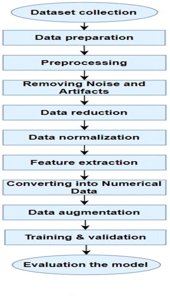
The load-bearing machinery is the custom DCNN pipeline: a preprocessing stage that removes reflective artifacts via a local-mean threshold, manually discards blurry and low-contrast images, normalizes and augments the data, and then a convolutional architecture with max pooling, binary cross-entropy loss, the Adam optimizer, and a final sigmoid layer. Interpretability comes from saliency maps, which mark pixel-level importance derived from the model's gradients, and attention maps, which show the regions the network focuses on; these are overlaid on the input images so a clinician can see where the model is looking. These maps are the mechanism claimed to add transparency without an accuracy penalty.
What would settle it
Re-run evaluation on the original unfiltered HAM10000 images, including the ones removed as blurry or artifact-heavy, and check whether accuracy and AUC stay near the reported values; they should if the filter was not responsible for the result.
Extended reading notes
Core claim
The central claim is that a custom DCNN, trained on HAM10000 after reflective-artifact removal, manual exclusion of blurry and low-contrast images, class balancing, and PCA-based augmentation, and using a sigmoid output for binary classification, outperforms five transfer-learning baselines on the same data. The authors report testing accuracy around 90 percent, an ROC AUC of 0.847, and faster per-epoch training than the baselines. They further argue that overlaying saliency maps and attention maps on input images highlights the pixels the model weighs most heavily, giving clinicians a visual explanation for each benign-versus-malignant decision, and claim this interpretability comes without materially reducing accuracy.
Load-bearing premise
The whole accuracy result depends on the assumption that deleting images because they look blurry, low-contrast, or artifact-heavy does not also delete a meaningful slice of the patient population; if it does, the reported accuracy does not transfer to new, uncurated images.
Editorial extensions
If this is right
- If the reported results generalize, a binary DCNN classifier can serve as an automated second reader for benign-versus-malignant screening where dermatologists are scarce.
- The saliency and attention maps give clinicians a concrete artifact to inspect: a diagnosis can be questioned when the highlighted region falls outside the lesion.
- The runtime comparison implies the proposed model is lighter than the transfer-learning baselines, so screening could run on ordinary laptops or modest cloud instances.
- The same preprocessing and augmentation recipe can be applied to other dermoscopic datasets, since the classifier itself is not tied to HAM10000.
Reading between the lines
- The paper leaves open whether the saliency and attention maps align with what a dermatologist would consider the lesion; comparing map highlights to expert-annotated borders would quantify the interpretability claim.
- Because the test set excluded visually imperfect images, the accuracy figures should not be read as a prediction of performance on uncurated smartphone images; that gap is an inference from the paper's own data-preparation section, not a claim it makes.
- The favorable runtime comparison suggests the DCNN might run on modest hardware, but the paper does not measure inference time in a clinical setting, so real-world latency remains an open question.
Editorial analysis
A structured set of objections, weighed in public.
Referee Report
Summary. The manuscript proposes a deep convolutional neural network (DCNN) for binary classification of benign versus malignant skin lesions from the HAM10000 dataset, compares it against AlexNet, ResNet, VGG-16, DenseNet, and MobileNet, and claims that the proposed model achieves the highest classification accuracy with lower execution time. The abstract and introduction further claim that the method enhances interpretability by combining saliency maps and attention maps to visualize features influencing model decisions. The body reports training/testing accuracy, precision, recall, F1-score, ROC AUC (0.847), and execution times for the proposed model and baselines, but it contains no saliency or attention maps, no procedure for generating them, and no interpretability evaluation.
Significance. If the advertised interpretability contribution were actually implemented and evaluated, the work could be relevant to computer-aided skin cancer diagnosis in settings with limited dermatologist access. Two aspects could have given the paper value: the explicit focus on a Bangladesh deployment scenario, and the effort to compare multiple transfer-learning baselines on the same data. However, as submitted, the paper does not deliver the interpretability method that motivates the title and abstract, and the accuracy claims are weakened by a non-representative test set and the absence of uncertainty quantification. The reported comparisons are therefore neither a valid generalization estimate nor evidence for the central interpretability claim.
major comments (3)
- [Abstract and Sections 1.2, 5-7] The central claim that the method 'enhance[s] the interpretability' of skin cancer classification through saliency and attention maps is unsupported by the manuscript body. The abstract and introduction promise a technique that visualizes critical features, but no equation, algorithm, layer description, or procedure for generating saliency or attention maps appears in Sections 4-7. Figures 6-9 show sample lesions, accuracy/loss curves, and an ROC curve only; no saliency or attention map is displayed, and no interpretability metric, localization benchmark, expert evaluation, or comparison against a non-interpretable baseline is provided. The paper therefore does not merely lack an evaluation of interpretability; it never states what the proposed interpretability method is.
- [Sections 4.2 and 4.3] The test-set construction invalidates the reported accuracy as an estimate of generalization. Section 4.2 states that 'some images were removed from the test and validation sets that are visually blurry and far-away but were still used in training,' and Section 4.3 reports that images with noise, poor contrast, nearby moles, and color lighting were manually removed, reducing the data from 10,015 to 7,115 images (about 28% discarded). Because the filtering rule is applied selectively to test and validation data, the reported test accuracy measures performance on a hand-picked subset rather than on the distribution of images encountered in a clinical or field deployment. The Bangladesh-specific deployment claim therefore has no supporting data.
- [Section 4.1 (Reflective noise removal) and Section 7] The preprocessing thresholds TR1 and TR2 in Eq. (1) are stated to be 'obtained by experimental' means, with no indication of a held-out tuning procedure or independent validation. Since these thresholds affect which images are retained and how artifacts are removed, tuning them on the same data used for evaluation introduces a form of information leakage that is not accounted for. Additionally, all reported performance numbers in Tables 2-3 and the AUC of 0.847 in Section 7 are point estimates without confidence intervals, error bars, or significance tests, so the claimed superiority of the proposed DCNN over the baselines is not statistically supported.
minor comments (5)
- [Section 4.2 (Data preparation) and Section 4.3 (Data reduction)] The sentence in Section 4.2 that mentions removing images from test and validation sets while keeping them in training is written as a statement of fact with a citation, but the methodological implications are not discussed. Please either remove this practice or justify it with a principled argument for why the filtered test set reflects the target deployment distribution.
- [Section 4 (Proposed methodology)] The subsection numbering is inconsistent: Sections 4.1, 4.2, and 4.3 appear multiple times, and the text after Section 4.3 reuses the same numbers (e.g., '4.1 Reflective noise and artifacts removal' after '4.3 Preprocessing'). This makes it difficult to refer to specific preprocessing steps. Renumber the subsections consecutively.
- [Equations in Section 6] Equations (6)-(9) for precision, recall, F1, and accuracy are correct, but there are no equations (3)-(5) after Eq. (2), and the references to 'Eq. (6)' in the surrounding text are not tied to the manuscript's equation numbering. Re-number the equations or add the missing intermediate equations.
- [Tables 2 and 3] The table columns are difficult to parse because the precision, recall, F1-score, training accuracy, and testing accuracy values are not clearly separated into rows per model. In Table 2, for example, the proposed DCNN row has six numeric values while the table has five columns of results. Please restructure the tables with one model per row and clear alignment of metrics.
- [Section 8 (Conclusion)] The conclusion states that 'there is no existing research that we are aware of that classified skin lesions as benign or malignant using the same dataset,' which is factually incorrect given the extensive literature on HAM10000 binary classification. Please remove or rephrase this claim and instead cite prior work on this dataset.
Circularity Check
No circular derivation found: reported accuracy is benchmarked on the external HAM10000 dataset, and the unsupported interpretability claim is an omission, not a circular reduction.
full rationale
The paper contains no derivation that reduces to its own inputs. The accuracy claim is an empirical comparison on the public HAM10000 dataset, and the transfer-learning baselines are external architectures trained on the same data. The preprocessing thresholds TR1 = 0.87 and TR2 = 0.096 are described as 'obtained by experimental' (Section 4.1), and the AUC of 0.847 is reported 'after multiple iterations of fine-tuning' (Section 7); these are ordinary model-selection choices, not fitted quantities renamed as predictions. The manual removal of blurry and far-away images from the test and validation sets while retaining them in training (Sections 4.2 and 4.3) undermines the validity of the reported generalization estimate, but it is a test-set curation problem, not a circular argument. The abstract and title promise a combination of saliency maps and attention maps, yet the body never specifies how these maps are computed, displays any map, or evaluates interpretability; this is a missing contribution and a falsifiability failure, not a self-referential derivation. No load-bearing self-citation appears: the cited work on data removal (Yan et al., 2023) and on interpretable skin-cancer classification (Mridha et al., 2023) is external to the authors. Therefore, by the strict definition used here, the paper has no significant circularity, and the appropriate score is 0.
Assumptions & free parameters
free parameters (4)
- Reflection threshold TR1 =
0.87
- Reflection threshold TR2 =
0.096
- Data augmentation ranges =
rotation 10, shifts 0.2, shear 0.2, zoom 0.2, channel shift 10
- Manual data retention rule =
6,136 benign and 979 malignant images retained from 10,015
assumptions (4)
- domain assumption HAM10000 ground truth labels are accurate for benign versus malignant classification.
- ad hoc to paper Removing 28 percent of the images preserves the input distribution relevant for Bangladeshi deployment.
- domain assumption Pre-trained ImageNet weights transfer to dermoscopic images.
- standard math Sigmoid output with binary cross-entropy is appropriate for the two-class setup.
Cite this review
Pith. "Pith review of Using Computer Vision for Skin Disease Diagnosis in Bangladesh Enhancing Interpretability and Transparency in Deep Learning Models for Skin Cancer Classification." pith.science (2026). https://pith.science/paper/OTCICNF3
@misc{pith2026250118161,
  author       = {Pith},
  title        = {Pith review of: Using Computer Vision for Skin Disease Diagnosis in Bangladesh Enhancing Interpretability and Transparency in Deep Learning Models for Skin Cancer Classification},
  year         = {2026},
  howpublished = {\url{https://pith.science/paper/OTCICNF3}},
  note         = {Machine review of arXiv:2501.18161}
}
read the original abstract
With over 2 million new cases identified annually, skin cancer is the most prevalent type of cancer globally and the second most common in Bangladesh, following breast cancer. Early detection and treatment are crucial for enhancing patient outcomes; however, Bangladesh faces a shortage of dermatologists and qualified medical professionals capable of diagnosing and treating skin cancer. As a result, many cases are diagnosed only at advanced stages. Research indicates that deep learning algorithms can effectively classify skin cancer images. However, these models typically lack interpretability, making it challenging to understand their decision-making processes. This lack of clarity poses barriers to utilizing deep learning in improving skin cancer detection and treatment. In this article, we present a method aimed at enhancing the interpretability of deep learning models for skin cancer classification in Bangladesh. Our technique employs a combination of saliency maps and attention maps to visualize critical features influencing the model's diagnoses.
Figures


Figures from the paper (6 more)


Reference graph
Works this paper leans on
-
[1]
According to this viewpoint, pre-processing is necessary for precise analysis
The pictures of skin cancer come in a variety of sizes and forms, making it difficult to identify the disease accurately. According to this viewpoint, pre-processing is necessary for precise analysis
-
[2]
Section 4 details the materials and procedures
The difficulties in detecting skin cancer are discussed in Section 3. Section 4 details the materials and procedures. In Section 5, we explain transfer learning using our suggested DCNN model. The training and performance of our suggested DCNN model are shown in Section 6. The results and discussion are presented in Section 7, and the conclusion and sugge...
work page 2020
-
[3]
In order to get a satisfactory outcome, a few inefficient signals that were not originally a component of the picture must be sacrificed. Therefore, all of this noise and artifacting should be eliminated during the pre-processing processes
-
[4]
Low contrast from nearby tissues might sometimes provide additional challenges and hinder accurate analysis of skin cancer. Page 5 of 18
-
[5]
Color lighting presents additional challenges because of its effects on color texture, light beams, and reflections
-
[6]
Although certain moles on the human body may never grow into cancer cells, they make it more difficult to effectively identify skin cancer from carcinogenic photos
-
[7]
Another issue with detecting skin cancer is the present bias, which alters the performance of the m odels to provide a better result. 4 PROPOSED METHODOLOGY In this part, we outline the subsequent phases and our methodology's flowchart, which is seen in Fig. 2. When classifying benign from malignant lesions, DCNN and transfer learning models are utilized....
work page 2022
-
[9]
A CAD system that uses the suggested DCNN model may efficiently identify skin lesions at an early stage. Additionally, the early identification of a malignant skin growth may greatly increase the chance of survival, particularly for people who lack access to medical care. Table 2 The performance comparison between our proposed DCNN model and transfer lear...
arXiv 2019
Reviewed August 10, 2026 · model on record in the stance chip above.
Discussion (0). Continue with ORCID to comment.