Distance-Aware Joint Spatio-Temporal Graph Contrastive Learning for Major Depressive Disorder Diagnosis
Pith reviewed 2026-06-30 16:15 UTC · model grok-4.3
The pith
HWSTCL jointly models spatio-temporal brain graphs from rs-fMRI using Hawkes-inspired kernels and distance-decay priors to improve MDD diagnosis.
A machine-rendered reading of the paper's core claim, the machinery that carries it, and where it could break.
Core claim
Reformulating DFC learning as joint spatio-temporal graph representation learning under a Hawkes-process-inspired temporal dependency prior, with reliability-refined edges from an exponential distance-decay prior and a kernel-weighted contrastive objective, produces coherent spatio-temporal representations that improve MDD diagnosis from rs-fMRI.
What carries the argument
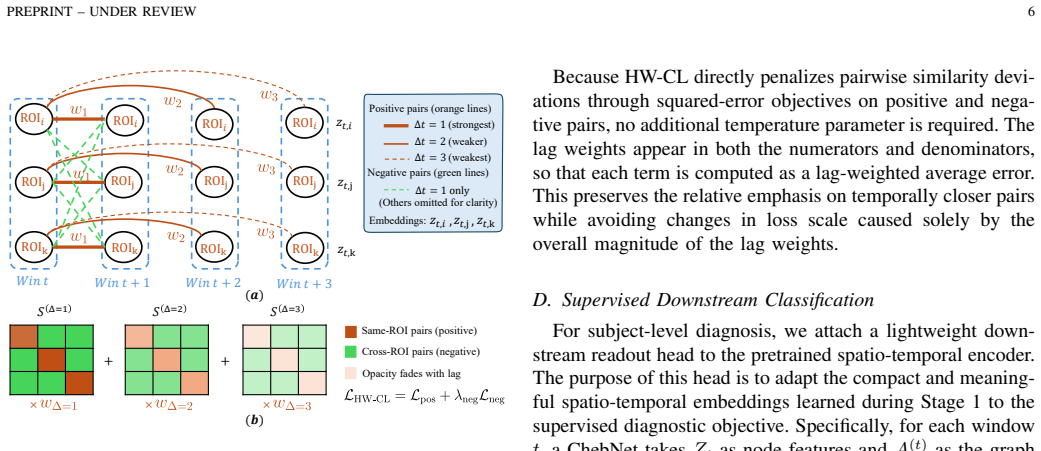
The reliability-refined joint spatio-temporal graph, formed by linking each region to itself across windows via a Hawkes-inspired exponential kernel after distance-decay edge refinement, which carries both spatial and temporal information during message passing.
If this is right
- Spatial and temporal brain network information propagate together during message passing instead of in separate stages.
- Each brain region maintains temporal consistency across windows while different regions show reduced redundant similarity.
- The method produces coherent spatio-temporal representations usable for MDD classification.
- The framework outperforms recent DFC-based baselines on the tested rs-fMRI dataset.
Where Pith is reading between the lines
- The same joint-graph construction could be tested on other time-varying neuroimaging modalities such as EEG or MEG for similar disorders.
- Removing the distance-decay prior alone would isolate whether long-range connection down-weighting is essential to the reported gains.
- The spectral node descriptors could be swapped for alternative frequency features to check robustness of the contrastive stage.
Load-bearing premise
The Hawkes-process-inspired exponential kernel and exponential distance-decay prior correctly capture the temporal dependencies and reliability of functional connections in rs-fMRI BOLD signals.
What would settle it
Performance on the benchmark dataset drops to baseline levels or below when the Hawkes kernel is replaced by a uniform or random temporal link model while keeping all other components fixed.
Figures

read the original abstract
Major depressive disorder (MDD) is a common neuropsychiatric condition whose accurate diagnosis from resting-state functional magnetic resonance imaging (rs-fMRI) remains difficult. Dynamic functional connectivity (DFC) captures time-varying interactions among brain regions and provides rich spatio-temporal information, yet current DFC-based methods face three limitations: sliding-window Pearson correlation yields noisy estimates sensitive to window length and motion artifacts; correlation-derived node features do not fully exploit frequency-domain properties of blood-oxygen-level-dependent (BOLD) signals; and most spatio-temporal graph models handle spatial structure and temporal dynamics in separate stages, restricting their ability to represent coupled brain network evolution. To overcome these issues, we reformulate DFC learning as joint spatio-temporal graph representation learning under a Hawkes-process-inspired temporal dependency prior and propose HWSTCL, a two-stage framework built on a reliability-refined joint spatio-temporal graph with a kernel-weighted pretraining objective. Within each temporal window, BOLD signals are encoded as spectral node descriptors and functional edges are refined by an exponential distance-decay prior that down-weights less reliable long-range connections. The joint graph is then formed by linking each region to itself across future windows through a Hawkes-inspired exponential kernel, allowing spatial and temporal information to be propagated together during message passing. A kernel-weighted contrastive objective further promotes temporal consistency for each region across windows while reducing redundant similarity between different regions. Experiments on a benchmark rs-fMRI dataset show that HWSTCL outperforms recent baselines and yields coherent spatio-temporal representations for MDD diagnosis.
Editorial analysis
A structured set of objections, weighed in public.
Referee Report
Summary. The paper proposes HWSTCL, a two-stage framework for MDD diagnosis from rs-fMRI that reformulates dynamic functional connectivity as joint spatio-temporal graph representation learning. It encodes BOLD signals as spectral node descriptors within windows, refines edges via an exponential distance-decay prior, links regions across windows using a Hawkes-process-inspired exponential kernel to form a reliability-refined joint graph, and applies a kernel-weighted contrastive pretraining objective to promote temporal consistency. Experiments on a benchmark rs-fMRI dataset are reported to show outperformance over recent baselines and coherent spatio-temporal representations.
Significance. If the priors are shown to be appropriate and performance gains are robust to ablations, the joint modeling of spatial and temporal dependencies via the reliability-refined graph could advance DFC-based MDD diagnosis by addressing noisy sliding-window estimates and separate-stage handling of space and time. The kernel-weighted contrastive objective and spectral descriptors represent a coherent integration of ideas from Hawkes processes and graph contrastive learning.
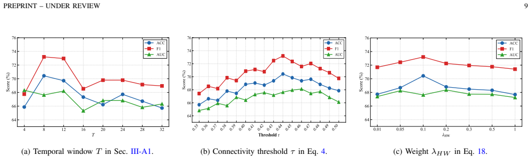
major comments (2)
- [Abstract and framework] Abstract and framework description: the central claim that the reliability-refined joint graph (exponential distance-decay within windows + Hawkes exponential kernel across windows) produces coherent spatio-temporal representations improving diagnosis rests on the assumption that these specific functional forms correctly encode BOLD temporal dependencies and connection reliability. No likelihood fit, spectral comparison, autocorrelation analysis, or ablation on kernel shape is referenced to support the exponential choice over alternatives; if misspecified, message passing and the contrastive loss propagate incorrect dependencies, so outperformance cannot be attributed to the joint modeling.
- [Experiments] Experiments section: the claim of outperformance over baselines is load-bearing for the contribution, yet the abstract supplies no details on statistical significance tests, cross-validation procedure, ablation results removing the distance-decay or Hawkes kernel, or error analysis; without these, it is impossible to verify whether gains derive from the proposed priors rather than other modeling choices.
minor comments (2)
- [Methods] Notation for the Hawkes kernel and distance-decay parameters should be defined explicitly with equations in the methods section to allow reproduction.
- [Abstract] The abstract mentions 'a benchmark rs-fMRI dataset' without naming it or providing basic statistics (e.g., number of subjects, scan length); this should be stated upfront.
Simulated Author's Rebuttal
Thank you for the opportunity to respond to the referee's comments on our manuscript arXiv:2605.24066. We address the major comments point by point below, proposing revisions where the manuscript can be strengthened.
read point-by-point responses
-
Referee: [Abstract and framework] Abstract and framework description: the central claim that the reliability-refined joint graph (exponential distance-decay within windows + Hawkes exponential kernel across windows) produces coherent spatio-temporal representations improving diagnosis rests on the assumption that these specific functional forms correctly encode BOLD temporal dependencies and connection reliability. No likelihood fit, spectral comparison, autocorrelation analysis, or ablation on kernel shape is referenced to support the exponential choice over alternatives; if misspecified, message passing and the contrastive loss propagate incorrect dependencies, so outperformance cannot be attributed to the joint modeling.
Authors: We thank the referee for this insightful comment. The exponential distance-decay is motivated by established findings that long-range functional connections are less reliable due to artifacts, while the Hawkes exponential kernel is selected for its ability to model decaying temporal influence in self-exciting processes, consistent with BOLD dynamics. We acknowledge that the original submission lacks direct empirical comparisons such as likelihood fits or kernel ablations. We will add an ablation study in the revised manuscript comparing exponential kernels to alternatives (e.g., Gaussian, power-law) and include literature-based justification to better support attribution of gains to the joint modeling. revision: partial
-
Referee: [Experiments] Experiments section: the claim of outperformance over baselines is load-bearing for the contribution, yet the abstract supplies no details on statistical significance tests, cross-validation procedure, ablation results, or error analysis; without these, it is impossible to verify whether gains derive from the proposed priors rather than other modeling choices.
Authors: We agree that the abstract would benefit from these details for transparency. The full experiments section reports 5-fold cross-validation, paired t-tests for significance (p<0.05), ablations removing the distance-decay and Hawkes components, and error analysis via per-class metrics. We will revise the abstract to concisely reference the cross-validation, significance testing, and ablation outcomes, and expand the experiments section to more explicitly link ablations to the contribution of the priors. revision: yes
Circularity Check
No circularity: modeling choices presented as explicit priors without reduction to fitted inputs or self-citation chains
full rationale
The paper's central framework adopts an exponential distance-decay prior within windows and a Hawkes-process-inspired exponential kernel across windows as modeling choices to refine the joint spatio-temporal graph. These are introduced directly in the abstract and framework description without any claim that they are derived predictions, fitted parameters renamed as outputs, or justified solely via self-citation. No equations reduce the claimed representations or contrastive objective to the inputs by construction, and no uniqueness theorems or ansatzes are smuggled through author-overlapping citations. The experimental outperformance on the benchmark dataset therefore rests on independent evaluation rather than definitional equivalence.
Axiom & Free-Parameter Ledger
axioms (2)
- domain assumption Hawkes process exponential kernel accurately captures temporal dependencies in dynamic functional connectivity.
- domain assumption Exponential distance-decay prior down-weights less reliable long-range connections in rs-fMRI.
Reference graph
Works this paper leans on
-
[1]
Major depressive disorder,
W. Marxet al., “Major depressive disorder,”Nature Reviews Disease Primers, vol. 9, no. 1, p. 44, 2023
2023
-
[2]
Identification of psychiatric disorder subtypes from functional connectivity patterns in resting-state electroencephalography,
Y . Zhanget al., “Identification of psychiatric disorder subtypes from functional connectivity patterns in resting-state electroencephalography,” Nature biomedical engineering, vol. 5, no. 4, pp. 309–323, 2021
2021
-
[3]
Graph autoencoders for embedding learning in brain networks and major depressive disorder identification,
F. Noman, C.-M. Ting, H. Kang, R. C.-W. Phan, and H. Ombao, “Graph autoencoders for embedding learning in brain networks and major depressive disorder identification,”IEEE Journal of Biomedical and Health Informatics, vol. 28, no. 3, pp. 1644–1655, 2024
2024
-
[4]
Graph neural network with modular attention for identifying brain disorders,
W. Si, G. Wang, L. Liu, L. Zhang, and L. Qiao, “Graph neural network with modular attention for identifying brain disorders,”Biomedical Signal Processing and Control, vol. 102, p. 107252, 2025
2025
-
[5]
Can sliding-window correlations reveal dynamic functional connectivity in resting-state fmri?
R. Hindrikset al., “Can sliding-window correlations reveal dynamic functional connectivity in resting-state fmri?”Neuroimage, vol. 127, pp. 242–256, 2016
2016
-
[6]
Enhancing major depressive disorder diagnosis with dynamic-static fusion graph neural networks,
T. Zhao and G. Zhang, “Enhancing major depressive disorder diagnosis with dynamic-static fusion graph neural networks,”IEEE Journal of Biomedical and Health Informatics, vol. 28, no. 8, pp. 4701–4710, 2024
2024
-
[7]
Spatio-temporal graph convolutional network for di- agnosis and treatment response prediction of major depressive disorder from functional connectivity,
Y . Konget al., “Spatio-temporal graph convolutional network for di- agnosis and treatment response prediction of major depressive disorder from functional connectivity,”Human brain mapping, vol. 42, no. 12, pp. 3922–3933, 2021
2021
-
[8]
Learning dynamic graph represen- tation of brain connectome with spatio-temporal attention,
B.-H. Kim, J. C. Ye, and J.-J. Kim, “Learning dynamic graph represen- tation of brain connectome with spatio-temporal attention,”Advances in Neural Information Processing Systems, vol. 34, pp. 4314–4327, 2021
2021
-
[9]
Spatio-temporal attention graph convolution network for functional connectome classi- fication,
W. Wang, Y . Kong, Z. Hou, C. Yang, and Y . Yuan, “Spatio-temporal attention graph convolution network for functional connectome classi- fication,” inIEEE International Conference on Acoustics, Speech and Signal Processing, 2022, pp. 1486–1490
2022
-
[10]
Temporal dynamic synchronous functional brain net- work for schizophrenia classification and lateralization analysis,
C. Zhuet al., “Temporal dynamic synchronous functional brain net- work for schizophrenia classification and lateralization analysis,”IEEE Transactions on Medical Imaging, vol. 43, no. 12, pp. 4307–4318, 2024
2024
-
[11]
Long-interval spatio-temporal graph convolution for brain disease diagnosis,
S. Liet al., “Long-interval spatio-temporal graph convolution for brain disease diagnosis,”IEEE Transactions on Instrumentation and Measure- ment, vol. 74, 2025
2025
-
[12]
Brain-wide functional connectivity artifactually inflates throughout functional magnetic reso- nance imaging scans,
C. Korponay, A. C. Janes, and B. B. Frederick, “Brain-wide functional connectivity artifactually inflates throughout functional magnetic reso- nance imaging scans,”Nature human behaviour, vol. 8, no. 8, pp. 1568– 1580, 2024
2024
-
[13]
Spectral brain graph neural network for prediction of anxiety in children with autism spectrum disorder,
P. Duanet al., “Spectral brain graph neural network for prediction of anxiety in children with autism spectrum disorder,” inIEEE Interna- tional Symposium on Biomedical Imaging, 2024, pp. 1–5. PREPRINT – UNDER REVIEW 11
2024
-
[14]
Spectral graph neural network-based multi-atlas brain network fusion for major depressive disorder diagnosis,
D.-J. Leeet al., “Spectral graph neural network-based multi-atlas brain network fusion for major depressive disorder diagnosis,”IEEE J. Biomed. Health Inform., vol. 28, no. 5, pp. 2967–2978, 2024
2024
-
[15]
Leveraging brain modularity prior for interpretable representation learning of fmri,
Q. Wanget al., “Leveraging brain modularity prior for interpretable representation learning of fmri,”IEEE Transactions on Biomedical Engineering, vol. 71, no. 8, pp. 2391–2401, 2024
2024
-
[16]
Fe-stgnn: Spatio-temporal graph neural network with functional and effective connectivity fusion for mci diagnosis,
D. Chen and L. Zhang, “Fe-stgnn: Spatio-temporal graph neural network with functional and effective connectivity fusion for mci diagnosis,” in Med. Image Comput. Comput. Assist. Interv., 2023, pp. 67–76
2023
-
[17]
Eigendecomposition-based spatial-temporal attention for brain cognitive states identification,
J. Lee, E. Kang, J. Maeng, and H.-I. Suk, “Eigendecomposition-based spatial-temporal attention for brain cognitive states identification,” in IEEE Int. Conf. Acoust., Speech, Signal Process., 2024, pp. 1921–1925
2024
-
[18]
Multi- scale spatial-temporal attention networks for functional connectome classification,
Y . Kong, X. Zhang, W. Wang, Y . Zhou, Y . Li, and Y . Yuan, “Multi- scale spatial-temporal attention networks for functional connectome classification,”IEEE transactions on medical imaging, vol. 44, no. 1, pp. 475–488, 2024
2024
-
[19]
Brainib: Interpretable brain network-based psychiatric diagnosis with graph information bot- tleneck,
K. Zheng, S. Yu, B. Li, R. Jenssen, and B. Chen, “Brainib: Interpretable brain network-based psychiatric diagnosis with graph information bot- tleneck,”IEEE Trans. Neural Netw. Learn. Syst., vol. 36, 2024
2024
-
[20]
Network modelling methods for fmri,
S. M. Smithet al., “Network modelling methods for fmri,”Neuroimage, vol. 54, pp. 875–891, 2011
2011
-
[21]
Spurious but systematic correlations in functional connectivity mri networks arise from subject motion,
J. D. Power, K. A. Barnes, A. Z. Snyder, B. L. Schlaggar, and S. E. Petersen, “Spurious but systematic correlations in functional connectivity mri networks arise from subject motion,”Neuroimage, vol. 59, no. 3, pp. 2142–2154, 2012
2012
-
[22]
Brain disorders? precisely,
T. R. Insel and B. N. Cuthbert, “Brain disorders? precisely,”Science, vol. 348, no. 6234, pp. 499–500, 2015
2015
-
[23]
Waving hello to noninvasive deep-brain stimulation,
A. M. Lozano, “Waving hello to noninvasive deep-brain stimulation,” New England Journal of Medicine, vol. 377, pp. 1096–1098, 2017
2017
-
[24]
Functional connectivity signatures of major depressive disorder: machine learning analysis of two multicenter neuroimaging studies,
S. Galloet al., “Functional connectivity signatures of major depressive disorder: machine learning analysis of two multicenter neuroimaging studies,”Molecular Psychiatry, vol. 28, no. 7, pp. 3013–3022, 2023
2023
-
[25]
A neuroimaging biomarker for striatal dysfunction in schizophrenia,
A. Liet al., “A neuroimaging biomarker for striatal dysfunction in schizophrenia,”Nature medicine, vol. 26, no. 4, pp. 558–565, 2020
2020
-
[26]
Self- supervised graph contrastive learning with diffusion augmentation for functional mri analysis and brain disorder detection,
X. Wang, Y . Fang, Q. Wang, P.-T. Yap, H. Zhu, and M. Liu, “Self- supervised graph contrastive learning with diffusion augmentation for functional mri analysis and brain disorder detection,”Medical image analysis, vol. 101, p. 103403, 2025
2025
-
[27]
A-gcl: Adversarial graph contrastive learning for fmri analysis to diagnose neurodevelopmental disorders,
S. Zhanget al., “A-gcl: Adversarial graph contrastive learning for fmri analysis to diagnose neurodevelopmental disorders,”Medical Image Analysis, vol. 90, p. 102932, 2023
2023
-
[28]
Contrastive multi- view composite graph convolutional networks based on contribution learning for autism spectrum disorder classification,
H. Zhu, J. Wang, Y .-P. Zhao, M. Lu, and J. Shi, “Contrastive multi- view composite graph convolutional networks based on contribution learning for autism spectrum disorder classification,”IEEE Transactions on Biomedical Engineering, vol. 70, no. 6, pp. 1943–1954, 2022
1943
-
[29]
Dclnet: Double collaborative learning network on stationary-dynamic functional brain network for brain disease classi- fication,
J. Zhouet al., “Dclnet: Double collaborative learning network on stationary-dynamic functional brain network for brain disease classi- fication,”IEEE Transactions on Image Processing, 2025
2025
-
[30]
Automated anatomical labelling atlas 3,
E. T. Rolls, C.-C. Huang, C.-P. Lin, J. Feng, and M. Joliot, “Automated anatomical labelling atlas 3,”Neuroimage, vol. 206, p. 116189, 2020
2020
-
[31]
Dparsf: a matlab toolbox for
C. Yan and Y . Zang, “Dparsf: a matlab toolbox for” pipeline” data analysis of resting-state fmri,”Frontiers in systems neuroscience, vol. 4, p. 1377, 2010
2010
-
[32]
Brain templates and atlases,
A. C. Evans, A. L. Janke, D. L. Collins, and S. Baillet, “Brain templates and atlases,”Neuroimage, vol. 62, no. 2, pp. 911–922, 2012
2012
-
[33]
The direct consortium and the rest-meta-mdd project: towards neuroimaging biomarkers of major depressive disorder,
X. Chenet al., “The direct consortium and the rest-meta-mdd project: towards neuroimaging biomarkers of major depressive disorder,”Psy- choradiology, vol. 2, pp. 32–42, 2022
2022
-
[34]
Braingnn: Interpretable brain graph neural network for fmri analysis,
X. Liet al., “Braingnn: Interpretable brain graph neural network for fmri analysis,”Medical Image Analysis, vol. 74, p. 102233, 2021
2021
-
[35]
Interpretable graph neural networks for connectome-based brain disorder analysis,
H. Cui, W. Dai, Y . Zhu, X. Li, L. He, and C. Yang, “Interpretable graph neural networks for connectome-based brain disorder analysis,” inMed. Image Comput. Comput. Assist. Interv., 2022, pp. 375–385
2022
-
[36]
Brainnetcnn: Convolutional neural networks for brain networks; towards predicting neurodevelopment,
J. Kawaharaet al., “Brainnetcnn: Convolutional neural networks for brain networks; towards predicting neurodevelopment,”NeuroImage, vol. 146, pp. 1038–1049, 2017
2017
-
[37]
Coordinate-based network mapping of brain structure in major depressive disorder in younger and older adults: a systematic review and meta-analysis,
P. Zhukovsky, J. A. Anderson, G. Coughlan, B. H. Mulsant, A. Cipriani, and A. N. V oineskos, “Coordinate-based network mapping of brain structure in major depressive disorder in younger and older adults: a systematic review and meta-analysis,”American Journal of Psychiatry, vol. 178, no. 12, pp. 1119–1128, 2021
2021
-
[38]
The multifaceted role of the ventromedial prefrontal cortex in emotion, decision making, social cognition, and psychopathology,
J. Hiser and M. Koenigs, “The multifaceted role of the ventromedial prefrontal cortex in emotion, decision making, social cognition, and psychopathology,”Biological psychiatry, vol. 83, no. 8, pp. 638–647, 2018
2018
-
[39]
Disrupted functional connectivity of the cerebellum with default mode and frontoparietal networks in young adults with major depressive disorder,
X. Wanget al., “Disrupted functional connectivity of the cerebellum with default mode and frontoparietal networks in young adults with major depressive disorder,”Psychiatry research, vol. 324, p. 115192, 2023
2023
-
[40]
Subcortical brain alterations in major depressive disorder: findings from the enigma major depressive disorder working group,
L. Schmaalet al., “Subcortical brain alterations in major depressive disorder: findings from the enigma major depressive disorder working group,”Molecular psychiatry, vol. 21, no. 6, pp. 806–812, 2016
2016
-
[41]
Adhd classification with gcn via joint feature learning among nodes and edges,
X. Wang, Y . Tang, Y . Gao, X. Meng, Y . Chen, and A. Jiang, “Adhd classification with gcn via joint feature learning among nodes and edges,”IEEE Transactions on Medical Imaging, 2026
2026
-
[42]
Fglfa: A federated graph learning-based cross-network layer feature alignment model for major depressive disorder identification,
Z. Jiao, X. Ding, Z. Xia, C. Liu, and Y . Zhang, “Fglfa: A federated graph learning-based cross-network layer feature alignment model for major depressive disorder identification,”IEEE Journal of Biomedical and Health Informatics, 2025
2025
discussion (0)
Sign in with ORCID, Apple, or X to comment. Anyone can read and Pith papers without signing in.