Adjoint-based Perfusion Estimation from Dynamic Contrast-Enhanced Ultrasound: Advection-Diffusion and Two-Compartment Models
Pith reviewed 2026-06-27 21:10 UTC · model grok-4.3
The pith
Continuous adjoint equations enable efficient recovery of spatially varying blood flow velocities and perfusion parameters from dynamic contrast-enhanced ultrasound data.
A machine-rendered reading of the paper's core claim, the machinery that carries it, and where it could break.
Core claim
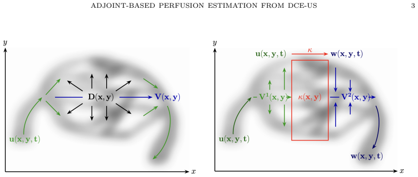
The central claim is that the parameter identification problem for perfusion velocities and exchange rates can be solved by Tikhonov-regularized least-squares minimization whose gradient is supplied by continuous adjoint equations for either the parabolic advection-diffusion model or the hyperbolic two-compartment model; the resulting reconstruction algorithms recover plausible parameter fields from both synthetic and in-vivo dynamic contrast-enhanced ultrasound data.
What carries the argument
Continuous adjoint equations that furnish the gradient of the Tikhonov-regularized misfit functional for the inverse perfusion problem.
If this is right
- Gradient computation becomes feasible at the cost of one additional PDE solve rather than many finite-difference perturbations.
- The two-compartment formulation can be treated with the same adjoint machinery as the simpler advection-diffusion model.
- Regularized reconstructions remain stable enough to produce usable maps from noisy in-vivo ultrasound time series.
- The same numerical discretization and optimization framework applies to both models, allowing direct comparison of their outputs.
Where Pith is reading between the lines
- The method could be adapted to other time-resolved imaging modalities that supply concentration time courses.
- If the models prove adequate, the recovered maps might be used to predict drug delivery or oxygen transport inside tumors.
- Extending the framework to time-varying parameters or to three-dimensional domains would be a direct next numerical step.
Load-bearing premise
The chosen advection-diffusion and two-compartment models correctly describe how the contrast agent moves through the tissue.
What would settle it
A controlled experiment in which recovered velocity and perfusion maps are compared against independent ground-truth measurements obtained by another modality on the same tissue; systematic mismatch would falsify the claim that the recovered fields are physiologically meaningful.
Figures


read the original abstract
Tumor perfusion and vascular properties are important determinants of a cancer's response to therapy. In this paper, we discuss the estimation of spatially varying blood flow velocities and perfusion parameters from time-resolved contrast agent concentration data. We compare a standard parabolic advection-diffusion model against a two-compartment model governed by a coupled system of hyperbolic advection-reaction equations, which is physiologically more sound. To address the inherent ill-posedness of this parameter identification problem, we employ Tikhonov regularization and derive continuous adjoint equations necessary for efficient, gradient-based minimization. We discuss the numerical discretization of the state and adjoint systems using state-of-the-art schemes, and demonstrate the efficacy of the proposed reconstruction algorithms through numerical experiments on synthetic data and in vivo dynamic contrast-enhanced ultrasound measurements.
Editorial analysis
A structured set of objections, weighed in public.
Referee Report
Summary. The paper develops adjoint-based reconstruction algorithms to estimate spatially varying blood flow velocities and perfusion parameters from time-resolved DCE-US concentration data. It compares a standard parabolic advection-diffusion model to a two-compartment model governed by coupled hyperbolic advection-reaction equations, employs Tikhonov regularization to handle ill-posedness, derives the corresponding continuous adjoint equations for gradient-based minimization, discusses state-of-the-art numerical discretizations, and demonstrates the methods on synthetic data generated from the forward models plus in vivo DCE-US measurements.
Significance. If the modeling assumptions are valid, the work provides an efficient computational framework for recovering physiologically relevant perfusion maps from DCE-US, which could support non-invasive evaluation of tumor response to therapy. The derivation of continuous adjoints and the direct comparison of the two transport models are technical strengths that enable scalable optimization and model selection. The inclusion of in vivo data adds practical relevance, though overall significance remains conditional on quantitative validation of reconstruction accuracy.
major comments (2)
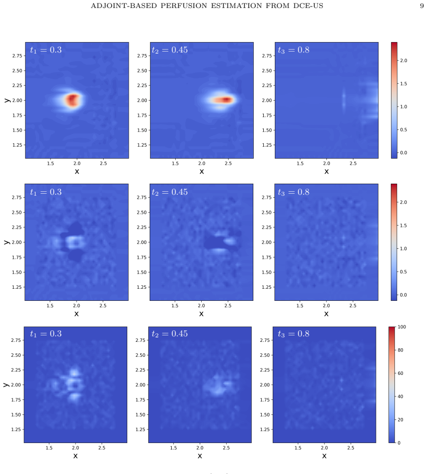
- [Numerical Experiments] Numerical Experiments section: synthetic tests are generated from the identical forward models used in the inversion, confirming only that the optimizer recovers parameters under exact model match; this provides no evidence on robustness to model mismatch and therefore does not support the efficacy claim for the in vivo reconstructions.
- [Abstract and Results] Abstract and Results: no quantitative error metrics (e.g., relative L2 errors on recovered velocity or perfusion fields, convergence rates under mesh refinement, or regularization-parameter sensitivity) are supplied for either the synthetic or in vivo cases, leaving the central claim of algorithmic efficacy without measurable support.
minor comments (1)
- [Numerical Discretization] The discretization subsection would benefit from explicit statements of the CFL or stability conditions used for the hyperbolic two-compartment scheme.
Simulated Author's Rebuttal
We thank the referee for the constructive comments. We address each major comment below.
read point-by-point responses
-
Referee: [Numerical Experiments] Numerical Experiments section: synthetic tests are generated from the identical forward models used in the inversion, confirming only that the optimizer recovers parameters under exact model match; this provides no evidence on robustness to model mismatch and therefore does not support the efficacy claim for the in vivo reconstructions.
Authors: We agree that the synthetic experiments only verify the method under exact model match and therefore do not test robustness to mismatch. The in vivo experiments apply the method to real DCE-US data, where model mismatch is necessarily present, and produce physiologically plausible maps. To directly address the concern we will add a new subsection with synthetic mismatch experiments (data generated from one model, inverted with the other) in the revised manuscript. revision: yes
-
Referee: [Abstract and Results] Abstract and Results: no quantitative error metrics (e.g., relative L2 errors on recovered velocity or perfusion fields, convergence rates under mesh refinement, or regularization-parameter sensitivity) are supplied for either the synthetic or in vivo cases, leaving the central claim of algorithmic efficacy without measurable support.
Authors: We agree that quantitative metrics are needed to support the efficacy claims. In the revision we will add relative L2 errors on recovered fields for all synthetic cases, mesh-refinement convergence rates, and regularization-parameter sensitivity studies. For the in vivo data we will report data-misfit residuals and quantitative comparisons between the two models. revision: yes
Circularity Check
No significant circularity detected
full rationale
The paper derives continuous adjoint equations for Tikhonov-regularized gradient-based minimization of perfusion parameters in advection-diffusion and two-compartment models. Synthetic experiments test recovery when data is generated from the identical forward models, which is standard validation and does not reduce any output to an input by construction. In vivo results apply the same numerical scheme under the stated modeling assumptions without any self-definitional loops, fitted inputs renamed as predictions, or load-bearing self-citations. The derivation chain consists of independent PDE analysis and discretization steps that remain self-contained against external benchmarks.
Axiom & Free-Parameter Ledger
axioms (1)
- domain assumption The advection-diffusion and two-compartment equations adequately describe contrast-agent transport in tissue
Reference graph
Works this paper leans on
-
[1]
Epidemiology of primary and secondary liver cancers
A. Ananthakrishnan, V. Gogineni, and K. Saeian. “Epidemiology of primary and secondary liver cancers”. In:Seminars in Interventional Radiology23 (2006), pp. 47–63
2006
-
[2]
Anderson.Compartmental Modeling and Tracer Kinetics
David H. Anderson.Compartmental Modeling and Tracer Kinetics. Springer, 1983
1983
-
[3]
Minimization of Functions Having Lipschitz Continuous First Partial Deriva- tives
Larry Armijo. “Minimization of Functions Having Lipschitz Continuous First Partial Deriva- tives”. In:Pacific Journal of Mathematics16.1 (1966), pp. 1–3
1966
-
[4]
An optimal control approach to optical flow computation
A. Borz` ı, K. Ito, and K. Kunisch. “An optimal control approach to optical flow computation”. In:International Journal for Numerical Methods in Fluids40.1-2 (2002), pp. 231–240.doi: https://doi.org/10.1002/fld.273
-
[5]
Numerical solution of the Navier-Stokes equations
Alexandre Joel Chorin. “Numerical solution of the Navier-Stokes equations”. In:Math. Comp. 22 (1968), pp. 745–762.doi:10.1090/S0025-5718-1968-0242392-2
-
[6]
Parameter Estimation in the 1-D Transport Equation with Advection
C. Coles. “Parameter Estimation in the 1-D Transport Equation with Advection”. In:PERG- AMON Computers and Mathematics with Applications44 (2002), pp. 1493–1502
2002
-
[7]
Reconstruction of a Velocity Field for a 3D Advection Diffusion Equation
Yi-Xin Dou and Bo Han. “Reconstruction of a Velocity Field for a 3D Advection Diffusion Equation”. In:International Journal of Thermal Sciences50 (2011), pp. 2340–2354
2011
-
[8]
Ahmed El Kaffas et al. “Spatial Characterization of Tumor Perfusion Properties from 3D DCE- US Perfusion Maps Are Early Predictors of Cancer Treatment Response”. In:Scientific Reports 10.1 (Apr. 24, 2020), p. 6996.issn: 2045-2322.doi:10.1038/s41598-020-63810-1
-
[9]
European Cancer Information System (ECIS).Fact Sheet Liver Cancer in the EU.https:// ecis.jrc.ec.europa.eu/sites/default/files/2024-10/2024-10-03_Factsheet_Liver- Cancer.pdf. 2024
2024
-
[10]
2026.url: https://doi.org/10.5281/zenodo.19063183(visited on 03/17/2026)
Sophie Externbrink.Parameter Identification for a Tracer Model (BaseVersion). 2026.url: https://doi.org/10.5281/zenodo.19063183(visited on 03/17/2026)
-
[11]
Implicit–explicit (IMEX) Runge–Kutta methods for non-hydrostatic atmospheric models
D. J. Gardner, J. E. Guerra, F. P. Hamon, D. R. Reynolds, P. A. Ullrich, and C. S. Woodward. “Implicit–explicit (IMEX) Runge–Kutta methods for non-hydrostatic atmospheric models”. In:Geoscientific Model Development11.4 (2018), pp. 1497–1515
2018
-
[12]
Goodman.Speckle phenomena in optics: theory and applications
Joseph W. Goodman.Speckle phenomena in optics: theory and applications. SPIE Digital Library, 2020.doi:10.1117/3.2548484
-
[13]
Guyton and J
A. Guyton and J. Hall.Textbook of Medical Physiology. W.B. Saunders, 1996
1996
-
[14]
A survey of nonlinear conjugate gradient methods
William W. Hager and Hongchao Zhang. “A survey of nonlinear conjugate gradient methods”. In:Pacific Journal of Optimization2 (2006), pp. 35–58. REFERENCES 17
2006
-
[15]
Identification of the advection- diffusion equation for predicting green water propagation
J.V. Hern´ andez-Fontes, L. Torres, E. Mendoza, and M. Escudero. “Identification of the advection- diffusion equation for predicting green water propagation”. In:Elsevier Ocean Engineering214 (2020)
2020
-
[16]
Dynamic Contrast-Enhanced Ultrasound Modeling of an Analog to Pseudo- Diffusivity in Intravoxel Incoherent Motion Magnetic Resonance Imaging
Dimitre Hristov, Lauri Mustonen, Rie von Eyben, Sebastian G¨ otschel, Michael Minion, and Ahmed El Kaffas. “Dynamic Contrast-Enhanced Ultrasound Modeling of an Analog to Pseudo- Diffusivity in Intravoxel Incoherent Motion Magnetic Resonance Imaging”. In:IEEE Transac- tions On Medical Imaging10 (2022)
2022
-
[17]
Global epidemiology of NAFLD- related HCC: trends, predictions, risk factors and prevention
Daniel Q. Huang, Hashem B. El-Serag, and Rohit Loomba. “Global epidemiology of NAFLD- related HCC: trends, predictions, risk factors and prevention”. eng. In:Nature Reviews. Gas- troenterology & Hepatology18.4 (2021), pp. 223–238.issn: 1759-5045.doi:10.1038/s41575- 020-00381-6
-
[18]
Polarization of Tumor-Associated Macrophages: A Novel Strategy for Vascular Normalization and Antitumor Immunity
Yuhui Huang, Matija Snuderl, and Rakesh K. Jain. “Polarization of Tumor-Associated Macrophages: A Novel Strategy for Vascular Normalization and Antitumor Immunity”. In:Cancer Cell19.1 (2011), pp. 1–2
2011
-
[19]
Clinical Intravoxel Incoherent Motion and Diffusion MR Imaging: Past, Present, and Future
M. Iima and D. Le Bihan. “Clinical Intravoxel Incoherent Motion and Diffusion MR Imaging: Past, Present, and Future”. In:Radiology278.1 (2016), pp. 13–32
2016
-
[20]
Johnson.Lecture notes in Introduction To Numerical Methods
Steven G. Johnson.Lecture notes in Introduction To Numerical Methods. Apr. 2019
2019
-
[21]
P. R. Peebles Jr.Probability, Random Variables and Random Signal Principles. McGraw-Hill, Inc., 2001
2001
-
[22]
Accuracy and efficiency of adjoint state based parameter identification for fractional advection diffusion equation with space-dependent coefficients
Boris Maryshev, Alain Cartalade, Christelle Latrille, and Marie-Christine N´ eel. “Accuracy and efficiency of adjoint state based parameter identification for fractional advection diffusion equation with space-dependent coefficients”. In:IEEE Xplore(2016)
2016
-
[23]
Robust estimation of ultrasound pulses using outlier-resistant de-noising
O. Michailovich and D. Adam. “Robust estimation of ultrasound pulses using outlier-resistant de-noising”. In:IEEE Transactions on Medical Imaging22.3 (2003), pp. 368–381.doi:10. 1109/TMI.2003.809603
arXiv 2003
-
[24]
The ”Fingerprint
Sneha R. Raoa, Sarah E. Shelton, and Paul A. Dayton. “The ”Fingerprint” of Cancer Extends Beyond Solid Tumor Boundaries: Assessment With a Novel Ultrasound Imaging Approach”. In:IEEE Trans Biomedical Engineering63.5 (2015), pp. 1082–1086
2015
-
[25]
Global burden of primary liver cancer in 2020 and predictions to 2040
Harriet Rumgay, Melina Arnold, Jacques Ferlay, Olufunmilayo Lesi, Citadel J. Cabasag, J´ erˆ ome Vignat, Mathieu Laversanne, Katherine A. McGlynn, and Isabelle Soerjomataram. “Global burden of primary liver cancer in 2020 and predictions to 2040”. In:Journal of Hepatology 77.6 (2022), pp. 1598–1606.issn: 0168-8278.doi:10.1016/j.jhep.2022.08.021
-
[26]
How to Identify the Right Patients for the Right Treatment in Metastatic Colorectal Cancer (mCRC)
Z. Saridaki, N. Asimakopoulou, I. Boukovinas, and J. Souglakos. “How to Identify the Right Patients for the Right Treatment in Metastatic Colorectal Cancer (mCRC)”. In:Curr Colorec- tal Cancer Rep.11.4 (2015), pp. 151–159
2015
-
[27]
Smoothing and Differentiation of Data by Simplified Least Squares Procedures
Abraham Savitzky and M. J. E. Golay. “Smoothing and Differentiation of Data by Simplified Least Squares Procedures”. In:Analytical Chemistry36.8 (1964), pp. 1627–1639
1964
-
[28]
Essentially Non-oscillatory and Weighted Essentially Non-oscillatory Schemes for Hyperbolic Conservation Laws
Chi-Wang Shu. “Essentially Non-oscillatory and Weighted Essentially Non-oscillatory Schemes for Hyperbolic Conservation Laws”. In:ICASE1.97–65 (1997)
1997
-
[29]
Tracer kinetic modelling in MRI: estimating perfusion and capillary permeability
S.P. Sourbron and D.L. Buckley. “Tracer kinetic modelling in MRI: estimating perfusion and capillary permeability”. In:Physics in Medicine and Biology57.2 (2011), pp. 1–33
2011
-
[30]
A Tracer-Kinetic Field Theory for Medical Imaging
Steven Sourbron. “A Tracer-Kinetic Field Theory for Medical Imaging”. In:IEEE Transactions On Medical Imaging33.4 (2014)
2014
-
[31]
Update on the Role of Imaging in Management of Metastatic Colorectal Cancer 1
S. Harsha Tirumani, K. Won Kim, M. Nishino, S.A. Howard, K.M. Krajewski, J.P. Jagan- nathan, J.M. Cleary, N.H. Ramaiya, and A.B. Shinagare. “Update on the Role of Imaging in Management of Metastatic Colorectal Cancer 1”. In:Radio Graphics34.1 (2014), pp. 1908– 1928
2014
-
[32]
Thodsawit Tiyarattanachai, Simona Turco, John R. Eisenbrey, Corinne E. Wessner, Alexan- dra Medellin-Kowalewski, Stephanie Wilson, Andrej Lyshchik, Aya Kamaya, and Ahmed El Kaffas. “A Comprehensive Motion Compensation Method for In-Plane and Out-of-Plane Mo- tion in Dynamic Contrast-Enhanced Ultrasound of Focal Liver Lesions”. In:Ultrasound in Medicine an...
work page doi:10.1016/j 2022
-
[33]
Fredi Tr¨ oltzsch.Optimal Control of Partial Differential Equations: Theory, Methods and Ap- plications. Vol. 112. Graduate Studies in Mathematics. AMS, 2010.doi:10.1090/gsm/112
-
[34]
Convective-Dispersion Modeling in 3D Contrast- Ultrasound Imaging for the Localization of Prostate Cancer
R. R. Wildeboer, R. J. G. van Sloun, S. G. Schalk, C. K. Mannaerts, J. C. van der Linden, P. Huang, H. Wijkstra, and M. Mischi. “Convective-Dispersion Modeling in 3D Contrast- Ultrasound Imaging for the Localization of Prostate Cancer”. In:IEEE Transactions on Medical Imaging37.12 (2018), pp. 2593–2602. 18 REFERENCES
2018
-
[35]
Convergence Conditions for Ascent Methods
Philip Wolfe. “Convergence Conditions for Ascent Methods”. In:SIAM Review11.2 (1969), pp. 226–235
1969
-
[36]
Cooperative Filtering and Parameter Identification for Advection–Diffusion Processes Using a Mobile Sensor Network
Jie You, Ziqiao Zhang, Fumin Zhang, and Wencen Wu. “Cooperative Filtering and Parameter Identification for Advection–Diffusion Processes Using a Mobile Sensor Network”. In:IEEE Transactions on Control Systems Technology31.2 (2023), pp. 527–542. AppendixA.Derivation of Adjoint System and Gradient for the Advection-Diffusion Model We follow the formal Lagra...
2023
discussion (0)
Sign in with ORCID, Apple, or X to comment. Anyone can read and Pith papers without signing in.