REVIEW 3 major objections 5 minor 30 references
HistoSmith: Single-Stage Histology Image-Label Generation via Conditional Latent Diffusion for Enhanced Cell Segmentation and Classification
T0 review · 3 major / 5 minor · reviewed 2026-08-07 · deepseek-v4-flash
Pith's one-line read HistoSmith is a single-stage latent diffusion model that jointly generates histology images and their cell labels, and augmenting real training data with its synthetic pairs improves cell instance segmentation and classification by 1.9%…
desk verdict HistoSmith is a sensible latent-diffusion extension of joint image-label generation, but the evaluation never isolates the generative mechanism from the simple addition of ~3,850 training patches. read the letter →
The pith
A machine-rendered reading of the paper's core claim, the machinery that carries it, and where it could break.
The reading
What carries the argument
The load-bearing machinery is a joint latent diffusion model: a VQ-VAE encodes the concatenation of image and masks into a discrete latent space, and a time-conditional U-Net denoises samples in that space, while a dual-head decoder reconstructs the image with distance map and the semantic cell-type mask. A 10-dimensional conditioning vector encoding staining type, normalized cell counts for five cell groups, and one-hot tissue type is injected with the time embedding, steering generation toward requested tissue and cellular composition. The joint representation is what lets image and labels be generated together from one noise sample, and the post-processing chain (thresholding, morphological operations, majority voting) turns the predicted distance map and semantic mask into instance label maps.
What would settle it
Train the downstream CISCA model on the real dataset augmented with the same number of real patches (or on the real dataset for enough additional epochs to match the extra training data), and compare segmentation and classification metrics with the reported D∪Dhat results; if the control matches or exceeds them, the specific contribution of HistoSmith's generated pairs is not supported.
Extended reading notes
Core claim
On the paper's own terms, the discovery is that one latent diffusion model can jointly learn cellular layout, classification masks, and histology appearance, so that sampling from the learned distribution yields coherent image-label pairs without a separate layout-generation stage. When these pairs are mixed into the real training data, the CISCA cell instance segmentation and classification model outperforms training on real data alone across most metrics on both datasets: average gains of 1.9% on CoNIC and 3.4% on CytoDArk0, with notable per-class improvements for neutrophils (+5.4%), eosinophils (+1.4%), hippocampus (+2.8%), and visual cortex (+2.6%). The authors also show that generation is controllable: conditioning vectors that specify cell counts produce corresponding counts in the synthetic images, though the correspondence weakens for rare classes and saturates when counts are pushed beyond the observed range.
Load-bearing premise
The evaluation assumes the reported gains come from HistoSmith's synthetic image-label pairs rather than from simply adding about 3,850 extra training patches, because the comparison is only between training on the real set and training on the real set plus the generated set.
Editorial extensions
If this is right
- Histology datasets can be augmented on demand with coherent image-label pairs, reducing the amount of expert manual annotation needed to train cell segmentation and classification models.
- Class imbalance can be addressed by conditioning the generator on higher counts of rare cell types, as shown by the +5.4% average gain for neutrophils in CoNIC.
- A single HistoSmith model can serve multiple tissues and stains, since it was trained jointly on H&E colon and Nissl-stained brain tissue with the same conditioning scheme.
- Controllable cell quantities allow targeted synthesis of specific cellular compositions, enabling stress-testing or rebalancing of training sets.
- The gain on visual cortex, a tissue type not present in the CytoDArk0 training set, suggests that synthetic augmentation can improve generalization to new tissue types.
Reading between the lines
- Editorial inference: a natural next experiment is a size-matched control, where the same number of real training patches is added instead of HistoSmith pairs, to isolate whether the gains come from the generated pairs specifically or from having more training data.
- Editorial inference: the saturation behavior reported for neutrophil counts suggests practical users should sample conditioning values inside roughly 1/5 to 9/5 of the observed maximum; the paper does not frame this as a deployment rule, but it follows directly from its Bland-Altman analysis.
- Editorial inference: because the conditioning vector encodes only two stain types, four tissue types, and five cell groups, extending HistoSmith to new domains would likely require retraining or a more flexible conditioning encoder rather than simple parameter changes.
- Editorial inference: the correlation results imply that cell-count control is most reliable for abundant classes (r=0.98 for cerebellum and auditory cortex) and weaker for rare ones (r=0.39 for neutrophils), so applying the method to class imbalance should involve quality checks on the generated rare-class patches.
Signed reviews
Editorial analysis
A structured set of objections, weighed in public.
Referee Report
Summary. The paper proposes HistoSmith, a latent diffusion model that jointly generates histology image patches and their cell instance segmentation/classification labels (distance map, semantic mask, label map) in a single stage, conditioned on a 10-dimensional vector encoding staining type, cell counts, and tissue type. It is trained on a merged dataset of CoNIC H&E colon patches and CytoDArk0 Nissl brain patches, then used to create a synthetic dataset Dhat (3,500 colon and 350 brain patches, roughly doubling the training set) that is added to D for training the CISCA model. Experiments report average gains of 1.9% on CoNIC and 3.4% on CytoDArk0 in cell segmentation and classification metrics, with larger gains for rare cell types and for visual cortex, a tissue absent from the CytoDArk0 training set. The paper also analyzes conditioning fidelity via Bland-Altman plots, correlations, density comparisons, and DINOv2-based image quality metrics.
Significance. If the central claim were fully supported, HistoSmith would be a practically useful contribution: single-stage joint generation simplifies the pipeline relative to two-model diffusion approaches, the conditioning formulation allows steering toward rare cell types, and the evaluation spans two staining modalities and held-out test sets, including a tissue type absent from training. The conditioning analysis in Fig. 4 is unusually honest and informative, and the use of public datasets and external test partitions is a strength. However, the empirical evidence is currently insufficient to attribute the downstream improvements to the generative mechanism, because the only comparison changes training-set size and label distribution simultaneously.
major comments (3)
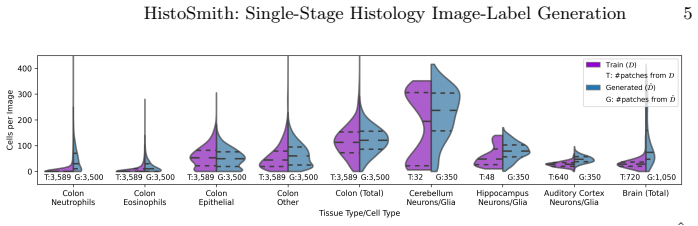
- [Section 5, Table 1] The load-bearing claim that HistoSmith's synthetic image-label pairs drive the reported improvements is not established by the current experimental design. The only comparison is CISCA trained on D versus CISCA trained on D ∪ Dhat, but these differ simultaneously in training-set size (Dhat roughly doubles D, per Section 3) and in label distribution (α = 2 for all cell types and α = 20 for neutrophils and eosinophils, shifting cell counts upward per Fig. 3). A model trained on more patches, especially with more instances of rare classes, could plausibly improve regardless of whether the extra patches are synthetic or real. The authors should add a size-matched control: train CISCA on D augmented with the same number of real training patches (sampled from the training split, with class balance and cell-count distribution matched to Dhat), and a longer-training baseline on D alone to match the number of optimization steps used for D ∪ Dhat. Without such controls, the causal attribution to the generative mechanism remains plausible but unsupported.
- [Section 3, Fig. 3] The deliberate shift in the synthetic label distribution confounds the interpretation of the gains for underrepresented cell types. Since Dhat increases the average cell count per patch and over-represents neutrophils and eosinophils, the observed improvements on these classes may reflect exposure to more instances of rare classes rather than the realism or diversity of generated images. A real-patch control with matched class-balanced oversampling (e.g., real patches selected for high neutrophil/eosinophil counts) would separate these effects. Without it, the statement in Section 4 that HistoSmith 'effectively enhances generalization' is an overreach.
- [Section 4, Fig. 4] The paper's own controllability analysis limits the scope of the 'parameter-driven generative process' contribution. Fig. 4(f) reports r = 0.39 for neutrophils, with systematic underestimation of the requested count and a distribution of generated counts shifted toward the training distribution nD; Fig. 4(c) shows that pushing the conditioning across the full range causes generated cells to transition into 'other' and 'neurons/glia' and degrades image realism. This should be stated as a quantitative operating range (e.g., the range [nmax/5, 9nmax/5] mentioned in the text, or an explicit validity interval for each cell type) rather than the unconditional claim in the abstract that the model 'effectively generates realistic histology images with controllable cell quantities.'
minor comments (5)
- [Table 1] The column headings are inconsistent between the CoNIC and CytoDArk0 blocks: CoNIC lists 'Dice P R DQ SQ PQ R2 mPQ+' while CytoDArk0 lists only 'Dice P R DQ SQ PQ R2'. The definitions of R2 and mPQ+ should be stated precisely, and the table should use identical column sets for both datasets or clearly mark unavailable metrics.
- [Section 3] The composition of the 350 generated brain patches is not specified. Since the conditioning vector includes one-hot codes for auditory cortex, cerebellum, and hippocampus but not visual cortex, the paper should state which brain tissues were generated and how the visual cortex test-set improvement is meant to be interpreted.
- [Section 2] The sentence 'Cell counts are normalized by the maximum value observed in a training patch' is ambiguous: it is unclear whether this is the maximum within each patch, the global maximum over all training patches, or the per-cell-type maximum. Please clarify.
- [Section 3 and supplemental materials] The code link at the end of Section 3 points to 'https://github.com/vadori/cytoark', which appears to be the CytoArk repository rather than the HistoSmith repository. The link should be checked and updated.
- [Introduction and abstract] The claim of being the 'first LDM-based approach for histology dataset augmentation' should be sharpened relative to the closely related work in [14], which also performs joint diffusion-based co-synthesis of histology image-label pairs; the distinguishing feature appears to be the latent-space formulation and the absence of external layout conditioning, and this should be stated explicitly.
Circularity Check
No significant circularity: reported gains are empirical, evaluated on external held-out test data, and not derived from fitted inputs or self-citations.
full rationale
The paper's claim is an empirical result: HistoSmith is trained on CoNIC and CytoDArk0, used to synthesize an augmented dataset D-hat, and then CISCA is trained on D versus D∪D-hat and evaluated on the held-out test portions of the same public benchmarks. No equation in the paper defines the reported improvements in terms of the conditioning vector, the generated counts, or any fitted constant; the gains are measured against real test masks, so they are not true by construction. The conditioning choices (alpha=2 and alpha=20 for minority cell types) are deliberate design interventions for class-balance augmentation, not parameters fitted to the test labels. Self-citations to the authors' CISCA model and CytoDArk0 dataset supply the evaluation tool and benchmark data, but both are externally available and falsifiable outside this paper's fitted values, so they do not constitute load-bearing circular evidence. The most serious concern is confounding: D-hat roughly doubles the training set size and shifts the class and cell-count distributions, so the specific contribution of synthetic realism over merely adding more data is not isolated by a size-matched control or repeated-seed runs. This is an experimental-validity limitation, not a circularity under the definitions used here, because the observed improvement is not equivalent to any input by construction.
Assumptions & free parameters
free parameters (2)
- alpha scaling of conditioning cell counts =
1 to 2 for all cell types; 20 for neutrophils and eosinophils
- number of synthetic patches =
3,500 colon; 350 brain
assumptions (4)
- domain assumption A two-head VQ-VAE decoder preserves alignment between generated images and generated masks.
- ad hoc to paper Training on D versus D union Dhat is a fair comparison for attributing improvement to synthetic data.
- domain assumption The conditioning vector can steer cell type and quantity as intended.
- domain assumption Generated labels are accurate enough to train a downstream segmentation model.
Cite this review
Pith. "Pith review of HistoSmith: Single-Stage Histology Image-Label Generation via Conditional Latent Diffusion for Enhanced Cell Segmentation and Classification." pith.science (2026). https://pith.science/paper/WHLQVYLF
@misc{pith2026250208754,
  author       = {Pith},
  title        = {Pith review of: HistoSmith: Single-Stage Histology Image-Label Generation via Conditional Latent Diffusion for Enhanced Cell Segmentation and Classification},
  year         = {2026},
  howpublished = {\url{https://pith.science/paper/WHLQVYLF}},
  note         = {Machine review of arXiv:2502.08754}
}
read the original abstract
Precise segmentation and classification of cell instances are vital for analyzing the tissue microenvironment in histology images, supporting medical diagnosis, prognosis, treatment planning, and studies of brain cytoarchitecture. However, the creation of high-quality annotated datasets for training remains a major challenge. This study introduces a novel single-stage approach (HistoSmith) for generating image-label pairs to augment histology datasets. Unlike state-of-the-art methods that utilize diffusion models with separate components for label and image generation, our approach employs a latent diffusion model to learn the joint distribution of cellular layouts, classification masks, and histology images. This model enables tailored data generation by conditioning on user-defined parameters such as cell types, quantities, and tissue types. Trained on the Conic H&E histopathology dataset and the Nissl-stained CytoDArk0 dataset, the model generates realistic and diverse labeled samples. Experimental results demonstrate improvements in cell instance segmentation and classification, particularly for underrepresented cell types like neutrophils in the Conic dataset. These findings underscore the potential of our approach to address data scarcity challenges.
Figures


Figures from the paper (2 more)


Reference graph
Works this paper leans on
-
[1]
NeuroImage37(4), 1061–1065 (2007)
Amunts, K., Schleicher, A., Zilles, K.: Cytoarchitecture of the cerebral cor- tex—more than localization. NeuroImage37(4), 1061–1065 (2007)
work page 2007
-
[2]
Chen, Y., Yan, Z., Zhu, Y.: A comprehensive survey for generative data augmen- tation. Neurocomputing p. 128167 (2024)
work page 2024
-
[3]
Advances in neural information processing systems34, 8780–8794 (2021)
Dhariwal, P., Nichol, A.: Diffusion models beat gans on image synthesis. Advances in neural information processing systems34, 8780–8794 (2021)
2021
-
[4]
Falcone, C., Mevises, N.Y., Hong, T., Dufour, B., Chen, X., Noctor, S.C., Martínez Cerdeño, V.: Neuronal and glial cell number is altered in a cortical layer- specific manner in autism. Autism25(8), 2238–2253 (2021)
work page 2021
-
[5]
Medical image analy- sis 92, 103047 (2024)
Graham, S., Vu, Q.D., Jahanifar, M., Weigert, M., Schmidt, U., Zhang, W., Zhang, J., Yang, S., Xiang, J., Wang, X., et al.: Conic challenge: Pushing the frontiers of nuclear detection, segmentation, classification and counting. Medical image analy- sis 92, 103047 (2024)
2024
-
[6]
Medical Image Analysis58, 101563 (2019)
Graham, S., Vu, Q.D., Raza, S.E.A., Azam, A., Tsang, Y.W., Kwak, J.T., Rajpoot, N.: Hover-net: Simultaneous segmentation and classification of nuclei in multi- tissue histology images. Medical Image Analysis58, 101563 (2019)
work page 2019
-
[7]
Brain, Behavior, & Immunity- Health 32, 100662 (2023)
Graïc, J.M., Finos, L., Vadori, V., Cozzi, B., Luisetto, R., Gerussi, T., Gatto, M., Doria, A., Grisan, E., Corain, L., et al.: Cytoarchitectureal changes in hippocampal subregions of the nzb/w f1 mouse model of lupus. Brain, Behavior, & Immunity- Health 32, 100662 (2023)
work page 2023
-
[8]
Frontiers in Neuroanatomy17, 1330384 (2024)
Graïc,J.M.,Corain,L.,Finos,L.,Vadori,V.,Grisan,E.,Gerussi,T.,Orekhova,K., Centelleghe, C., Cozzi, B., Peruffo, A.: Age-related changes in the primary auditory cortex of newborn, adults and aging bottlenose dolphins (tursiops truncatus) are located in the upper cortical layers. Frontiers in Neuroanatomy17, 1330384 (2024)
work page 2024
Show all 30 references
-
[9]
Advances in neural information processing systems30 (2017)
Heusel,M.,Ramsauer,H.,Unterthiner,T.,Nessler,B.,Hochreiter,S.:Ganstrained by a two time-scale update rule converge to a local nash equilibrium. Advances in neural information processing systems30 (2017)
2017
-
[10]
Advances in neural information processing systems33, 6840–6851 (2020)
Ho, J., Jain, A., Abbeel, P.: Denoising diffusion probabilistic models. Advances in neural information processing systems33, 6840–6851 (2020)
2020
-
[11]
arXiv preprint arXiv:2208.11970 (2022)
Luo, C.: Understanding diffusion models: A unified perspective. arXiv preprint arXiv:2208.11970 (2022)
2022 arXiv
-
[12]
Journal of machine learning research 9(11) (2008)
Van der Maaten, L., Hinton, G.: Visualizing data using t-sne. Journal of machine learning research 9(11) (2008)
2008
-
[13]
Frontiers in Neuroanatomy 18, 1463632 (2024)
Meystre, J., Jacquemier, J., Burri, O., Zsolnai, C., Frank, N., Vieira, J.P., Shi, Y., Perin, R., Keller, D., Markram, H.: Cell density quantification of high resolution nissl images of the juvenile rat brain. Frontiers in Neuroanatomy 18, 1463632 (2024)
2024
-
[14]
In: European Conference on Computer Vision
Min, S., Oh, H.J., Jeong, W.K.: Co-synthesis of histopathology nuclei image-label pairs using a context-conditioned joint diffusion model. In: European Conference on Computer Vision. pp. 146–162. Springer (2025)
2025
-
[15]
In: International Conference on Machine Learning
Naeem, M.F., Oh, S.J., Uh, Y., Choi, Y., Yoo, J.: Reliable fidelity and diversity metrics for generative models. In: International Conference on Machine Learning. pp. 7176–7185. PMLR (2020)
2020
-
[16]
Medical Image Analysis p
Nunes, J.D., Montezuma, D., Oliveira, D., Pereira, T., Cardoso, J.S.: A survey on cell nuclei instance segmentation and classification: Leveraging context and attention. Medical Image Analysis p. 103360 (2024)
2024
-
[17]
In: Proceedings of the IEEE/CVF conference on computer vision and pattern recognition
Rombach, R., Blattmann, A., Lorenz, D., Esser, P., Ommer, B.: High-resolution image synthesis with latent diffusion models. In: Proceedings of the IEEE/CVF conference on computer vision and pattern recognition. pp. 10684–10695 (2022) 10 Vadori et al
2022
-
[18]
In: International workshop on machine learning in medical imaging
Salehi, S.S.M., Erdogmus, D., Gholipour, A.: Tversky loss function for image seg- mentation using 3d fully convolutional deep networks. In: International workshop on machine learning in medical imaging. pp. 379–387. Springer (2017)
2017
-
[19]
Advances in neural information processing systems 29 (2016)
Salimans, T., Goodfellow, I., Zaremba, W., Cheung, V., Radford, A., Chen, X.: Improved techniques for training gans. Advances in neural information processing systems 29 (2016)
2016
-
[20]
In: Medical Image Computing and Computer Assisted Intervention– MICCAI 2018: 21st International Conference, Granada, Spain, September 16-20, 2018, Proceedings, Part II 11
Schmidt, U., Weigert, M., Broaddus, C., Myers, G.: Cell detection with star-convex polygons. In: Medical Image Computing and Computer Assisted Intervention– MICCAI 2018: 21st International Conference, Granada, Spain, September 16-20, 2018, Proceedings, Part II 11. pp. 265–273....
2018
-
[21]
Nature Reviews Bioengineering 1(12), 930–949 (2023)
Song, A.H., Jaume, G., Williamson, D.F., Lu, M.Y., Vaidya, A., Miller, T.R., Mah- mood, F.: Artificial intelligence for digital and computational pathology. Nature Reviews Bioengineering 1(12), 930–949 (2023)
2023
-
[22]
Advances in Neural Information Processing Systems36 (2024)
Stein, G., Cresswell, J., Hosseinzadeh, R., Sui, Y., Ross, B., Villecroze, V., Liu, Z., Caterini, A.L., Taylor, E., Loaiza-Ganem, G.: Exposing flaws of generative model evaluation metrics and their unfair treatment of diffusion models. Advances in Neural Information Processing...
2024
-
[23]
Nature Methods18(1), 100–106 (2021)
Stringer, C., Wang, T., Michaelos, M., Pachitariu, M.: Cellpose: a generalist algo- rithm for cellular segmentation. Nature Methods18(1), 100–106 (2021)
2021
-
[24]
Cell Reports Methods3(9) (2023)
Tasnadi, E., Sliz-Nagy, A., Horvath, P.: Structure preserving adversarial generation of labeled training samples for single-cell segmentation. Cell Reports Methods3(9) (2023)
2023
-
[25]
In: Proceedings of the IEEE/CVF Conference on Computer Vision and Pattern Recog- nition
Toker, A., Eisenberger, M., Cremers, D., Leal-Taixé, L.: Satsynth: Augmenting image-mask pairs through diffusion models for aerial semantic segmentation. In: Proceedings of the IEEE/CVF Conference on Computer Vision and Pattern Recog- nition. pp. 27695–27705 (2024)
2024
-
[26]
arXiv preprint arXiv:2409.04175 (2024)
Vadori, V., Graïc, J.M., Peruffo, A., Vadori, G., Finos, L., Grisan, E.: Cisca and cytodark0: a cell instance segmentation and classification method for histo (patho) logical image analyses and a new, open, nissl-stained dataset for brain cytoarchi- tecture studies. arXiv prep...
2024 arXiv
-
[27]
Vadori, V., Graïc, J.M., Peruffo, A., Vadori, G., Finos, L., Grisan, E.: Cytodark0 (Sep 2024), https://doi.org/10.5281/zenodo.13694738
2024 doi
-
[28]
arXiv preprint arXiv:2502.02471 (2025)
Vadori, V., Peruffo, A., GraÃŊc, J.M., Finos, L., Grisan, E.: Mind the gap: Evalu- ating patch embeddings from general-purpose and histopathology foundation mod- els for cell segmentation and classification. arXiv preprint arXiv:2502.02471 (2025)
2025 arXiv
-
[29]
Advances in neural information processing systems30 (2017)
Van Den Oord, A., Vinyals, O., et al.: Neural discrete representation learning. Advances in neural information processing systems30 (2017)
2017
-
[30]
In: International Conference on Med- ical Image Computing and Computer-Assisted Intervention
Yu, X., Li, G., Lou, W., Liu, S., Wan, X., Chen, Y., Li, H.: Diffusion-based data augmentation for nuclei image segmentation. In: International Conference on Med- ical Image Computing and Computer-Assisted Intervention. pp. 592–602. Springer (2023)
2023
Reviewed August 7, 2026 · model on record in the stance chip above.
Discussion (0). Continue with ORCID to comment.