Unraveling the vascular fate of deformable circulating tumor cells via a hierarchical computational model
Pith reviewed 2026-05-24 20:42 UTC · model grok-4.3
The pith
Deformability of circulating tumor cells controls whether they adhere to vessel walls in large or small microcapillaries.
A machine-rendered reading of the paper's core claim, the machinery that carries it, and where it could break.
Core claim
Varying CTC shear modulus relative to RBCs produces three adhesion regimes (firm adhesion, rolling, crawling) in large vessels and a reversal in small vessels where only soft CTCs rapidly establish firm adhesion while rigid ones cannot.
What carries the argument
Lattice-Boltzmann plasma solver coupled to Immersed Boundary Method for deformable cells, with CTC shear modulus set stiffer than, equal to, or softer than RBCs.
Load-bearing premise
The model assumes that varying only the CTC shear modulus across three discrete values captures the dominant biological differences without additional factors such as receptor expression.
What would settle it
High-resolution imaging of labeled CTCs flowing through artificial or in-vivo microcapillaries of two distinct diameters that fails to reproduce the predicted reversal in adhesion location with stiffness.
Figures


read the original abstract
Distant spreading of primary lesions is modulated by the vascular dynamics of circulating tumor cells (CTCs) and their ability to establish metastatic niches. While the mechanisms regulating CTC homing in specific tissues are yet to be elucidated, it is well documented that CTCs possess different size, biological properties and deformability. A computational model is presented to predict the vascular transport and adhesion of CTCs in whole blood. A Lattice-Boltzmann method, which is employed to solve the Navier-Stokes equation for the plasma flow, is coupled with an Immersed Boundary Method. The vascular dynamics of a CTC is assessed in large and small microcapillaries. The CTC shear modulus k ctc is varied returning CTCs that are stiffer, softer and equally deformable as compared to RBCs. In large microcapillaries, soft CTCs behave similarly to RBCs and move away from the vessel walls; whereas rigid CTCs are pushed laterally by the fast moving RBCs and interact with the vessel walls. Three adhesion behaviors are observed, firm adhesion, rolling and crawling over the vessel walls, depending on the CTC stiffness. On the contrary, in small microcapillaries, rigid CTCs are pushed downstream by a compact train of RBCs and cannot establish any firm interaction with the vessel walls; whereas soft CTCs are squeezed between the vessel wall and the RBC train and rapidly establish firm adhesion. These findings document the relevance of cell deformability in CTC vascular adhesion and provide insights on the mechanisms regulating metastasis formation in different vascular districts.
Editorial analysis
A structured set of objections, weighed in public.
Referee Report
Summary. The manuscript presents a computational model coupling the Lattice-Boltzmann method for plasma flow with the Immersed Boundary Method to simulate the transport and adhesion of circulating tumor cells (CTCs) in large and small microcapillaries. The CTC shear modulus k_ctc is varied to produce cells stiffer than, softer than, or equal in deformability to red blood cells (RBCs). The simulations report that in large vessels rigid CTCs are pushed to the walls and exhibit firm adhesion, rolling or crawling, while soft CTCs behave like RBCs and avoid walls; in small vessels the pattern reverses, with soft CTCs squeezed into firm adhesion and rigid CTCs advected downstream without adhesion. The central claim is that these deformability-dependent behaviors document the relevance of CTC mechanical properties to vascular adhesion and metastasis in different vascular districts.
Significance. If the reported behaviors are robust, the work isolates the effect of a single mechanical parameter (k_ctc) on CTC-wall interactions in whole-blood flow and thereby supplies mechanistic insight into why metastasis patterns differ across vessel sizes. The use of an established LBM-IBM coupling is a methodological strength; the parameter variation is performed explicitly rather than fitted to the target outcome.
major comments (3)
- [Abstract] Abstract: the stated claims rest entirely on qualitative descriptions of simulation outcomes (e.g., “move away from the vessel walls,” “rapidly establish firm adhesion”) with no accompanying quantitative metrics such as adhesion probability, contact time, force magnitudes, or statistical measures across multiple realizations. This absence directly limits the evidential support for the relevance conclusion.
- [Methods and Results] Methods/Results: no validation against experimental data on CTC adhesion, no error analysis or convergence checks on the LBM-IBM discretization, and no tabulated values or ranges for k_ctc, RBC parameters, or adhesion-receptor strengths are provided. These omissions are load-bearing because the central claim equates the observed qualitative differences with biological relevance.
- [Results] Results: the model holds all parameters except k_ctc fixed; the manuscript does not test whether the reported adhesion behaviors persist when surface-receptor density or ligand-receptor kinetics are also varied, leaving open whether deformability is sufficient to explain the claimed vascular-district differences.
minor comments (2)
- [Abstract] Notation: “k ctc” appears with an extraneous space in the abstract; consistent subscript formatting should be used throughout.
- [Abstract] The title refers to a “hierarchical computational model,” yet the abstract and provided text do not define the hierarchy or its relation to the LBM-IBM coupling; a clarifying sentence would improve readability.
Simulated Author's Rebuttal
We thank the referee for the constructive comments. We respond to each major point below, indicating where revisions will be made.
read point-by-point responses
-
Referee: [Abstract] Abstract: the stated claims rest entirely on qualitative descriptions of simulation outcomes (e.g., “move away from the vessel walls,” “rapidly establish firm adhesion”) with no accompanying quantitative metrics such as adhesion probability, contact time, force magnitudes, or statistical measures across multiple realizations. This absence directly limits the evidential support for the relevance conclusion.
Authors: We agree that quantitative metrics would strengthen the presentation. In the revised manuscript, we will add adhesion probabilities, average contact times, force magnitudes, and statistical measures across multiple realizations to support the described behaviors. revision: yes
-
Referee: [Methods and Results] Methods/Results: no validation against experimental data on CTC adhesion, no error analysis or convergence checks on the LBM-IBM discretization, and no tabulated values or ranges for k_ctc, RBC parameters, or adhesion-receptor strengths are provided. These omissions are load-bearing because the central claim equates the observed qualitative differences with biological relevance.
Authors: We will add tabulated parameter values and ranges for k_ctc, RBC parameters, and adhesion-receptor strengths, along with convergence checks and error analysis for the LBM-IBM discretization. Direct experimental validation of CTC adhesion is not available within this computational study, though the underlying LBM-IBM framework draws from literature-validated RBC models; we will expand the discussion of model assumptions and limitations. revision: partial
-
Referee: [Results] Results: the model holds all parameters except k_ctc fixed; the manuscript does not test whether the reported adhesion behaviors persist when surface-receptor density or ligand-receptor kinetics are also varied, leaving open whether deformability is sufficient to explain the claimed vascular-district differences.
Authors: The study deliberately isolates CTC deformability by varying only k_ctc. This demonstrates that mechanical differences alone produce the vessel-size-dependent adhesion patterns. Varying receptor density or kinetics is a natural extension but not required to establish the reported sufficiency of deformability for the observed behaviors. revision: no
Circularity Check
No significant circularity identified
full rationale
The paper describes a computational simulation study coupling Lattice-Boltzmann flow solution with Immersed Boundary cell modeling. All reported outcomes (wall interactions, adhesion modes in large vs. small vessels) are generated by explicitly varying the input parameter k_ctc across discrete values while holding other quantities fixed; no equation or result is obtained by fitting to the target behaviors, no self-referential definition equates an output to its input, and no load-bearing premise rests on self-citation chains. The derivation chain consists of direct numerical experiments whose conclusions follow from the stated parametric choices without reduction to the inputs by construction.
Axiom & Free-Parameter Ledger
free parameters (1)
- CTC shear modulus k_ctc
axioms (1)
- domain assumption Lattice-Boltzmann method coupled with Immersed Boundary Method accurately models cell-fluid interactions and adhesion in microcapillaries
Lean theorems connected to this paper
-
IndisputableMonolith/Cost/FunctionalEquation.leanwashburn_uniqueness_aczel unclear?
unclearRelation between the paper passage and the cited Recognition theorem.
The CTC shear modulus k_ctc is varied returning CTCs that are stiffer, softer and equally deformable as compared to RBCs... Three adhesion behaviors are observed – firm adhesion, rolling and crawling over the vessel walls – depending on the CTC stiffness.
-
IndisputableMonolith/Foundation/RealityFromDistinction.leanreality_from_one_distinction unclear?
unclearRelation between the paper passage and the cited Recognition theorem.
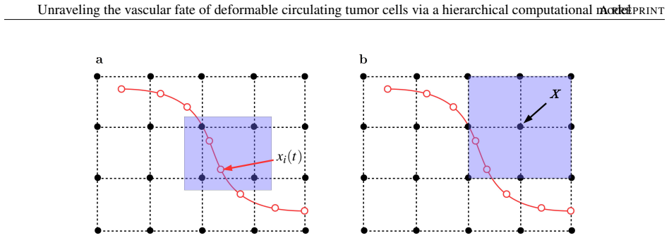
A Lattice-Boltzmann method... coupled with an Immersed Boundary Method... cell membranes are discretized as an ensemble of linear elastic springs
What do these tags mean?
- matches
- The paper's claim is directly supported by a theorem in the formal canon.
- supports
- The theorem supports part of the paper's argument, but the paper may add assumptions or extra steps.
- extends
- The paper goes beyond the formal theorem; the theorem is a base layer rather than the whole result.
- uses
- The paper appears to rely on the theorem as machinery.
- contradicts
- The paper's claim conflicts with a theorem or certificate in the canon.
- unclear
- Pith found a possible connection, but the passage is too broad, indirect, or ambiguous to say the theorem truly supports the claim.
Reference graph
Works this paper leans on
-
[1]
D. X. Nguyen, P.D. Bos, and J. Massagué. Metastasis: from dissemination to organ-specific colonization. Nat. Rev. Cancer, 9(4):274–284, 2009
work page 2009
- [2]
-
[3]
J. A. Joyce and J. W. Pollard. Microenvironmental regulation of metastasis. Nat. Rev. Cancer, 9(4):239–252, 2009
work page 2009
-
[4]
H. Mollica, C. Coclite, M.E. Miali, R.C. Pereira, L. Paleari, C. Manneschi, A. DeCensi, and P. Decuzzi. Deciphering the relative contribution of vascular inflammation and blood rheology in metastatic spreading. Biomicrofluidics, 12(4):042205, 2018
work page 2018
-
[5]
M.R. King, K.G. Phillips, A. Mitrugno, T.R. Lee, A.M. de Guillebon, S. Chandrasekaran andM.J. McGuire, R.T. Carr, S.M. Baker-Groberg, R.A. Riggand, A. Kolatkar, M. Luttgen, K. Bethel, P. Kuhn, P. Decuzzi, and O.J. McCarty. A physical sciences network characterization of circulating tumor cell aggregate transport. Am J Physiol Cell Physiol, 308(10):C792–C802, 2015
work page 2015
-
[6]
S. J. Tan, L. Yobas, G. Y . Lee, C. N. Ong, and C. T. Lim. Microdevice for the isolation and enumeration of cancer cells from blood. Biomed Microdevices, pages 883–892, 2009
work page 2009
-
[7]
E. Sollier, D. E. Go, J. Che, D. R. Gossett, S. O’Byrne, W. M. Weaver, N. Kummer, M. Rettig, J. Goldman, N. Nickols, S. McCloskey, R. P. Kulkarni, and D. Di Carlo. Size-selective collection of circulating tumor cells using vortex technology. Lab Chip, 14:63–77, 2014
work page 2014
- [8]
- [9]
-
[10]
T. W. Remmerbach, F. Wottawah, J. Dietrich, B. Lincoln, C. Wittekind, and J. Guck. Oral cancer diagnosis by mechanical phenotyping. Cancer ResEur Biophys J, 69:1728–32, 2009
work page 2009
-
[11]
S. E. Cross, Y . S. Jin, J. Rao, and J. K. Gimzewski. Nanomechanical analysis of cells from cancer patients.Nat Nanotechnol, 2:780–3, 2007
work page 2007
-
[12]
J. Shaw Bagnall, S. Byun, S. Begum, D. T. Miyamoto, V . C. Hecht, S. Maheswaran, S. L. Stott, M. Toner, R. O. Hynes, and S. R. Manalis. Deformability of tumor cells versus blood cells. Sci Rep, 5:18542, 2015
work page 2015
-
[13]
J. Zhang, P.C. Johnson, and A.S. Popel. Effects of erythrocyte deformability and aggregation on the cell free layer and apparent viscosity of microscopic blood flows. Microvasc. Res., 77(3):265–272, 2009
work page 2009
-
[14]
R. Skalak. Strain energy function of red blood cell membranes. Biophys J, 13(3):245–264, 2009
work page 2009
- [15]
-
[16]
U.D. Schiller, T. Krüger, and O. Henrich. Mesoscopic modelling and simulation of soft matter. Soft Matter, 14(1):9–26, 2017
work page 2017
-
[18]
D.A. Fedosov, B. Caswell, and G.E. Karniadakis. A multiscale red blood cell model with accurate mechanics. Biophysical Journal, 98(10):2215–2225, 2010
work page 2010
-
[19]
D.A. Fedosov, B. Caswell, A.S. Popel, and G.E. Karniadakis. Blood flow and cell-free layer in microvessels. Microcirculation, 17(8):615–628, 2010. 18 Unraveling the vascular fate of deformable circulating tumor cells via a hierarchical computational modelA PREPRINT
work page 2010
-
[20]
D.A. Fedosov and G. Gompper. White blood cell margination in microcirculation. Soft Matter, 10(8):2961–70, 2014
work page 2014
-
[21]
D.A. Fedosov, M. Peltmäki, and G. Gompper. Deformation and dynamics of red blood cells in flow through cylindrical microchannels. Soft Matter, 10(24):4258–67, 2014
work page 2014
-
[22]
C.S. Peskin. The immersed boundary method. Acta Numerica, 11(3-4):479–511, 2002
work page 2002
- [23]
-
[24]
On the near-wall accumulation of injectable particles in the microcirculation: Smaller is not better
Tae-Rin Lee, Myunghwan Choi, Adrian M Kopacz, Seok-Hyun Yun, Wing Liu, and Paolo Decuzzi. On the near-wall accumulation of injectable particles in the microcirculation: Smaller is not better. Scientific reports, 3:2079, 06 2013
work page 2079
-
[25]
Ying Li, Wylie Stroberg, Tae-Rin Lee, Hansung Kim, Han Man, Dean Ho, Paolo Decuzzi, and Wing Liu. Multiscale modeling and uncertainty quantification in nanoparticle-mediated drug/gene delivery.Computational Mechanics, 53:511–537, 03 2014
work page 2014
-
[26]
G. Falcucci, S. Ubertini, D. Chiappini, and S. Succi. Modern lattice boltzmann methods for multiphase microflows. IMA Journal of Applied Mathematics, 76(5):712–725, 2011
work page 2011
-
[27]
S. Succi. Lattice boltzmann across scales: from turbulence to dna translocation. European Physical Journal B, 64(3-4):471–479, 2008
work page 2008
- [28]
-
[29]
C. Sun, C. Migliorini, and L.L. Munn. Red blood cells initiate leukocyte rolling in postcapillary expansions: a lattice boltzmann analysis. Biophys J, 85(1):208–22, 2003
work page 2003
-
[30]
A. Coclite, H. Mollica, S. Ranaldo, G. Pascazio, M. D. de Tullio, and P. Decuzzi. Predicting different adhesive regimens of circulating particles at blood capillary walls. Microfluidics and Nanofluidics, 21(11):168, 2017
work page 2017
- [31]
-
[32]
H. Ye, Z. Shen, and Y . Li. Shear rate dependent margination of sphere-like, oblate-like and prolate-like micro-particles within blood flow. Soft Matter, 14(36):7401–7419, 2018
work page 2018
-
[33]
S. Gekle. Strongly accelerated margination of active particles in blood flow. Biophysical Journal, 110(2):514 – 520, 2016
work page 2016
-
[34]
K.A. Rejniak. Circulating tumor cells: When a solid tumor meets a fluid microenvironment. Front Oncol, 2(111), 2012
work page 2012
-
[35]
W.W. Yan, Y . Liu, and B.M. Fu. Effects of curvature and cell-cell interaction on cell adhesion in microvessels. Biomech. Model. Mechanobiol, 9(5):629–40, 2010
work page 2010
-
[36]
L.L Xiao, Y . Liu, S. Chen, and B.M. Fu. Effects of flowing rbcs on adhesion of a circulating tumor cell in microvessels. Biomech. Model. Mechanobiol, 16(2):597–610, 2017
work page 2017
-
[37]
S. Succi. The lattice Boltzmann equation: for fluid dynamics and beyond. Oxford University Press, 2001
work page 2001
-
[38]
Simulating engineering flows through complex porous media via the lattice boltzmann method
Vesselin Krassimirov Krastev and Giacomo Falcucci. Simulating engineering flows through complex porous media via the lattice boltzmann method. Energies, 11(4), 2018
work page 2018
-
[39]
D. A. Hammer and S.M. Apte. Simulation of cell rolling and adhesion on surfaces in shear flow: general results and analysis of selectin-mediated neutrophil adhesion. Biophys. J., 63(1):35–57, 1992
work page 1992
-
[40]
M.R. King and D. A. Hammer. Multiparticle adhesive dynamics: Hydrodynamic recruitment of rolling leukocytes. Proc. Natl. Acad. Sci. U.S.A., 98(26):14919 – 14924, 2001
work page 2001
-
[41]
H. Ye, Z. Shen, and Y . Li. Cell stiffness governs its adhesion dynamics on substrate under shear flow. Journal of IEEE Transactions on Nanotechnology, 17(3):407–411, 2017
work page 2017
-
[42]
Y .H. Qian, D. Dhumieres, and P. Lallemand. Lattice bgk models for navier-stokes equation.Europhysics Letters, 17(6):479–484, 1992
work page 1992
-
[43]
T. Krüger. Effect of tube diameter and capillary number on platelet margination and near-wall dynamics. Rheol. Acta., 55(6):511–526, 2016
work page 2016
-
[44]
J. Sigüenza, S. Mendez, D. Ambard, F. Dubois, F. Jourdan, R. Mozul, and F. Nicoud. Validation of an immersed thick boundary method for simulating fluid–structure interactions of deformable membranes. Journal of Computational Physics, 322(1):723–746, 2016. 19 Unraveling the vascular fate of deformable circulating tumor cells via a hierarchical computational...
work page 2016
- [45]
-
[46]
J. Li, M. Dao, C. T. Lim, and S. Suresh. Spectrin-level modeling of the cytoskeleton and optical tweezers stretching of the erythrocyte. Biophysical Journal, 88(1):3707–3719, 2005
work page 2005
- [47]
-
[48]
J.L. McWhirter, H. Noguchi, and G. Gompper. Nonlinear elastic and viscoelastic deformation of the human red blood cell with optical tweezers. Proceedings of the National Academy of Sciences of the United States of America, 106(15):6039–6043, 2009
work page 2009
-
[49]
N. A. Mody, O. Lomakin, T. A. Doggett, T. G. Diacovo, and M. R. King. Mechanics of transient platelet adhesion to von willebrand factor under flow. Biophys. J., 88(2):1432–1443, 2005
work page 2005
-
[50]
W. Wang, N. A. Mody, and M. R. King. Multiscale model of platelet translocation and collision. J. Comput. Phys., 244:223–235, 2005
work page 2005
-
[51]
E. Lac, D. Barthes-Biesel, N.A. Pelekasis, and J. Tsamopoulos. Spherical capsuls in three- dimensional unbounded stokes flow: Effect of the membrane constitutive law and onset of buckling. Journal of Fluid Mechanics, 516:303–334, 2004
work page 2004
-
[52]
Y . Sui, YT. Chew, P. Roy XB., Chen, HT., and Low. Transient deformation of elastic capsules in shear flow: effect of membrane bending stiffness. Phys Rev E Stat Nonlin Soft Matter Phys, 75(6), 2007
work page 2007
-
[53]
C. Pozrikidis. Numerical simulation of the flow-induced deformation of red blood cells. Annals of Biomedical Engineering, 31(10):1194–1205, 2003
work page 2003
-
[54]
D. Peer, J.M Karp, S. Hong, O.C. Farokhzad, R. Margalit, and R. Langer. Nanocarriers as an emerging platform for cancer therapy. Nat Nanotechnol, 2(12):751–60, 2007
work page 2007
-
[55]
N. Guz, M. Dokukin, V . Kalaparthi, and I. Sokolov. If cell mechanics can be described by elastic modulus: Study of different models and probes used in indentation experiments. Biophysical Journal, 107:564–575, 2014
work page 2014
- [56]
-
[57]
N. Takeishi, Y . Imai, T. Yamaguchi, and T. Ishikawa. Flow of a circulating tumor cell and red blood cells in microvessels. Phys. Rev. E, 92(063011), 2015. 20
work page 2015
discussion (0)
Sign in with ORCID, Apple, or X to comment. Anyone can read and Pith papers without signing in.