Lightweight Physics-Aware Zero-Shot Ultrasound Plane-Wave Denoising
Pith reviewed 2026-05-22 00:12 UTC · model grok-4.3
The pith
Splitting steering angles into two subsets creates pseudo-pairs that train a two-layer network to denoise ultrasound images on the test sample alone.
A machine-rendered reading of the paper's core claim, the machinery that carries it, and where it could break.
Core claim
Partitioning the steering angles into two disjoint subsets produces compounded images whose underlying tissue structures remain consistent while the incoherent artifacts and noise differ; these images then serve as pseudo-pairs for self-supervised residual learning inside a two-layer convolutional network that is trained and applied directly on the test sample.
What carries the argument
The physics-aware pairing strategy that treats angle-subset reconstructions as pseudo-pairs for residual learning in a lightweight CNN.
If this is right
- Fewer steering angles can be used while still achieving higher image quality, supporting higher frame rates for moving targets.
- The method adapts to different anatomical sites and acquisition settings without domain-specific retraining or paired datasets.
- Training finishes quickly because the network contains only two convolutional layers.
- No external clean reference images or large training collections are required at any stage.
Where Pith is reading between the lines
- The same split-and-pair idea might apply to other modalities that combine multiple acquisitions whose signal is stable but whose noise changes with acquisition parameters.
- In fast-moving organs the approach could reduce motion blur more effectively than simply increasing the number of angles in standard compounding.
- Because the network is so small, the denoising step could be inserted into existing ultrasound scanners with only modest added computation.
Load-bearing premise
Tissue structures remain consistent across the two angle-subset reconstructions while the incoherent artifacts and noise vary with steering angle selection.
What would settle it
If visual or quantitative comparison shows that the denoised output loses anatomical detail or reduces contrast relative to the original low-angle compounded image in phantom or in-vivo tests, the claim would be falsified.
Figures


read the original abstract
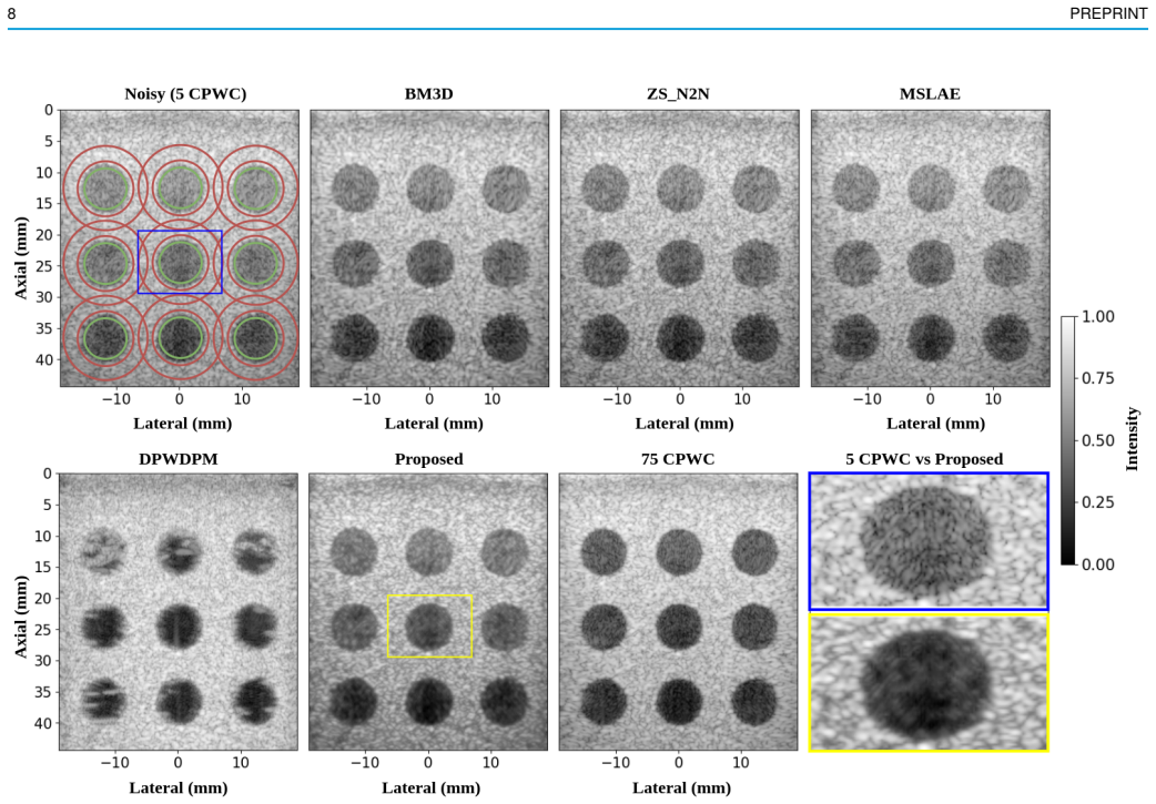
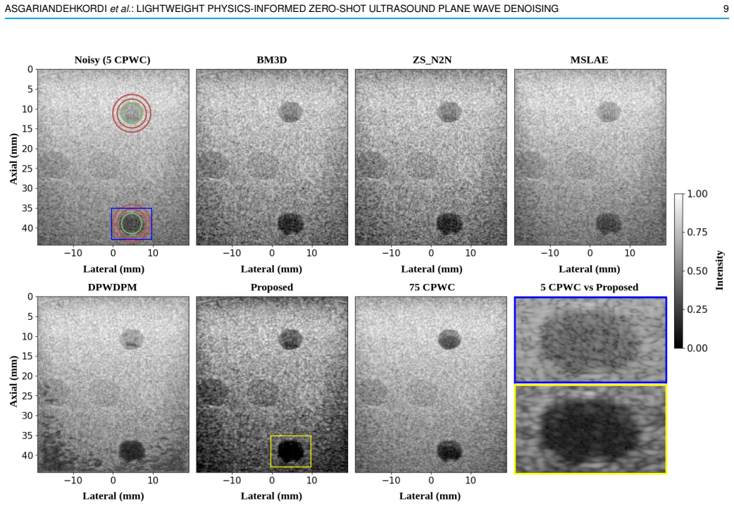
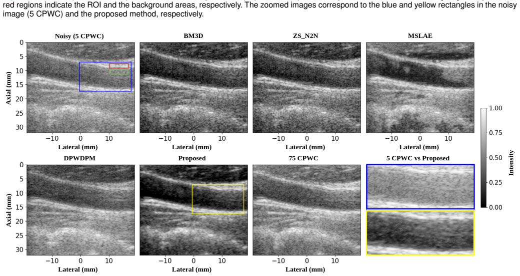
Ultrasound Coherent Plane-Wave Compounding (CPWC) enhances image contrast by combining echoes from multiple steered transmissions. While increasing the number of steering angles generally improves image quality, it significantly reduces frame rate and may introduce blurring artifacts in fast-moving targets. In addition, compounded images remain susceptible to noise, particularly when acquired using a limited number of transmissions. In this work, we propose a lightweight physics-aware zero-shot denoising framework for low-angle CPWC ultrasound imaging that improves image quality without requiring external training datasets or clean reference images. The proposed approach partitions the available steering angles into two disjoint subsets, each used to reconstruct compounded images with different angle-dependent artifacts and noise characteristics. These reconstructed images are then used as pseudo-pairs within a self-supervised residual learning framework to train a lightweight convolutional neural network directly on the test sample. Because the underlying tissue structures remain consistent across the subsets while the incoherent artifacts vary with steering angle selection, the proposed physics-aware pairing strategy enables the network to distinguish anatomical information from inconsistent noise and artifacts. Unlike supervised approaches, the proposed method does not require domain-specific fine-tuning or paired datasets, making it adaptable across different anatomical regions and acquisition settings. Furthermore, the proposed framework employs an efficient architecture composed of only two convolutional layers, enabling fast and computationally inexpensive training.
Editorial analysis
A structured set of objections, weighed in public.
Referee Report
Summary. The manuscript proposes a lightweight physics-aware zero-shot denoising framework for ultrasound coherent plane-wave compounding (CPWC) images acquired with a limited number of steering angles. The method partitions the steering angles into two disjoint subsets to reconstruct two compounded images with different artifacts, which serve as pseudo-pairs to train a 2-layer CNN using self-supervised residual learning directly on the test sample. The key idea is that tissue structures are consistent across subsets while noise and artifacts are inconsistent, allowing the network to learn to remove the latter without external data or clean references.
Significance. If the core assumption holds and the method is validated with quantitative results, this could provide a practical, training-free denoising solution for high-frame-rate ultrasound imaging, particularly useful in dynamic scenarios where full compounding is not feasible. The emphasis on a very lightweight architecture (only two convolutional layers) is a notable strength for computational efficiency. The approach builds on self-supervised learning ideas but adapts them specifically to the physics of plane-wave compounding.
major comments (2)
- [Abstract] The central claim relies on the assumption that 'the underlying tissue structures remain consistent across the subsets while the incoherent artifacts vary with steering angle selection' (Abstract). However, in CPWC beamforming, different angle subsets produce distinct effective point-spread functions and speckle realizations, which could introduce systematic differences in edge sharpness, contrast, and local intensity. This risks the residual-learning objective treating anatomical features as noise. The manuscript should provide simulation or phantom experiments to verify that the chosen partitions preserve structure at the network's receptive field scale.
- [Abstract] The abstract describes the method and its intended mechanism but provides no quantitative results, error bars, ablation studies, or comparisons to baselines. The soundness of the approach and the validity of the domain assumption cannot be assessed from the given text; experimental validation is required to support the claim of improved image quality.
minor comments (1)
- [Abstract] The description is clear but could benefit from a brief mention of how the two subsets are chosen (e.g., even/odd angles or random partition) to make the method reproducible.
Simulated Author's Rebuttal
We thank the referee for the constructive comments on our manuscript. We address each major comment point by point below and have made revisions to strengthen the presentation of our assumptions and results.
read point-by-point responses
-
Referee: [Abstract] The central claim relies on the assumption that 'the underlying tissue structures remain consistent across the subsets while the incoherent artifacts vary with steering angle selection' (Abstract). However, in CPWC beamforming, different angle subsets produce distinct effective point-spread functions and speckle realizations, which could introduce systematic differences in edge sharpness, contrast, and local intensity. This risks the residual-learning objective treating anatomical features as noise. The manuscript should provide simulation or phantom experiments to verify that the chosen partitions preserve structure at the network's receptive field scale.
Authors: We agree that angle-dependent variations in the effective point-spread function and speckle statistics represent a valid concern that could affect the residual-learning objective. Our physics-aware pairing strategy is motivated by the fact that specular and diffuse tissue backscattering remains largely invariant to small steering-angle changes, whereas grating-lobe and reverberation artifacts are strongly angle-dependent. To directly verify that structural content is preserved at the scale of the two-layer network’s receptive field, we have added new simulation and tissue-mimicking phantom experiments in the revised manuscript. These experiments quantify structural similarity (SSIM and edge-preservation metrics) between the two subset-compounded images and demonstrate that anatomical features are retained while artifact patterns differ, thereby supporting the validity of the self-supervised residual target. revision: yes
-
Referee: [Abstract] The abstract describes the method and its intended mechanism but provides no quantitative results, error bars, ablation studies, or comparisons to baselines. The soundness of the approach and the validity of the domain assumption cannot be assessed from the given text; experimental validation is required to support the claim of improved image quality.
Authors: We acknowledge that the original abstract emphasized the methodological contribution without summarizing numerical outcomes. In the revised version we have updated the abstract to report the principal quantitative findings, including mean PSNR and SSIM gains with standard deviations across multiple acquisitions, as well as brief comparisons against both conventional compounding and other zero-shot baselines. Full ablation studies, error-bar analyses, and statistical significance tests remain in the results section but are now referenced concisely in the abstract so that readers can immediately gauge performance. revision: yes
Circularity Check
No significant circularity; procedural construction is self-contained
full rationale
The paper proposes a zero-shot denoising method that partitions available steering angles into two disjoint subsets, reconstructs separate CPWC images from each, and uses those images as pseudo-pairs to train a two-layer CNN via residual learning directly on the test sample. The key premise that tissue structures remain consistent while artifacts vary is stated explicitly as an enabling physical assumption rather than derived from any equation or fitted parameter within the paper. No load-bearing step reduces a claimed result to its own inputs by construction, no self-citation chain is invoked to justify uniqueness or an ansatz, and the residual-learning objective operates on the generated pairs without the output being statistically forced by the partitioning choice itself. The framework therefore constitutes an independent procedural construction once the angle split is selected.
Axiom & Free-Parameter Ledger
axioms (1)
- domain assumption Underlying tissue structures remain consistent across the two angle-subset reconstructions while the incoherent artifacts and noise vary with steering angle selection
Lean theorems connected to this paper
-
IndisputableMonolith/Cost/FunctionalEquation.leanwashburn_uniqueness_aczel unclear?
unclearRelation between the paper passage and the cited Recognition theorem.
Because the underlying tissue structures remain consistent across the subsets while the incoherent artifacts vary with steering angle selection, the proposed physics-aware pairing strategy enables the network to distinguish anatomical information from inconsistent noise and artifacts.
-
IndisputableMonolith/Foundation/AlexanderDuality.leanalexander_duality_circle_linking unclear?
unclearRelation between the paper passage and the cited Recognition theorem.
we partition the available steering angles into two subsets, reconstruct two compounded images that share the same tissue response but exhibit distinct angle-dependent artifacts
What do these tags mean?
- matches
- The paper's claim is directly supported by a theorem in the formal canon.
- supports
- The theorem supports part of the paper's argument, but the paper may add assumptions or extra steps.
- extends
- The paper goes beyond the formal theorem; the theorem is a base layer rather than the whole result.
- uses
- The paper appears to rely on the theorem as machinery.
- contradicts
- The paper's claim conflicts with a theorem or certificate in the canon.
- unclear
- Pith found a possible connection, but the passage is too broad, indirect, or ambiguous to say the theorem truly supports the claim.
Forward citations
Cited by 1 Pith paper
-
Pyramid Self-contrastive Learning Framework for Test-time Ultrasound Image Denoising
A2A achieves one-shot ultrasound denoising via pyramid self-contrastive learning on sub-aperture signals to disentangle anatomy from noise, yielding large SNR and CNR gains in simulations and in vivo scans.
Reference graph
Works this paper leans on
-
[1]
Dehazing ultrasound using diffusion models,
T. S. Stevens, F. C. Meral, J. Yu, I. Z. Apostolakis, J.-L. Robert, and R. J. van Sloun, “Dehazing ultrasound using diffusion models,” IEEE Transactions on Medical Imaging , pp. 1–1, 2024
work page 2024
-
[2]
Y . Liu, N. Jiang, Z. Dai, and M. Zhang, “Advancing single-plane wave ultrasound imaging with implicit multiangle acoustic synthesis via deep learning,” IEEE Trans. Ultrason. Ferroelectr . Freq. Control, vol. 72, no. 4, pp. 479–497, Apr. 2025, doi: 10.1109/TUFFC.2025.3541113. ASGARIANDEHKORDI et al.: LIGHTWEIGHT PHYSICS-INFORMED ZERO-SHOT UL TRASOUND PLAN...
-
[3]
Adaptive subarray coherence based post-filter using array gain in medical ultrasound imaging,
L. Eslami and B. Mohammadzadeh Asl, “Adaptive subarray coherence based post-filter using array gain in medical ultrasound imaging,” Ultrasonics, vol. 126, p. 106808, 2022
work page 2022
-
[4]
Deep learning in ultrasound imaging,
R. J. G. van Sloun, R. Cohen, and Y . C. Eldar, “Deep learning in ultrasound imaging,” Proceedings of the IEEE , vol. 108, no. 1, pp. 11– 29, 2020
work page 2020
-
[5]
Deep neural networks for ultrasound beamforming,
A. C. Luchies and B. C. Byram, “Deep neural networks for ultrasound beamforming,” IEEE Transactions on Medical Imaging , vol. 37, no. 9, pp. 2010–2021, Sept. 2018, doi: 10.1109/TMI.2018.2809641
-
[6]
Ultrasound beamforming using MobileNetV2,
S. Goudarzi, A. Asif, and H. Rivaz, “Ultrasound beamforming using MobileNetV2,” in Proc. IEEE Int. Ultrason. Symp. (IUS) , Sep. 2020, pp. 1–4
work page 2020
-
[7]
Beamforming-integrated neural networks for ultrasound imaging,
D. Xiao and A. C. H. Yu, “Beamforming-integrated neural networks for ultrasound imaging,” Ultrasonics, vol. 145, p. 107474, 2025, doi: 10.1016/j.ultras.2024.107474
-
[8]
Noise2V oid—Learning denoising from single noisy images,
A. Krull, T.-O. Buchholz, and F. Jug, “Noise2V oid—Learning denoising from single noisy images,” presented at the IEEE/CVF Conf. Comput. Vis. Pattern Recognit. , 2019, pp. 2129–2137
work page 2019
-
[9]
Noise2Self: Blind denoising by self- supervision,
J. Batson and L. Royer, “Noise2Self: Blind denoising by self- supervision,” presented at the Int. Conf. Mach. Learn. , 2019, pp. 524– 533
work page 2019
-
[10]
Neighbor2neighbor: Self- supervised denoising from single noisy images,
T. Huang, S. Li, X. Jia, H. Lu, and J. Liu, “Neighbor2neighbor: Self- supervised denoising from single noisy images,” in Proc. IEEE/CVF Conf. Comput. Vis. Pattern Recognit. (CVPR) , 2021, pp. 14781–14790
work page 2021
-
[11]
Mitigating aberration-induced noise: A deep learning-based aberration- to-aberration approach,
M. Sharifzadeh, S. Goudarzi, A. Tang, H. Benali, and H. Rivaz, “Mitigating aberration-induced noise: A deep learning-based aberration- to-aberration approach,” IEEE Transactions on Medical Imaging , pp. 1– 1, 2024
work page 2024
-
[12]
Deep ultrasound denoising without clean data,
S. Goudarzi and H. Rivaz, “Deep ultrasound denoising without clean data,” in Medical Imaging 2023: Ultrasonic Imaging and Tomography , vol. 12470, pp. 131–136, SPIE, Apr. 2023
work page 2023
-
[13]
H. Cho, S. Park, J. Kang, and Y . Yoo, “Deep coherence learning: An unsupervised deep beamformer for high-quality single-plane-wave imaging in medical ultrasound,” Ultrasonics, vol. 143, p. 107408, 2024
work page 2024
-
[14]
Self-supervised ultrasound image denoising based on weighted joint loss,
C. Yu, F. Ren, S. Bao, Y . Yang, and X. Xu, “Self-supervised ultrasound image denoising based on weighted joint loss,” Digital Signal Process- ing, vol. 162, p. 105151, 2025
work page 2025
-
[15]
Zero-Shot Noise2Noise: Efficient Image Denoising Without Any Data,
O. Mansour, P. I. Baykal, A. Katyal, D. Erdogmus, and V . K. Goyal, “Zero-Shot Noise2Noise: Efficient Image Denoising Without Any Data,” in IEEE/CVF Conf. Comput. Vis. Pattern Recognit. (CVPR) , 2023, pp. 1–10
work page 2023
-
[16]
Nonlocal means- based speckle filtering for ultrasound images,
P. Coupe, P. Hellier, C. Kervrann, and C. Barillot, “Nonlocal means- based speckle filtering for ultrasound images,” in IEEE Trans. Image Process., vol. 18, no. 10, pp. 2221–2229, Oct. 2009
work page 2009
-
[17]
BM3D-based ultrasound image denoising via brushlet thresholding,
Y . Gan, E. Angelini, A. Laine, and C. Hendon, “BM3D-based ultrasound image denoising via brushlet thresholding,” in Proc. IEEE Int. Symp. Biomed. Imaging (ISBI) , Brooklyn, NY , USA, 2015, pp. 667–670, doi: 10.1109/ISBI.2015.7163961
-
[18]
G. Montaldo, M. Tanter, J. Bercoff, N. Benech, and M. Fink, “Coherent plane-wave compounding for very high frame rate ultra- sonography and transient elastography,” IEEE Trans. Ultrason. Ferro- electr . Freq. Control , vol. 56, no. 3, pp. 489–506, Mar. 2009, doi: 10.1109/TUFFC.2009.1067
-
[19]
H. Rivaz, R. Zellars, G. Hager, G. Fichtinger, and E. Boctor, “9c-1 beam steering approach for speckle characterization and out-of-plane motion estimation in real tissue,” in Proc. IEEE Ultrason. Symp. , Oct. 2007, pp. 781–784
work page 2007
-
[20]
Coherence factor of speckle from a multi-row probe,
K. Hollman, K. Rigby, and M. O’Donnell, “Coherence factor of speckle from a multi-row probe,” presented at the IEEE Ultrason. Symp. , 1999, pp. 1257–1260
work page 1999
-
[21]
Adaptive imaging using the generalized coher- ence factor,
P.-C. Li and M.-L. Li, “Adaptive imaging using the generalized coher- ence factor,” IEEE Trans. Ultrason., Ferroelectr ., Freq. Control, vol. 50, no. 2, pp. 128–141, 2003
work page 2003
-
[22]
C. Yang, Y . Jiao, T. Jiang, Y . Xu, and Y . Cui, “A united sign coherence factor beamformer for coherent plane-wave compounding with improved contrast,” Applied Sciences , vol. 10, no. 7, p. 2250, 2020
work page 2020
-
[23]
Dynamic coherence factor based on the standard deviation for coherent plane-wave compounding,
Y . Wang, C. Zheng, and H. Peng, “Dynamic coherence factor based on the standard deviation for coherent plane-wave compounding,” Comput. Biol. Med. , vol. 108, pp. 249–262, 2019
work page 2019
-
[24]
Denoising plane wave ultrasound images us- ing diffusion probabilistic models,
H. Asgariandehkordi et al., “Denoising plane wave ultrasound images us- ing diffusion probabilistic models,” IEEE Trans. Ultrason., Ferroelectr ., Freq. Control, vol. 71, no. 11, pp. 1526–1539, Nov. 2024
work page 2024
-
[25]
Improved Transcranial Plane- Wave Imaging With Learned Speed-of-Sound Maps,
Y . Yang, H. Duan, and Y . Zheng, “Improved Transcranial Plane- Wave Imaging With Learned Speed-of-Sound Maps,” IEEE Trans. Med. Imaging , vol. 43, no. 6, pp. 2191–2201, Jun. 2024, doi: 10.1109/TMI.2024.3358307
-
[26]
Z. Zhou et al., “High spatial-temporal resolution reconstruction of plane- wave ultrasound images with a multichannel multiscale convolutional neural network,” IEEE Trans. Ultrason., Ferroelectr ., Freq. Control, vol. 65, no. 11, pp. 1983–1996, Nov. 2018
work page 1983
-
[27]
Reconstruction for plane-wave ultrasound imag- ing using modified U-Net-based beamformer,
L. S. Nguon et al., “Reconstruction for plane-wave ultrasound imag- ing using modified U-Net-based beamformer,” Comput. Med. Imaging Graph., vol. 98, p. 102073, 2022
work page 2022
-
[28]
S. Goudarzi and H. Rivaz, “Deep reconstruction of high-quality ul- trasound images from raw plane-wave data: A simulation and in vivo study,” Ultrasonics, vol. 125, Sep. 2022, Art. no. 106778
work page 2022
-
[29]
J.-Y . Lu, P.-Y . Lee, and C.-C. Huang, “Improving image quality for single-angle plane-wave ultrasound imaging with convolutional neural network beamformer,” IEEE Trans. Ultrason., Ferroelectr ., Freq. Con- trol, vol. 69, no. 8, pp. 1326–1336, 2022
work page 2022
-
[30]
Deep convolutional neural network for ultrasound image enhancement,
D. Perdios et al., “Deep convolutional neural network for ultrasound image enhancement,” presented at the IEEE Int. Ultrason. Symp. (IUS) , Oct. 2018, pp. 1–4
work page 2018
-
[31]
Cnn-based projected gradient descent for consistent ct image reconstruction,
H. Gupta et al., “Cnn-based projected gradient descent for consistent ct image reconstruction,” IEEE Transactions on Medical Imaging , vol. 37, no. 6, pp. 1440–1453, 2018
work page 2018
-
[32]
Mimicknet, mimicking clinical image post- processing under black-box constraints,
O. Huang, W. Long, N. Bottenus, M. Lerendegui, G. E. Trahey, S. Farsiu, and M. L. Palmeri, “Mimicknet, mimicking clinical image post- processing under black-box constraints,” IEEE Transactions on Medical Imaging , vol. 39, no. 6, pp. 2277–2286, 2020
work page 2020
-
[33]
Displacement estimation in ultrasound elastography using pyramidal convolutional neural network,
A. K. Z. Tehrani and H. Rivaz, “Displacement estimation in ultrasound elastography using pyramidal convolutional neural network,” IEEE Transactions on Ultrasonics, Ferroelectrics, and Frequency Control , vol. 67, no. 12, pp. 2629–2639, 2020
work page 2020
-
[34]
Cnn-based image reconstruction method for ultrafast ultrasound imag- ing,
D. Perdios, M. V onlanthen, F. Martinez, M. Arditi, and J.-P. Thiran, “Cnn-based image reconstruction method for ultrafast ultrasound imag- ing,” IEEE Transactions on Ultrasonics, Ferroelectrics, and Frequency Control, vol. 69, no. 4, pp. 1154–1168, 2022
work page 2022
-
[35]
Z. Zhou et al., “Ultrafast plane wave imaging with line-scan-quality using an ultrasound-transfer generative adversarial network,” IEEE J. Biomed. Health Inform. , vol. 24, no. 4, pp. 943–956, Apr. 2020
work page 2020
-
[36]
Plane-wave image recon- struction via generative adversarial network and attention mechanism,
J. Tang, B. Zou, C. Li, S. Feng, and H. Peng, “Plane-wave image recon- struction via generative adversarial network and attention mechanism,” IEEE Trans. Instrum. Meas. , vol. 70, art. no. 4505115, pp. 1–15, 2021, doi: 10.1109/TIM.2021.3087819
-
[37]
Fast multi-focus ultrasound image recovery using generative adversarial networks,
S. Goudarzi, A. Asif, and H. Rivaz, “Fast multi-focus ultrasound image recovery using generative adversarial networks,” IEEE Trans. Comput. Imag., vol. 6, pp. 1272–1284, 2020
work page 2020
-
[38]
Denoising diffusion probabilistic models,
J. Ho, A. Jain, and P. Abbeel, “Denoising diffusion probabilistic models,” in Proc. NeurIPS, vol. 33, pp. 6840–6851, 2020
work page 2020
-
[39]
Improved denoising diffusion proba- bilistic models,
A. Q. Nichol and P. Dhariwal, “Improved denoising diffusion proba- bilistic models,” in Proc. Int. Conf. Mach. Learn. (ICML) , 2021, pp. 8162–8171
work page 2021
-
[40]
Tackling the generative learning trilemma with denoising diffusion GANs,
Z. Xiao, K. Kreis, and A. Vahdat, “Tackling the generative learning trilemma with denoising diffusion GANs,” presented at the Int. Conf. Learn. Represent. (ICLR) , 2022
work page 2022
-
[41]
H. Chung, B. Sim, and J. C. Ye, “Come-closer-diffuse-faster: Accelerat- ing conditional diffusion models for inverse problems through stochastic contraction,” in Proc. IEEE/CVF Conf. Comput. Vis. Pattern Recognit. (CVPR), 2022, pp. 12413–12422
work page 2022
-
[42]
Flow Straight and Fast: Learning to Generate and Transfer Data with Rectified Flow
X. Liu, C. Gong, and Q. Liu, “Flow straight and fast: Learning to generate and transfer data with rectified flow,” arXiv preprint arXiv:2209.03003, 2022
work page internal anchor Pith review Pith/arXiv arXiv 2022
-
[43]
Super-resolution reconstruction of ultrasound image using a modified diffusion model,
T. Liu et al., “Super-resolution reconstruction of ultrasound image using a modified diffusion model,” Phys. Med. Biol. , vol. 69, no. 12, Art. no. 125026, Jun. 2024
work page 2024
-
[44]
Diffusion reconstruction of ultrasound images with informative uncertainty,
Y . Zhang, C. Huneau, J. Idier, and D. Mateus, “Diffusion reconstruction of ultrasound images with informative uncertainty,” arXiv preprint arXiv:2310.20618, 2023
-
[45]
High Volume Rate 3D Ultrasound Reconstruction with Diffusion Models
T. S. Stevens, O. Nolan, O. Somphone, J. L. Robert, and R. J. van Sloun, “High volume rate 3D ultrasound reconstruction with diffusion models,” arXiv preprint arXiv:2505.22090 , 2025
work page internal anchor Pith review Pith/arXiv arXiv 2025
-
[46]
Ultra- sound image-to-video synthesis via latent dynamic diffusion models,
T. Chen, Y . Shi, Z. Zheng, B. Yan, J. Hu, X. X. Zhu, and L. Mou, “Ultra- sound image-to-video synthesis via latent dynamic diffusion models,” in Proc. Int. Conf. Med. Image Comput. Comput.-Assist. Interv. (MICCAI) , Cham, Switzerland: Springer Nature, Oct. 2024, pp. 764–774
work page 2024
-
[47]
Diffusion as sound propagation: Physics-inspired model for ultrasound image generation,
M. Dom ´ınguez, L. Mou, T. Chen, B. Yan, and X. X. Zhu, “Diffusion as sound propagation: Physics-inspired model for ultrasound image generation,” in Proc. Int. Conf. Med. Image Comput. Comput.-Assist. Interv. (MICCAI), Cham, Switzerland: Springer, 2024
work page 2024
-
[48]
DiG: Scalable and efficient diffusion models with gated linear attention,
L. Zhu, J. Liu, Z. Liu, H. Zha, and S. Yan, “DiG: Scalable and efficient diffusion models with gated linear attention,” in Proc. IEEE/CVF Conf. Comput. Vis. Pattern Recognit. (CVPR) , 2025. 12 PREPRINT
work page 2025
-
[49]
Noise2Noise: Learning image restoration without clean data,
J. Lehtinen, J. Munkberg, J. Hasselgren, S. Laine, T. Karras, M. Aittala, and T. Aila, “Noise2Noise: Learning image restoration without clean data,” in Proc. Int. Conf. Mach. Learn. (ICML) , 2018, pp. 2965–2974
work page 2018
-
[50]
J. Zhang, Q. He, Y . Xiao, H. Zheng, C. Wang, and J. Luo, “Ultrasound image reconstruction from plane wave radio-frequency data by self- supervised deep neural network,” Medical Image Analysis , vol. 70, p. 102018, 2021
work page 2021
-
[51]
Deblurring masked image modeling for ultrasound image analysis,
Q. Kang, Q. Lao, J. Gao, J. Liu, H. Yi, B. Ma, X. Zhang, and K. Li, “Deblurring masked image modeling for ultrasound image analysis,” Medical Image Analysis , vol. 97, p. 103256, 2024. doi: 10.1016/j.media.2024.103256
-
[52]
Beyond a Gaussian Denoiser: Residual Learning of Deep CNN for Image Denoising,
Zhang, K., Zuo, W., Chen, Y ., Meng, D., Zhang, L. “Beyond a Gaussian Denoiser: Residual Learning of Deep CNN for Image Denoising,” in IEEE Transactions on Image Processing , 26(7), 3142-3155 (2017)
work page 2017
-
[53]
Exploring simple siamese representation learning,
Chen, X., He, K., “Exploring simple siamese representation learning,” in Proceedings of the IEEE/CVF Conference on Computer Vision and Pattern Recognition (CVPR) , pp. 15750–15758 (2021)
work page 2021
-
[54]
Plane-wave imaging challenge in medical ultrasound,
H. Liebgott, A. Rodriguez-Molares, F. Cervenansky, J. Jensen, and O. Bernard, “Plane-wave imaging challenge in medical ultrasound,” in 2016 IEEE International Ultrasonics Symposium , pp. 1–4, 2016
work page 2016
-
[55]
The generalized contrast-to-noise ratio: A formal definition for lesion detectability,
Rodriguez-Molares, A., et al., “The generalized contrast-to-noise ratio: A formal definition for lesion detectability,” in IEEE Transactions on Ultrasonics, Ferroelectrics, and Frequency Control , 67(4), 745–759 (2020)
work page 2020
discussion (0)
Sign in with ORCID, Apple, or X to comment. Anyone can read and Pith papers without signing in.