KongNet: A Multi-headed Deep Learning Model for Detection and Classification of Nuclei in Histopathology Images
Pith reviewed 2026-05-18 02:59 UTC · model grok-4.3
The pith
A multi-headed neural network with a shared encoder and cell-type-specialised decoders delivers leading performance in nuclei detection and classification across varied histopathology images.
A machine-rendered reading of the paper's core claim, the machinery that carries it, and where it could break.
Core claim
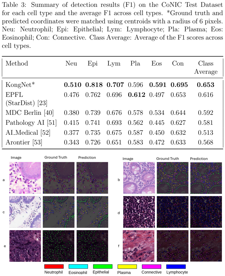
The central claim is that the specialised multi-decoder design is highly effective for nuclei detection and classification across diverse tissue and stain types. KongNet uses a shared encoder with parallel cell-type-specialised decoders; each decoder jointly predicts centroids, masks, and contours with SCSE attention modules and a composite loss. Through this multi-task setup the model took first place on MONKEY Challenge track 1 and second on track 2, its lightweight variant won the 2025 MIDOG Challenge, pre-trained KongNet ranked top three on PUMA without further tuning, and it set new state-of-the-art results on PanNuke and CoNIC.
What carries the argument
The multi-decoder architecture of a shared encoder feeding parallel, cell-type-specialised decoders that each jointly predict nuclei centroids, segmentation masks, and contours using SCSE attention and a composite loss.
Load-bearing premise
The observed performance gains stem primarily from the multi-decoder architecture and multi-task learning rather than from dataset-specific tuning, challenge evaluation protocols, or undisclosed training details.
What would settle it
A head-to-head experiment in which a single-decoder baseline trained with the same data, augmentations, and loss reaches equal or higher scores on the MONKEY, MIDOG, and PUMA challenges would show the specialised multi-decoder design is not required for the reported gains.
Figures


read the original abstract
Accurate detection and classification of nuclei in histopathology images are critical for diagnostic and research applications. We present KongNet, a multi-headed deep learning architecture featuring a shared encoder and parallel, cell-type-specialised decoders. Through multi-task learning, each decoder jointly predicts nuclei centroids, segmentation masks, and contours, aided by Spatial and Channel Squeeze-and-Excitation (SCSE) attention modules and a composite loss function. We validate KongNet in three Grand Challenges. The proposed model achieved first place on track 1 and second place on track 2 during the MONKEY Challenge. Its lightweight variant (KongNet-Det) secured first place in the 2025 MIDOG Challenge. KongNet pre-trained on the MONKEY dataset and fine-tuned on the PUMA dataset ranked among the top three in the PUMA Challenge without further optimisation. Furthermore, KongNet established state-of-the-art performance on the publicly available PanNuke and CoNIC datasets. Our results demonstrate that the specialised multi-decoder design is highly effective for nuclei detection and classification across diverse tissue and stain types. The pre-trained model weights along with the inference code have been publicly released to support future research.
Editorial analysis
A structured set of objections, weighed in public.
Referee Report
Summary. The paper presents KongNet, a multi-headed deep learning architecture with a shared encoder and parallel cell-type-specialised decoders. Each decoder performs multi-task learning to jointly predict nuclei centroids, segmentation masks, and contours, incorporating SCSE attention modules and a composite loss function. The model is validated on three grand challenges, achieving first place on MONKEY track 1, first place for its lightweight variant in MIDOG 2025, top-3 on PUMA after pre-training on MONKEY, and state-of-the-art results on the public PanNuke and CoNIC datasets. The authors conclude that the specialised multi-decoder design is highly effective across diverse tissue and stain types and release the pre-trained weights and inference code.
Significance. If the reported rankings and SOTA results are driven by the multi-decoder architecture rather than tuning or evaluation specifics, the work offers a practical advance in computational pathology for nuclei analysis. The public release of model weights and code is a clear strength that supports reproducibility and extension by the community.
major comments (2)
- [Experimental validation and results sections] The central claim that the parallel cell-type-specialised decoders (with joint centroid/mask/contour prediction and SCSE modules) are the decisive factor behind the MONKEY, MIDOG, and PUMA rankings and PanNuke/CoNIC SOTA results is not isolated by ablation. No experiment replaces the multi-decoder head with a single shared decoder (or removes the multi-task losses) while holding the encoder, training schedule, augmentations, and loss weights fixed; without this control the attribution remains unverified.
- [Results on public datasets and challenges] Baseline comparisons and statistical details are absent from the reported challenge outcomes. The manuscript does not include direct comparisons against standard single-decoder or multi-task baselines on the same splits, nor does it report confidence intervals, p-values, or cross-validation statistics that would allow assessment of whether the observed margins are significant.
minor comments (2)
- [Methods] The composite loss function and the weighting of its terms are described at a high level; explicit equations or hyperparameter values would improve reproducibility.
- [Figure 1 or equivalent] Figure captions and architecture diagrams would benefit from clearer labeling of the parallel decoder branches and the exact output heads (centroid, mask, contour) to match the textual description.
Simulated Author's Rebuttal
We thank the referee for the constructive feedback on our manuscript. We address each major comment below, proposing specific revisions to strengthen the experimental validation and statistical reporting while maintaining the integrity of the reported challenge results.
read point-by-point responses
-
Referee: [Experimental validation and results sections] The central claim that the parallel cell-type-specialised decoders (with joint centroid/mask/contour prediction and SCSE modules) are the decisive factor behind the MONKEY, MIDOG, and PUMA rankings and PanNuke/CoNIC SOTA results is not isolated by ablation. No experiment replaces the multi-decoder head with a single shared decoder (or removes the multi-task losses) while holding the encoder, training schedule, augmentations, and loss weights fixed; without this control the attribution remains unverified.
Authors: We agree that the manuscript would benefit from an explicit ablation isolating the multi-decoder design. The current experiments demonstrate strong performance across diverse challenges and datasets, but do not include a controlled replacement of the parallel specialised decoders with a single shared decoder under identical conditions. In the revised manuscript we will add this ablation on the PanNuke and CoNIC datasets, reporting the performance difference while keeping the encoder, training schedule, augmentations and loss weights fixed. revision: yes
-
Referee: [Results on public datasets and challenges] Baseline comparisons and statistical details are absent from the reported challenge outcomes. The manuscript does not include direct comparisons against standard single-decoder or multi-task baselines on the same splits, nor does it report confidence intervals, p-values, or cross-validation statistics that would allow assessment of whether the observed margins are significant.
Authors: We acknowledge that baseline comparisons and statistical details are missing. For the public PanNuke and CoNIC datasets we will add direct comparisons against standard single-decoder and multi-task baselines on the same splits together with confidence intervals. For the challenge results, the official leaderboards provide fixed test sets and we lack access to other participants' predictions, which limits our ability to compute p-values or perform cross-validation; we will instead discuss the observed margins relative to the published challenge rankings. revision: partial
- Statistical significance testing (p-values or formal hypothesis tests) for the MONKEY, MIDOG and PUMA challenge rankings, because the test sets are hidden and detailed predictions from other teams are not available.
Circularity Check
No circularity: empirical results on external benchmarks
full rationale
The manuscript describes an empirical CNN architecture (shared encoder plus parallel cell-type-specialised decoders with SCSE and multi-task losses) and reports its performance on independent public challenges and datasets. No derivation, uniqueness theorem, or first-principles prediction is offered that reduces by construction to quantities defined inside the paper; the central claim rests on externally verifiable rankings rather than on any fitted parameter renamed as a prediction or on a self-citation chain.
Axiom & Free-Parameter Ledger
free parameters (1)
- composite loss weights
axioms (1)
- domain assumption Challenge evaluation protocols and public datasets are representative of diverse tissue and stain types encountered in practice.
Lean theorems connected to this paper
-
IndisputableMonolith/Cost/FunctionalEquation.leanwashburn_uniqueness_aczel unclear?
unclearRelation between the paper passage and the cited Recognition theorem.
KongNet builds upon the established encoder-decoder paradigm... multi-headed design with specialised decoders for each cell type... SCSE attention... composite loss function L = Σ λk · LClass k + LInterclass
-
IndisputableMonolith/Foundation/AlphaCoordinateFixation.leanalpha_pin_under_high_calibration unclear?
unclearRelation between the paper passage and the cited Recognition theorem.
We integrate Spatial and Channel Squeeze-and-Excitation (SCSE) modules... PixelShuffle Upsampling... SiLU Activation
What do these tags mean?
- matches
- The paper's claim is directly supported by a theorem in the formal canon.
- supports
- The theorem supports part of the paper's argument, but the paper may add assumptions or extra steps.
- extends
- The paper goes beyond the formal theorem; the theorem is a base layer rather than the whole result.
- uses
- The paper appears to rely on the theorem as machinery.
- contradicts
- The paper's claim conflicts with a theorem or certificate in the canon.
- unclear
- Pith found a possible connection, but the passage is too broad, indirect, or ambiguous to say the theorem truly supports the claim.
Forward citations
Cited by 1 Pith paper
-
NucEval: A Robust Evaluation Framework for Nuclear Instance Segmentation
NucEval is a unified evaluation framework for nuclear instance segmentation that modifies standard metrics to handle vague regions, normalize scores, manage overlaps, and account for border uncertainty.
Reference graph
Works this paper leans on
-
[1]
The evolving tumor microenvironment: From cancer initiation to metastatic outgrowth,
K. E. de Visser and J. A. Joyce, “The evolving tumor microenvironment: From cancer initiation to metastatic outgrowth,”Cancer Cell, vol. 41, no. 3, pp. 374–403, 2023
work page 2023
-
[2]
The tumor microenvironment shows a hierarchy of cell-cell interactions dominated by fibroblasts,
S. Mayer, T. Milo, A. Isaacson, C. Halperin, S. Miyara, Y. Stein, C. Lior, M. Pevsner-Fischer, E. Tzahor, A. Mayo, U. Alon, and R. Scherz- Shouval, “The tumor microenvironment shows a hierarchy of cell-cell interactions dominated by fibroblasts,”Nature Communications, vol. 14, p. 5810, Sep 2023
work page 2023
-
[3]
Role of tumor microenvironment in cancer pro- gression and therapeutic strategy,
Q. Wang, X. Shao, Y. Zhang, M. Zhu, F. X. C. Wang, J. Mu, J. Li, H. Yao, and K. Chen, “Role of tumor microenvironment in cancer pro- gression and therapeutic strategy,”Cancer Medicine, vol. 12, no. 10, pp. 11149–11165, 2023
work page 2023
-
[4]
J. Saltz, R. Gupta, and L. H. et al., “Spatial organization and molecu- lar correlation of tumor-infiltrating lymphocytes using deep learning on pathology images,”Cell Reports, vol. 23, no. 1, pp. 181–193.e7, 2018
work page 2018
-
[5]
Tumour- infiltrating lymphocytes: from prognosis to treatment selection,
K. Brummel, A. L. Eerkens, M. de Bruyn, and H. W. Nijman, “Tumour- infiltrating lymphocytes: from prognosis to treatment selection,”British Journal of Cancer, vol. 128, pp. 451–458, Feb 2023
work page 2023
-
[6]
K. El Bairi, H. R. Haynes, E. Blackley, S. Fineberg, J. Shear, S. Turner, J. R. de Freitas, D. Sur, L. C. Amendola, M. Gharib, A. Kallala, I. Arun, F. Azmoudeh-Ardalan, L. Fujimoto, L. F. Sua, S.-W. Liu, H.-C. Lien, P. Kirtani, M. Balancin, H. El Attar, P. Guleria, W. Yang, E. Shash, I.-C. Chen, V. Bautista, J. F. Do Prado Moura, B. L. Rapoport, C. Cas- t...
work page 2021
-
[7]
C. Valenza, B. Taurelli Salimbeni, C. Santoro, D. Trapani, G. An- tonarelli, and G. Curigliano, “Tumor infiltrating lymphocytes across breast cancer subtypes: Current issues for biomarker assessment,”Can- cers (Basel), vol. 15, p. 767, Jan. 2023
work page 2023
-
[8]
A 2018 Reference Guide to the Banff Classification of Renal Allograft Pathology,
C. Roufosse, N. Simmonds, M. C.-v. Groningen, M. Haas, K. J. Henrik- sen, C. Horsfield, A. Loupy, M. Mengel, A. Perkowska-Ptasiska, M. Ra- bant, L. C. Racusen, K. Solez, and J. U. Becker, “A 2018 Reference Guide to the Banff Classification of Renal Allograft Pathology,”Trans- plantation, vol. 102, no. 11, pp. 1795–1814, 2018
work page 2018
-
[9]
Structured description of the monkey challenge,
L. Studer, “Structured description of the monkey challenge,” Sept. 2024
work page 2024
-
[10]
M. Schuiveling, H. Liu, D. Eek, G. Breimer, K. Suijkerbuijk, W. Blokx, and M. Veta, “A novel dataset for nuclei and tissue segmentation in melanoma with baseline nuclei segmentation and tissue segmentation benchmarks,”GigaScience, vol. 14, 01 2025
work page 2025
-
[11]
Mitosis domain generalization challenge 2025,
J. Ammeling, M. Aubreville, S. Banerjee, C. A. Bertram, K. Breininger, D. Hirling, P. Horvath, N. Stathonikos, and M. Veta, “Mitosis domain generalization challenge 2025,” Mar. 2025
work page 2025
-
[12]
Pannuke dataset extension, insights and baselines,
J. Gamper, N. A. Koohbanani, K. Benes, S. Graham, M. Jahanifar, S. A. Khurram, A. Azam, K. Hewitt, and N. Rajpoot, “Pannuke dataset extension, insights and baselines,” 2020
work page 2020
-
[13]
S. Graham, Q. D. Vu, M. Jahanifar, M. Weigert, U. Schmidt, W. Zhang, J. Zhang, S. Yang, J. Xiang, X. Wang, J. L. Rumberger, E. Baumann, P. Hirsch, L. Liu, C. Hong, A. I. Aviles-Rivero, A. Jain, H. Ahn, Y. Hong, H. Azzuni, M. Xu, M. Yaqub, M.-C. Blache, B. Pi´ egu, B. Vernay, T. Scherr, M. B¨ ohland, K. L¨ offler, J. Li, W. Ying, C. Wang, D. Snead, S. E. A...
work page 2024
-
[14]
You only look once: Unified, real-time object detection,
J. Redmon, S. Divvala, R. Girshick, and A. Farhadi, “You only look once: Unified, real-time object detection,” in2016 IEEE Conference on Computer Vision and Pattern Recognition (CVPR), pp. 779–788, 2016. 40
work page 2016
-
[15]
S. S. Debsarkar, B. Aronow, and V. S. Prasath, “Advancements in auto- mated nuclei segmentation for histopathology using you only look once- driven approaches: A systematic review,”Computers in Biology and Medicine, vol. 190, p. 110072, 2025
work page 2025
-
[16]
K. Sirinukunwattana, S. E. A. Raza, Y.-W. Tsang, D. R. J. Snead, I. A. Cree, and N. M. Rajpoot, “Locality sensitive deep learning for detection and classification of nuclei in routine colon cancer histology images,” IEEE Transactions on Medical Imaging, vol. 35, no. 5, pp. 1196–1206, 2016
work page 2016
-
[17]
Deconvolving convolutional neural network for cell detection,
S. E. A. Raza, K. AbdulJabbar, M. Jamal-Hanjani, S. Veeriah, J. L. Quesne, C. Swanton, and Y. Yuan, “Deconvolving convolutional neural network for cell detection,” in2019 IEEE 16th International Symposium on Biomedical Imaging (ISBI 2019), pp. 891–894, 2019
work page 2019
-
[18]
Multi- class cell detection using modified self-attention,
T. Sugimoto, H. Ito, Y. Teramoto, A. Yoshizawa, and R. Bise, “Multi- class cell detection using modified self-attention,” in2022 IEEE/CVF Conference on Computer Vision and Pattern Recognition Workshops (CVPRW), pp. 1854–1862, 2022
work page 2022
-
[19]
nnu-net: a self-configuring method for deep learning-based biomedical image segmentation,
F. Isensee, P. F. Jaeger, S. A. A. Kohl, J. Petersen, and K. H. Maier-Hein, “nnu-net: a self-configuring method for deep learning-based biomedical image segmentation,”Nature Methods, vol. 18, pp. 203–211, Feb 2021
work page 2021
-
[20]
Faster r-cnn: Towards real- time object detection with region proposal networks,
S. Ren, K. He, R. Girshick, and J. Sun, “Faster r-cnn: Towards real- time object detection with region proposal networks,” inAdvances in Neural Information Processing Systems(C. Cortes, N. Lawrence, D. Lee, M. Sugiyama, and R. Garnett, eds.), vol. 28, Curran Associates, Inc., 2015
work page 2015
-
[21]
R. Lomans, V. Angerilli, J. Spronck, L. L. Kodach, I. Gullo, F. Carneiro, R. S. van der Post, and F. Ciompi, “Deep learning for multiclass tumor cell detection in histopathology slides of hereditary diffuse gastric can- cer,”iScience, vol. 28, Aug 2025
work page 2025
-
[22]
S. Graham, Q. D. Vu, S. E. A. Raza, A. Azam, Y. W. Tsang, J. T. Kwak, and N. Rajpoot, “Hover-net: Simultaneous segmentation and 41 classification of nuclei in multi-tissue histology images,”Medical Image Analysis, vol. 58, p. 101563, 2019
work page 2019
-
[23]
Nuclei instance segmentation and classi- fication in histopathology images with stardist,
M. Weigert and U. Schmidt, “Nuclei instance segmentation and classi- fication in histopathology images with stardist,” in2022 IEEE Interna- tional Symposium on Biomedical Imaging Challenges (ISBIC), pp. 1–4, 2022
work page 2022
-
[24]
E. Baumann, B. Dislich, J. L. Rumberger, I. D. Nagtegaal, M. R. Mar- tinez, and I. Zlobec, “Hover-next: A fast nuclei segmentation and classi- fication pipeline for next generation histopathology,” inMedical Imaging with Deep Learning, 2024
work page 2024
-
[26]
Transnuseg: A lightweight multi-task transformer for nuclei segmentation,
Z. He, M. Unberath, J. Ke, and Y. Shen, “Transnuseg: A lightweight multi-task transformer for nuclei segmentation,” inMedical Image Computing and Computer Assisted Intervention – MICCAI 2023 (H. Greenspan, A. Madabhushi, P. Mousavi, S. Salcudean, J. Dun- can, T. Syeda-Mahmood, and R. Taylor, eds.), (Cham), pp. 206–215, Springer Nature Switzerland, 2023
work page 2023
-
[27]
Cellvit: Vision trans- formers for precise cell segmentation and classification,
F. H¨ orst, M. Rempe, L. Heine, C. Seibold, J. Keyl, G. Baldini, S. Ugurel, J. Siveke, B. Gr¨ unwald, J. Egger, and J. Kleesiek, “Cellvit: Vision trans- formers for precise cell segmentation and classification,”Medical Image Analysis, vol. 94, p. 103143, 2024
work page 2024
-
[28]
Nulite – lightweight and fast model for nuclei instance segmentation and classification,
C. Tommasino, C. Russo, and A. M. Rinaldi, “Nulite – lightweight and fast model for nuclei instance segmentation and classification,” 2024
work page 2024
-
[29]
Cell nuclei detection and classi- fication in whole slide images with transformers,
O. Pina, E. Dorca, and V. Vilaplana, “Cell nuclei detection and classi- fication in whole slide images with transformers,” 2025
work page 2025
-
[30]
T. Goldsborough, B. Philps, A. O’Callaghan, F. Inglis, L. Leplat, A. Filby, H. Bilen, and P. Bankhead, “Instanseg: an embedding-based instance segmentation algorithm optimized for accurate, efficient and portable cell segmentation,” 2024. 42
work page 2024
-
[31]
Ocelot: Over- lapped cell on tissue dataset for histopathology,
J. Ryu, A. V. Puche, J. Shin, S. Park, B. Brattoli, J. Lee, W. Jung, S. I. Cho, K. Paeng, C.-Y. Ock, D. Yoo, and S. Pereira, “Ocelot: Over- lapped cell on tissue dataset for histopathology,” inProceedings of the IEEE/CVF Conference on Computer Vision and Pattern Recognition (CVPR), 2023
work page 2023
-
[32]
N. Torbati, A. Meshcheryakova, D. Mechtcheriakova, and A. Mahbod, “A multi-stage auto-context deep learning framework for tissue and nu- clei segmentation and classification in h&e-stained histological images of advanced melanoma,” 2025
work page 2025
-
[33]
A comprehensive multi-domain dataset for mitotic figure detection,
M. Aubreville, F. Wilm, N. Stathonikos, K. Breininger, T. A. Dono- van, S. Jabari, M. Veta, J. Ganz, J. Ammeling, P. J. Van Diest, R. Klopfleisch, and C. A. Bertram, “A comprehensive multi-domain dataset for mitotic figure detection,”Scientific Data, vol. 10, p. 484, July 2023
work page 2023
-
[34]
M. Aubreville, C. A. Bertram, T. A. Donovan, C. Marzahl, A. Maier, and R. Klopfleisch, “A completely annotated whole slide image dataset of canine breast cancer to aid human breast cancer research,”Scientific Data, vol. 7, p. 417, Nov. 2020
work page 2020
-
[35]
C. A. Bertram, M. Aubreville, C. Marzahl, A. Maier, and R. Klopfleisch, “A large-scale dataset for mitotic figure assessment on whole slide images of canine cutaneous mast cell tumor,”Scientific Data, vol. 6, p. 274, Nov 2019
work page 2019
-
[36]
M. Jahanifar, “Mitosis subtyping dataset.”https://doi.org/10. 5281/zenodo.15390543, 2025. [Data set]
work page 2025
-
[37]
Nuclick: A deep learning framework for interactive segmentation of microscopic images,
N. Alemi Koohbanani, M. Jahanifar, N. Zamani Tajadin, and N. Ra- jpoot, “Nuclick: A deep learning framework for interactive segmentation of microscopic images,”Medical Image Analysis, vol. 65, p. 101771, 2020
work page 2020
-
[38]
TIAToolbox as an end- to-end library for advanced tissue image analytics,
J. Pocock, S. Graham, Q. D. Vu, M. Jahanifar, S. Deshpande, G. Had- jigeorghiou, A. Shephard, R. M. S. Bashir, M. Bilal, W. Lu, D. Epstein, F. Minhas, N. M. Rajpoot, and S. E. A. Raza, “TIAToolbox as an end- to-end library for advanced tissue image analytics,”Communications Medicine, vol. 2, p. 120, sep 2022. 43
work page 2022
-
[39]
Size-dependent positioning of hu- man chromosomes in interphase nuclei,
H. B. Sun, J. Shen, and H. Yokota, “Size-dependent positioning of hu- man chromosomes in interphase nuclei,”Biophysical Journal, vol. 79, no. 1, pp. 184–190, 2000
work page 2000
-
[40]
Panoptic segmentation with highly imbalanced seman- tic labels,
J. L. Rumberger, E. Baumann, P. Hirsch, A. Janowczyk, I. Zlobec, and D. Kainmueller, “Panoptic segmentation with highly imbalanced seman- tic labels,” in2022 IEEE International Symposium on Biomedical Imag- ing Challenges (ISBIC), pp. 1–4, 2022
work page 2022
-
[41]
Efficientnetv2: Smaller models and faster training,
M. Tan and Q. Le, “Efficientnetv2: Smaller models and faster training,” 04 2021
work page 2021
-
[42]
Concurrent spatial and chan- nel squeeze & excitation in fully convolutional networks,
A. G. Roy, N. Navab, and C. Wachinger, “Concurrent spatial and chan- nel squeeze & excitation in fully convolutional networks,” 2018
work page 2018
-
[43]
W. Shi, J. Caballero, F. Husz´ ar, J. Totz, A. P. Aitken, R. Bishop, D. Rueckert, and Z. Wang, “Real-time single image and video super- resolution using an efficient sub-pixel convolutional neural network,” in 2016 IEEE Conference on Computer Vision and Pattern Recognition (CVPR), pp. 1874–1883, 2016
work page 2016
-
[44]
Multi-task learning using un- certainty to weigh losses for scene geometry and semantics,
A. Kendall, Y. Gal, and R. Cipolla, “Multi-task learning using un- certainty to weigh losses for scene geometry and semantics,”2018 IEEE/CVF Conference on Computer Vision and Pattern Recognition, pp. 7482–7491, 2017
work page 2018
-
[45]
Thibautgoldsborough-instanseg-monkey-challenge
T. Goldsborough, “Thibautgoldsborough-instanseg-monkey-challenge.”
-
[46]
G. Deotale, A. Ambast, L. Ramchandani, D. K. Das, and T. Thomas, “Ensemble object detection methodology for automated detection of inflammatory cells in kidney biopsies,” inMedical Imaging with Deep Learning - Short Papers, 2025
work page 2025
- [47]
-
[48]
Biototem-monkey-challenge: Scripts for monkey challenge 2024
S. Xu, “Biototem-monkey-challenge: Scripts for monkey challenge 2024.”
work page 2024
-
[49]
Jun-sato-monkey-challenge-ouradiology: Ouradiology ap- proach in monkey challenge
J. Sato, “Jun-sato-monkey-challenge-ouradiology: Ouradiology ap- proach in monkey challenge.” 44
-
[50]
E. Candeloro, “Aimagelab-zip-monkey-challenge-ziplab: Aimagelab zip unimore solution for the monkey challenge of radboud university medical center.”
-
[51]
Keep it accurate and robust: An enhanced nuclei analysis framework,
W. Zhang, S. Yang, M. Luo, C. He, Y. Li, J. Zhang, X. Wang, and F. Wang, “Keep it accurate and robust: An enhanced nuclei analysis framework,”Comput. Struct. Biotechnol. J., vol. 24, pp. 699–710, Dec. 2024
work page 2024
-
[52]
A deep learning framework for nuclear segmentation and classification in histopathological images,
S. Yang, J. Xiang, and X. Wang, “A deep learning framework for nuclear segmentation and classification in histopathological images,” 2022
work page 2022
-
[53]
Class-controlled copy-paste based cell segmenta- tion for conic challenge,
H. Ahn and Y. Hong, “Class-controlled copy-paste based cell segmenta- tion for conic challenge,”bioRxiv, 2022
work page 2022
-
[54]
D. Anglada-Rotger, B. Jansat, F. Marques, and M. Pard` as, “Two heads are enough: Dualu-net, a fast and efficient architecture for nuclei in- stance segmentation,” inMedical Imaging with Deep Learning, 2025
work page 2025
-
[55]
Cracking the puma challenge in 24 hours with cellvit++ and nnu-net,
N. Shahamiri, M. Rempe, L. Heine, J. Kleesiek, and F. H¨ orst, “Cracking the puma challenge in 24 hours with cellvit++ and nnu-net,” 2025
work page 2025
-
[56]
Pan-cancer mitotic figures detection and domain generalization: Midog 2025 challenge,
Z. Shen, E. B¨ ar, M. Hawkins, K. Br¨ autigam, and C.-A. Collins-Fekete, “Pan-cancer mitotic figures detection and domain generalization: Midog 2025 challenge,” 2025
work page 2025
-
[57]
A bag of tricks for real-time mitotic figure detection,
C. Marzahl and B. Napora, “A bag of tricks for real-time mitotic figure detection,” 2025
work page 2025
-
[58]
Robust pan-cancer mitotic figure detection with yolov12,
R. Bourgade, G. Balezo, and T. Walter, “Robust pan-cancer mitotic figure detection with yolov12,” 2025
work page 2025
-
[59]
A single detect focused yolo framework for robust mitotic figure detection,
Y. Topuz, M. T. G¨ okcan, S. Yıldız, and S. Varlı, “A single detect focused yolo framework for robust mitotic figure detection,” 2025
work page 2025
-
[60]
Leveraging pathology foundation models for panoptic segmentation of melanoma in h&e images,
J. Lv, Y. Zhu, C. G. C. Tenorio, B. S. Chohan, M. Eastwood, and S. E. A. Raza, “Leveraging pathology foundation models for panoptic segmentation of melanoma in h&e images,” inMedical Image Under- standing and Analysis(S. Ali, D. C. Hogg, and M. Peckham, eds.), (Cham), pp. 58–72, Springer Nature Switzerland, 2026. 45
work page 2026
-
[61]
Albumentations: Fast and flexible image augmen- tations,
A. Buslaev, V. I. Iglovikov, E. Khvedchenya, A. Parinov, M. Druzhinin, and A. A. Kalinin, “Albumentations: Fast and flexible image augmen- tations,”Information, vol. 11, no. 2, 2020
work page 2020
-
[62]
Augment like there’s no tomorrow: Consistently performing neural networks for med- ical imaging,
J. Pohjonen, C. St¨ urenberg, A. F¨ ohr, R. Randen-Brady, L. Luomala, J. Lohi, E. Pitk¨ anen, A. Rannikko, and T. Mirtti, “Augment like there’s no tomorrow: Consistently performing neural networks for med- ical imaging,” 2022. 46 Supplementary Material A Supplementary Information A.1 Summary of MONKEY Challenge Submission We used MONKEY Dataset as the pri...
discussion (0)
Sign in with ORCID, Apple, or X to comment. Anyone can read and Pith papers without signing in.