Enhancing Histopathological Image Classification via Integrated HOG and Deep Features with Robust Noise Performance
Pith reviewed 2026-05-16 18:23 UTC · model grok-4.3
The pith
Integrating HOG features with deep features from a fine-tuned InceptionResNet-v2 network reaches 99.84% accuracy on five-class histopathological images.
A machine-rendered reading of the paper's core claim, the machinery that carries it, and where it could break.
Core claim
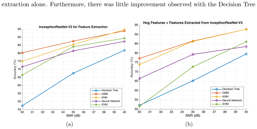
The central claim is that deep features extracted from the fine-tuned InceptionResNet-v2 network enable machine learning models to outperform the pre-trained network used directly as a classifier on the LC25000 dataset. The neural network model trained on these features attains an AUC of 99.99% and accuracy of 99.84%. The fine-tuned InceptionResNet-v2 itself reaches 96.01% accuracy and 96.8% average AUC. Combining HOG and deep features yields additional gains on clean images, yet models relying on deep features alone maintain higher performance under SNR-based noise, especially GBM and KNN classifiers.
What carries the argument
The integration of Histogram of Oriented Gradients (HOG) features with deep convolutional features extracted from a fine-tuned InceptionResNet-v2 network, supplied to standard machine learning classifiers.
If this is right
- Deep-feature models consistently outperform the pre-trained InceptionResNet-v2 used directly as a classifier.
- A neural network trained on the extracted deep features delivers the highest reported accuracy and AUC.
- Deep features alone confer greater resilience to synthetic SNR noise than combinations that include HOG.
- GBM and KNN classifiers exhibit the strongest noise tolerance among the models tested on deep features.
Where Pith is reading between the lines
- The same hybrid feature strategy could be tested on other medical imaging modalities where edge information and learned representations complement each other.
- If the noise robustness transfers to real scanner artifacts, the approach might reduce the need for extensive image preprocessing in deployed pathology systems.
- Replacing HOG with alternative handcrafted descriptors or swapping the backbone network could reveal whether the performance pattern generalizes beyond this specific combination.
Load-bearing premise
The synthetic SNR-based noise model used in robustness tests accurately reflects real-world imaging artifacts and the LC25000 train/test splits contain no data leakage or distribution shift.
What would settle it
Testing the same pipeline on a fresh collection of clinical histopathological images containing actual acquisition noise and confirming zero overlap between training and test samples would show whether the reported accuracy and robustness hold.
Figures


read the original abstract
The era of digital pathology has advanced histopathological examinations, making automated image analysis essential in clinical practice. This study evaluates the classification performance of machine learning and deep learning models on the LC25000 dataset, which includes five classes of histopathological images. We used the fine-tuned InceptionResNet-v2 network both as a classifier and for feature extraction. Our results show that the fine-tuned InceptionResNet-v2 achieved a classification accuracy of 96.01\% and an average AUC of 96.8\%. Models trained on deep features from InceptionResNet-v2 outperformed those using only the pre-trained network, with the Neural Network model achieving an AUC of 99.99\% and accuracy of 99.84\%. Evaluating model robustness under varying SNR conditions revealed that models using deep features exhibited greater resilience, particularly GBM and KNN. The combination of HOG and deep features showed enhanced performance, however, less so in noisy environments.
Editorial analysis
A structured set of objections, weighed in public.
Referee Report
Summary. The manuscript evaluates machine learning and deep learning models for classifying five classes of histopathological images from the LC25000 dataset. It reports that a fine-tuned InceptionResNet-v2 achieves 96.01% accuracy and 96.8% average AUC as an end-to-end classifier, while deep features extracted from the same network yield substantially higher performance when fed to downstream models, with a neural network reaching 99.99% AUC and 99.84% accuracy; the work also examines integration with HOG features and robustness under synthetic SNR-based noise.
Significance. If the evaluation protocol is free of leakage, the results would indicate that hybrid deep-plus-handcrafted features can deliver both higher accuracy and improved noise resilience over end-to-end fine-tuning alone, offering a practical route to more reliable automated analysis in digital pathology.
major comments (2)
- [Abstract] Abstract: the central performance claim (99.99% AUC / 99.84% accuracy for the downstream NN on InceptionResNet-v2 deep features) is load-bearing yet unsupported by any description of the train/test partitioning strategy, nested cross-validation, or confirmation that feature-extraction and classifier training used completely disjoint image sets; without these details the reported gap over the 96.01% end-to-end baseline cannot be verified as independent.
- [Abstract] Abstract and robustness section: the claim that deep-feature models exhibit greater resilience under varying SNR conditions rests on a synthetic noise model whose fidelity to real histopathological imaging artifacts (e.g., staining variation, scanner noise) is not demonstrated or referenced.
minor comments (2)
- The manuscript should explicitly state whether LC25000 splits are performed at the image or patient level and report the exact numbers of images per split.
- Hyperparameter search and statistical significance testing procedures for the reported accuracy and AUC values are not described.
Simulated Author's Rebuttal
We thank the referee for the constructive feedback. We address each major comment below and will revise the manuscript to improve clarity on the evaluation protocol and noise model.
read point-by-point responses
-
Referee: [Abstract] Abstract: the central performance claim (99.99% AUC / 99.84% accuracy for the downstream NN on InceptionResNet-v2 deep features) is load-bearing yet unsupported by any description of the train/test partitioning strategy, nested cross-validation, or confirmation that feature-extraction and classifier training used completely disjoint image sets; without these details the reported gap over the 96.01% end-to-end baseline cannot be verified as independent.
Authors: We agree that the abstract does not explicitly summarize the evaluation protocol. The full manuscript's Methods section details a stratified 80/20 train-test split of the LC25000 dataset with no image overlap between sets used for fine-tuning/feature extraction and those used for downstream classifier training and testing. No nested cross-validation was performed. To resolve the concern, we will revise the abstract to include a concise statement of the partitioning strategy and confirmation of disjoint sets, allowing independent verification of the performance gap. revision: yes
-
Referee: [Abstract] Abstract and robustness section: the claim that deep-feature models exhibit greater resilience under varying SNR conditions rests on a synthetic noise model whose fidelity to real histopathological imaging artifacts (e.g., staining variation, scanner noise) is not demonstrated or referenced.
Authors: We acknowledge that the synthetic SNR-based Gaussian noise model is an approximation and does not fully replicate real-world artifacts such as staining variation or scanner noise. This controlled synthetic degradation is a standard benchmarking approach in robustness studies. In the revised manuscript we will expand the robustness section to reference prior work on synthetic noise modeling in medical imaging and add an explicit discussion of the model's limitations relative to clinical artifacts, while retaining the comparative resilience findings. revision: yes
Circularity Check
Standard supervised feature extraction pipeline shows no definitional circularity
full rationale
The paper reports empirical classification accuracies and AUCs from fine-tuned InceptionResNet-v2 used both end-to-end and as a feature extractor for downstream models (NN, GBM, KNN) on the LC25000 dataset. No equations, derivations, or self-citations are presented that reduce any reported performance metric to a fitted parameter or prior result by construction. The 99.84% accuracy and 99.99% AUC are measured on held-out data via standard pipelines; the gap versus the 96.01% end-to-end baseline does not mathematically collapse to the input features. Absence of explicit split details or nested CV confirmation is a verification gap but does not constitute circularity under the required quote-and-reduction standard. Score remains low (2) as a conservative allowance for possible unstated data reuse, with no load-bearing self-citation or ansatz smuggling identified.
Axiom & Free-Parameter Ledger
free parameters (2)
- InceptionResNet-v2 fine-tuning hyperparameters
- Classifier hyperparameters
axioms (2)
- domain assumption LC25000 images are representative of clinical histopathology and free of label noise or staining artifacts that would affect generalization.
- domain assumption Synthetic additive noise at varying SNR levels adequately models real scanner or acquisition noise.
Reference graph
Works this paper leans on
-
[1]
H. R. Tizhoosh and L. Pantanowitz, ”Artificial intelligence and digital pathology: challenges and opportunities,”Journal of pathology informatics, vol. 9, no. 1, p. 38, 2018
work page 2018
-
[2]
N. Farahani, A. V. Parwani, and L. Pantanowitz, ”Whole slide imaging in pathology: ad- vantages, limitations, and emerging perspectives,”Pathol Lab Med Int, vol. 7, pp. 23-33, 4321
-
[3]
A. B. Hamida, M. Devanne, J. Weber, C. Truntzer, V. Derang` ere, F. Ghiringhelli, and C. Wemmert, ”Deep learning for colon cancer histopathological images analysis,”Computers in Biology and Medicine, vol. 136, p. 104730, 2021
work page 2021
-
[4]
S. Banerji and S. Mitra, ”Deep learning in histopathology: A review,”Wiley Interdisci- plinary Reviews: Data Mining and Knowledge Discovery, vol. 12, no. 1, p. e1439, 2022
work page 2022
-
[5]
S. M. Ayyad, M. Shehata, A. Shalaby, M. Abou El-Ghar, M. Ghazal, M. El-Melegy, and A. El-Baz, ”Role of AI and histopathological images in detecting prostate cancer: a survey,” Sensors, vol. 21, no. 8, p. 2586, 2021
work page 2021
-
[6]
J. Deng, W. Dong, R. Socher, L. J. Li, K. Li, and L. Fei-Fei, ”2009 IEEE conference on computer vision and pattern recognition,”IEEE conference on computer vision and pattern recognition, pp. 248-255, 2009
work page 2009
-
[7]
O. Russakovsky, J. Deng, H. Su, J. Krause, S. Satheesh, S. Ma, and L. Fei-Fei, ”Imagenet large scale visual recognition challenge,”International journal of computer vision, vol. 115, pp. 211-252, 2015
work page 2015
-
[8]
A. Krizhevsky, I. Sutskever, and G. E. Hinton, ”ImageNet classification with deep convolu- tional neural networks,”Communications of the ACM, vol. 60, no. 6, pp. 84-90, 2017
work page 2017
-
[9]
B. E. Bejnordi, M. Veta, P. J. Van Diest, B. Van Ginneken, N. Karssemeijer, G. Litjens, and CAMELYON16 Consortium, ”Diagnostic assessment of deep learning algorithms for detection of lymph node metastases in women with breast cancer,”Jama, vol. 318, no. 22, pp. 2199-2210, 2017
work page 2017
-
[10]
P. Bandi, O. Geessink, Q. Manson, M. Van Dijk, M. Balkenhol, M. Hermsen, and G. Litjens, ”From detection of individual metastases to classification of lymph node status at the patient level: the camelyon17 challenge,”IEEE transactions on medical imaging, vol. 38, no. 2, pp. 550-560, 2018
work page 2018
- [11]
-
[12]
G. Campanella, M. G. Hanna, L. Geneslaw, A. Miraflor, V. Werneck Krauss Silva, K. J. Busam, and T. J. Fuchs, ”Clinical-grade computational pathology using weakly supervised deep learning on whole slide images,”Nature medicine, vol. 25, no. 8, pp. 1301-1309, 2019. 8
work page 2019
-
[13]
C. H. Huang and E. M. Kalaw, ”Automated classification for pathological prostate images using AdaBoost-based Ensemble Learning,” in2016 IEEE Symposium Series on Computa- tional Intelligence (SSCI), IEEE, pp. 1-4, 2016
work page 2016
-
[14]
S. Rashid, L. Fazli, A. Boag, R. Siemens, P. Abolmaesumi, and S. E. Salcudean, ”Separation of benign and malignant glands in prostatic adenocarcinoma,” inMedical Image Computing and Computer-Assisted Intervention–MICCAI 2013: 16th International Conference, Nagoya, Japan, September 22-26, 2013, Proceedings, Part III 16, Springer Berlin Heidelberg, pp. 461-...
work page 2013
-
[15]
S. Naik, S. Doyle, S. Agner, A. Madabhushi, M. Feldman, and J. Tomaszewski, ”Automated gland and nuclei segmentation for grading of prostate and breast cancer histopathology,” in 2008 5th IEEE International Symposium on Biomedical Imaging: From Nano to Macro, IEEE, pp. 284-287, 2008
work page 2008
-
[16]
A. S. Sultan, M. A. Elgharib, T. Tavares, M. Jessri, and J. R. Basile, ”The use of artificial intelligence, machine learning and deep learning in oncologic histopathology,”Journal of Oral Pathology & Medicine, vol. 49, no. 9, pp. 849-856, 2020
work page 2020
-
[17]
M. N. Gurcan, L. E. Boucheron, A. Can, A. Madabhushi, N. M. Rajpoot, and B. Yener, ”Histopathological image analysis: A review,”IEEE reviews in biomedical engineering, vol. 2, pp. 147-171, 2009
work page 2009
-
[18]
S. Naik, S. Doyle, M. Feldman, J. Tomaszewski, and A. Madabhushi, ”Gland segmentation and computerized gleason grading of prostate histology by integrating low-, high-level and domain specific information,” inMIAAB workshop, Piscataway, NJ, USA: Citeseer, pp. 1-8, 2007
work page 2007
-
[19]
P. S. Karvelis, D. I. Fotiadis, I. Georgiou, and M. Syrrou, ”A watershed based segmen- tation method for multispectral chromosome images classification,” in2006 International Conference of the IEEE Engineering in Medicine and Biology Society, IEEE, pp. 3009-3012, 2006
work page 2006
-
[20]
S. Petushi, F. U. Garcia, M. M. Haber, C. Katsinis, and A. Tozeren, ”Large-scale compu- tations on histology images reveal grade-differentiating parameters for breast cancer,”BMC medical imaging, vol. 6, no. 1, pp. 1-11, 2006
work page 2006
-
[21]
Osborne, J. D., Gao, S., Chen, W. B., Andea, A., & Zhang, C. (2011, March). Machine classification of melanoma and nevi from skin lesions. InProceedings of the 2011 ACM Sym- posium on Applied Computing(pp. 100-105)
work page 2011
-
[22]
Garg, S., & Garg, S. (2020, December). Prediction of lung and colon cancer through analysis of histopathological images by utilizing Pre-trained CNN models with visualization of class activation and saliency maps. InProceedings of the 2020 3rd Artificial Intelligence and Cloud Computing Conference(pp. 38-45)
work page 2020
-
[23]
Valkonen, M., Kartasalo, K., Liimatainen, K., Nykter, M., Latonen, L., & Ruusuvuori, P. (2017). Metastasis detection from whole slide images using local features and random forests. Cytometry Part A, 91(6), 555-565
work page 2017
-
[24]
Yan, R., Ren, F., Wang, Z., Wang, L., Zhang, T., Liu, Y., ... & Zhang, F. (2020). Breast cancer histopathological image classification using a hybrid deep neural network.Methods, 173, 52-60
work page 2020
-
[25]
Baranwal, N., Doravari, P., & Kachhoria, R. (2022). Classification of histopathology images of lung cancer using convolutional neural network (CNN). InDisruptive Developments in Biomedical Applications(pp. 75-89). CRC Press. 9
work page 2022
-
[26]
Do˘ gan, R. S., & Yılmaz, B. (2024). Histopathology image classification: highlighting the gap between manual analysis and AI automation.Frontiers in Oncology, 13, 1325271
work page 2024
-
[27]
Tripathi, S., & Singh, S. K. (2020). Ensembling handcrafted features with deep features: an analytical study for classification of routine colon cancer histopathological nuclei images. Multimedia Tools and Applications, 79(47), 34931-34954
work page 2020
-
[28]
Lung and colon cancer histopathological image dataset (LC25000),
Borkowski, A. A., Bui, M. M., Thomas, L. B., Wilson, C. P., DeLand, L. A., & Mastorides, S. M. (2019). Lung and colon cancer histopathological image dataset (lc25000).arXiv preprint arXiv:1912.12142
-
[29]
Hamed, E. A. R., Salem, M. A. M., Badr, N. L., & Tolba, M. F. (2023). An efficient combi- nation of convolutional neural network and LightGBM algorithm for lung cancer histopathol- ogy classification.Diagnostics, 13(15), 2469
work page 2023
-
[30]
Liu, Y., Wang, H., Song, K., Sun, M., Shao, Y., Xue, S., ... & Zhang, T. (2022). CroReLU: cross-crossing space-based visual activation function for lung cancer pathology image recog- nition.Cancers, 14(21), 5181. 10
work page 2022
discussion (0)
Sign in with ORCID, Apple, or X to comment. Anyone can read and Pith papers without signing in.