Enhancing Ultra-low-field MRI with Segmentation-guided Adversarial Learning
Pith reviewed 2026-06-29 13:18 UTC · model grok-4.3
The pith
Tissue segmentation priors from ULF data condition CycleGAN and T-REX networks to produce 3 T-comparable MRIs from 64 mT scans.
A machine-rendered reading of the paper's core claim, the machinery that carries it, and where it could break.
Core claim
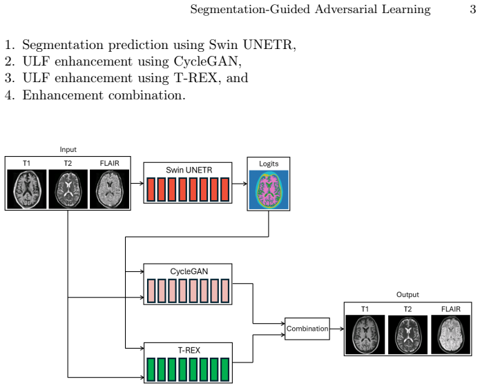
Training a Swin UNETR exclusively on challenge-provided ULF scans yields segmentation priors that condition a CycleGAN and a T-REX network; ensembling the outputs of these two conditioned models produces 3 T-like MRIs from 64 mT inputs that are comparable to high-field scans both quantitatively and qualitatively.
What carries the argument
Segmentation priors generated by Swin UNETR that condition and guide a CycleGAN and a T-REX enhancement network before their outputs are averaged.
If this is right
- ULF MRI can reach high-field visual and metric quality using only challenge data for all training steps.
- Anatomical conditioning from segmentation maps helps preserve tissue boundaries during synthesis.
- Averaging outputs from an adversarial model and a transformer residual model improves final image consistency.
- The pipeline operates without external high-field training data beyond the challenge references.
Where Pith is reading between the lines
- Portable low-cost scanners could become clinically viable in settings that lack access to 3 T systems if the conditioning remains stable across scanners.
- Any drop in segmentation accuracy on unseen patient populations would directly limit the reliability of the enhancement step.
- The same segmentation-conditioning pattern could be tested on other low-signal modalities such as portable ultrasound or low-dose CT.
Load-bearing premise
The tissue segmentation maps produced by Swin UNETR trained only on the supplied ULF data remain accurate enough to steer the enhancement networks without adding or amplifying artifacts.
What would settle it
A test set comparison in which the enhanced images receive markedly lower structural similarity scores or display new artifacts absent from the original 3 T reference scans would falsify the comparability claim.
Figures
read the original abstract
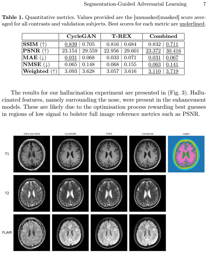
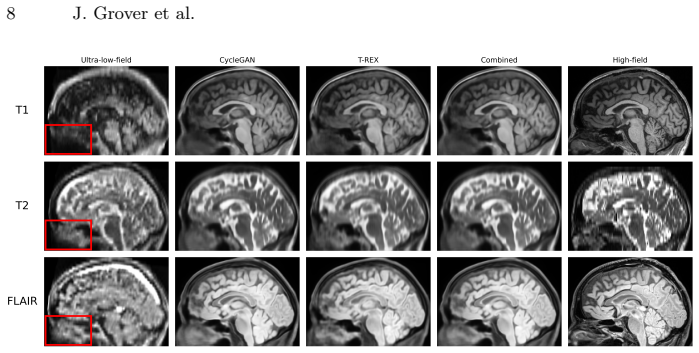
Ultra-low-field (ULF) MRI offers portable and low-cost imaging but suffers from poor image quality. To address this, we present our submission to the 2025 ULF Enhancement Challenge (ULF-EnC), where the goal is to synthesise high-field-like MRIs from 64 mT scans. Our pipeline enhances ULF MRI through a combination of anatomical conditioning and model ensembling. We first generate tissue segmentation priors using a Swin UNETR trained solely on challenge-provided data. These priors condition two independent enhancement networks - a CycleGAN and a transformer-based residual enhancement model (T-REX) - each trained to synthesise 3 T-like MRIs. Outputs from both models are combined using a weighted average. Our approach produces enhanced MRIs that were comparable to high-field scans both quantitatively and qualitatively.
Editorial analysis
A structured set of objections, weighed in public.
Referee Report
Summary. The manuscript presents a pipeline submitted to the 2025 ULF Enhancement Challenge for synthesizing 3 T-like images from 64 mT ultra-low-field MRI. Tissue segmentation priors are generated by a Swin UNETR trained exclusively on the challenge ULF data; these priors condition two separate enhancement networks (CycleGAN and a transformer residual model T-REX). The outputs are combined by weighted averaging. The central claim is that the resulting images achieve quantitative and qualitative comparability to high-field scans.
Significance. If the segmentation priors prove accurate and the comparability claim is supported by proper metrics and controls, the work would demonstrate a practical way to incorporate anatomical conditioning into adversarial and residual enhancement frameworks for portable MRI, potentially increasing the clinical utility of low-cost ULF systems.
major comments (1)
- [Abstract / Methods] Abstract / Methods (segmentation step): The central claim that the enhanced images are quantitatively and qualitatively comparable to high-field scans rests on the assumption that the Swin UNETR segmentation priors are sufficiently accurate to usefully condition both the CycleGAN and T-REX without introducing or amplifying artifacts. No Dice scores, Hausdorff distances, or other segmentation metrics are reported for the ULF-trained Swin UNETR, nor is an ablation presented that removes the segmentation conditioning. This omission directly undermines evaluation of whether the priors help or harm performance.
Simulated Author's Rebuttal
We thank the referee for the constructive feedback. We address the single major comment below and agree that additional evaluation of the segmentation component is warranted.
read point-by-point responses
-
Referee: [Abstract / Methods] Abstract / Methods (segmentation step): The central claim that the enhanced images are quantitatively and qualitatively comparable to high-field scans rests on the assumption that the Swin UNETR segmentation priors are sufficiently accurate to usefully condition both the CycleGAN and T-REX without introducing or amplifying artifacts. No Dice scores, Hausdorff distances, or other segmentation metrics are reported for the ULF-trained Swin UNETR, nor is an ablation presented that removes the segmentation conditioning. This omission directly undermines evaluation of whether the priors help or harm performance.
Authors: We agree that the absence of segmentation metrics and an ablation study limits the ability to isolate the contribution of the priors. The Swin UNETR was trained on challenge-provided ULF data that includes segmentation labels, so Dice scores, Hausdorff distances, and related metrics can be computed on the validation split. We will report these metrics in the revised manuscript. We will also add an ablation that removes the segmentation conditioning from both the CycleGAN and T-REX models while keeping all other training details fixed, allowing direct quantification of whether the priors improve or degrade the final synthesis quality. revision: yes
Circularity Check
No significant circularity; empirical ML pipeline evaluated on external challenge data
full rationale
The manuscript describes a practical image-enhancement pipeline (Swin UNETR segmentation priors conditioning CycleGAN + T-REX, followed by weighted ensembling) trained and tested exclusively on the 2025 ULF-EnC challenge dataset. No equations, fitted parameters renamed as predictions, self-citations, or uniqueness theorems appear in the abstract or described method. All performance claims are measured against held-out high-field reference scans supplied by the challenge, satisfying the criterion of external falsifiability. Consequently the work contains no load-bearing step that reduces to its own inputs by construction.
Axiom & Free-Parameter Ledger
axioms (1)
- domain assumption Tissue segmentation priors from Swin UNETR on challenge ULF data provide useful anatomical conditioning for enhancement without error propagation
Reference graph
Works this paper leans on
-
[1]
MONAI: An open-source framework for deep learning in healthcare
Cardoso, M.J., Li, W., Brown, R., Ma, N., Kerfoot, E., Wang, Y., Murrey, B., Myronenko, A., Zhao, C., Yang, D., et al.: Monai: An open-source framework for deep learning in healthcare. arXiv preprint arXiv:2211.02701 (2022)
work page internal anchor Pith review Pith/arXiv arXiv 2022
-
[2]
In: 2024 IEEE International Symposium on Biomedical Imaging (ISBI)
Dayarathna, S., Islam, K.T., Chen, Z.: Ultra low-field to high-field MRI translation using adversarial diffusion. In: 2024 IEEE International Symposium on Biomedical Imaging (ISBI). pp. 1–4. IEEE (2024)
2024
-
[3]
Medical image analysis92, 103046 (2024)
Dayarathna, S., Islam, K.T., Uribe, S., Yang, G., Hayat, M., Chen, Z.: Deep learn- ing based synthesis of MRI, CT and PET: Review and analysis. Medical image analysis92, 103046 (2024)
2024
-
[4]
In: 2025 IEEE/CVF Winter Conference on Applica- tions of Computer Vision (WACV)
Dayarathna, S., Islam, K.T., Zhuang, B., Yang, G., Cai, J., Law, M., Chen, Z.: Mccad: Multi-contrast MRI conditioned, adaptive adversarial diffusion model for high-fidelity MRI synthesis. In: 2025 IEEE/CVF Winter Conference on Applica- tions of Computer Vision (WACV). pp. 670–679. IEEE (2025)
2025
-
[5]
arXiv preprint arXiv:2402.17317 (2024)
Ferreira, A., Solak, N., Li, J., Dammann, P., Kleesiek, J., Alves, V., Egger, J.: How we won BRATS 2023 adult glioma challenge? just faking it! enhanced synthetic data augmentation and model ensemble for brain tumour segmentation. arXiv preprint arXiv:2402.17317 (2024)
-
[6]
arXiv preprint arXiv:2505.12228 (2025)
Gopinath, K., Sorby-Adams, A., Ramirez, J.W., Zemlyanker, D., Guo, J., Hunt, D., Mac Donald, C.L., Keene, C.D., Coalson, T., Glasser, M.F., et al.: From low field to high value: Robust cortical mapping from low-field MRI. arXiv preprint arXiv:2505.12228 (2025)
-
[7]
Communications Medicine4, 64 (2024) 10 J
Grover, J., Liu, P., Dong, B., Shan, S., Whelan, B., Keall, P., Waddington, D.: Super-resolution neural networks improve the spatiotemporal resolution of adap- tive MRI-guided radiation therapy. Communications Medicine4, 64 (2024) 10 J. Grover et al
2024
-
[8]
In: International MICCAI brainlesion workshop
Hatamizadeh, A., Nath, V., Tang, Y., Yang, D., Roth, H.R., Xu, D.: Swin unetr: Swin transformers for semantic segmentation of brain tumors in mri images. In: International MICCAI brainlesion workshop. pp. 272–284. Springer (2021)
2021
-
[9]
He,K.,Zhang,X.,Ren,S.,Sun,J.:Deepresiduallearningforimagerecognition.In: Proceedings of the IEEE conference on computer vision and pattern recognition. pp. 770–778 (2016)
2016
-
[10]
Radiology306(3), e220522 (2022)
Iglesias, J.E., Schleicher, R., Laguna, S., Billot, B., Schaefer, P., McKaig, B., Goldstein, J.N., Sheth, K.N., Rosen, M.S., Kimberly, W.T.: Quantitative brain morphometry of portable low-field-strength MRI using super-resolution machine learning. Radiology306(3), e220522 (2022)
2022
-
[11]
Scientific Reports13(1), 21183 (2023)
Islam, K.T., Zhong, S., Zakavi, P., Chen, Z., Kavnoudias, H., Farquharson, S., Durbridge,G.,Barth,M.,McMahon,K.L.,Parizel,P.M.,etal.:Improvingportable low-field MRI image quality through image-to-image translation using paired low- and high-field images. Scientific Reports13(1), 21183 (2023)
2023
-
[12]
In: Proceedings of the IEEE conference on computer vision and pattern recognition
Isola, P., Zhu, J.Y., Zhou, T., Efros, A.A.: Image-to-image translation with condi- tional adversarial networks. In: Proceedings of the IEEE conference on computer vision and pattern recognition. pp. 1125–1134 (2017)
2017
-
[13]
In: Eleventh Annual Meeting of the Organization for Human Brain Mapping, 2005
Jenkinson, M.: Bet2: Mr-based estimation of brain, skull and scalp surfaces. In: Eleventh Annual Meeting of the Organization for Human Brain Mapping, 2005. vol. 17, p. 167 (2005)
2005
-
[14]
Neuroimage62(2), 782–790 (2012)
Jenkinson, M., Beckmann, C.F., Behrens, T.E., Woolrich, M.W., Smith, S.M.: Fsl. Neuroimage62(2), 782–790 (2012)
2012
-
[15]
Nat Commun15, 2260 (2024)
Kwikima, U.: Looking towards the future of MRI in africa. Nat Commun15, 2260 (2024)
2024
-
[16]
In: Proceedings of the IEEE conference on computer vision and pattern recognition workshops
Lim,B.,Son,S.,Kim,H.,Nah,S.,MuLee,K.:Enhanceddeepresidualnetworksfor single image super-resolution. In: Proceedings of the IEEE conference on computer vision and pattern recognition workshops. pp. 136–144 (2017)
2017
-
[17]
Liu, Z., Lin, Y., Cao, Y., Hu, H., Wei, Y., Zhang, Z., Lin, S., Guo, B.: Swin transformer:Hierarchicalvisiontransformerusingshiftedwindows.In:Proceedings of the IEEE/CVF international conference on computer vision. pp. 10012–10022 (2021)
2021
-
[18]
Radiology315(1), e233529 (2025)
Lucas, A., Arnold, T.C., Okar, S.V., Vadali, C., Kawatra, K.D., Ren, Z., Cao, Q., Shinohara, R.T., Schindler, M.K., Davis, K.A., et al.: Multisequence 3-t image synthesis from 64-mt low-field-strength MRI using generative adversarial networks in multiple sclerosis. Radiology315(1), e233529 (2025)
2025
-
[19]
In: Proceedings of the IEEE international conference on computer vision
Mao, X., Li, Q., Xie, H., Lau, R.Y., Wang, Z., Paul Smolley, S.: Least squares gen- erative adversarial networks. In: Proceedings of the IEEE international conference on computer vision. pp. 2794–2802 (2017)
2017
-
[20]
NMR in Biomedicine36(7), e4917 (2023)
Obungoloch, J., Muhumuza, I., Teeuwisse, W., Harper, J., Etoku, I., Asiimwe, R., Tusiime,P.,Gombya,G.,Mugume,C.,Namutebi,M.H.,etal.:On-siteconstruction of a point-of-care low-field MRI system in africa. NMR in Biomedicine36(7), e4917 (2023)
2023
-
[21]
In: Proceedings of the IEEE/CVF conference on computer vision and pattern recognition
Park, T., Liu, M.Y., Wang, T.C., Zhu, J.Y.: Semantic image synthesis with spatially-adaptive normalization. In: Proceedings of the IEEE/CVF conference on computer vision and pattern recognition. pp. 2337–2346 (2019)
2019
-
[22]
Advances in neural information processing sys- tems32(2019) Segmentation-Guided Adversarial Learning 11
Paszke, A., Gross, S., Massa, F., Lerer, A., Bradbury, J., Chanan, G., Killeen, T., Lin, Z., Gimelshein, N., Antiga, L., et al.: Pytorch: An imperative style, high- performance deep learning library. Advances in neural information processing sys- tems32(2019) Segmentation-Guided Adversarial Learning 11
2019
-
[23]
In: International Conference on Medical image computing and computer-assisted intervention
Ronneberger, O., Fischer, P., Brox, T.: U-net: Convolutional networks for biomedi- cal image segmentation. In: International Conference on Medical image computing and computer-assisted intervention. pp. 234–241. Springer (2015)
2015
-
[24]
Scientific Reports5, 15177 (2015)
Sarracanie, M., LaPierre, C., Salameh, N., Waddington, D., Witzel, T., Rosen, M.: Low-cost high-performance MRI. Scientific Reports5, 15177 (2015)
2015
-
[25]
arXiv preprint arXiv:2411.06704 (2024)
Shimron, E., Shan, S., Grover, J., Koonjoo, N., Shen, S., Boele, T., Sorby-Adams, A.J., Kirsch, J.E., Rosen, M.S., Waddington, D.E.: Accelerating low-field MRI: From compressed sensing to deep learning reconstruction with cnns and trans- formers. arXiv preprint arXiv:2411.06704 (2024)
-
[26]
Smith,S.M.:Fastrobustautomatedbrainextraction.Humanbrainmapping17(3), 143–155 (2002)
2002
-
[27]
https://doi.org/10.5281/zenodo.15259777, https://doi.org/10.5281/zenodo.15259777
Tohidul Islam, K., Zhong, S., Peiris, H., Zakavi, P., Dayarathna, S., Chen, Z.: Enhancing ultra-low-field MRI with paired high- field MRI comparisons for brain imaging (ULF-EnC) (Apr 2025). https://doi.org/10.5281/zenodo.15259777, https://doi.org/10.5281/zenodo.15259777
-
[28]
Critical Care Explorations2(12), e0306 (2020)
Turpin, J., Unadkat, P., Thomas, J., Kleiner, N., Khazanehdari, S., Wanchoo, S., Samuel, K., Moclair, B.O., Black, K., Dehdashti, A.R., et al.: Portable magnetic resonance imaging for icu patients. Critical Care Explorations2(12), e0306 (2020)
2020
-
[29]
Advances in neural information pro- cessing systems30(2017)
Vaswani,A.,Shazeer,N.,Parmar,N.,Uszkoreit,J.,Jones,L.,Gomez,A.N.,Kaiser, Ł., Polosukhin, I.: Attention is all you need. Advances in neural information pro- cessing systems30(2017)
2017
-
[30]
IEEE transactions on medical imaging20(1), 45–57 (2002)
Zhang, Y., Brady, M., Smith, S.: Segmentation of brain mr images through a hidden markov random field model and the expectation-maximization algorithm. IEEE transactions on medical imaging20(1), 45–57 (2002)
2002
-
[31]
In: Proceedings of the IEEE interna- tional conference on computer vision
Zhu, J.Y., Park, T., Isola, P., Efros, A.A.: Unpaired image-to-image translation using cycle-consistent adversarial networks. In: Proceedings of the IEEE interna- tional conference on computer vision. pp. 2223–2232 (2017)
2017
discussion (0)
Sign in with ORCID, Apple, or X to comment. Anyone can read and Pith papers without signing in.