A unified deeplearning framework for contrast-phase-specific virtual monochromatic imaging
Reviewed by Pith T0 review T1 audit T2 compute T3 formal T4 kernel 2026-06-29 00:44 UTCgrok-4.3pith:WSIRJM7Precord.jsonopen to challenge →
The pith
A single deep learning model generates contrast-phase-specific 50 keV images from single-energy CT scans by using phase information as a prior.
A machine-rendered reading of the paper's core claim, the machinery that carries it, and where it could break.
Core claim
The central claim is that a unified deep learning framework with a novel prior conditioning architecture, trained on DECT-derived 70 keV and 50 keV image pairs for Angio, Arterial, Portal, and Delayed phases, synthesizes accurate 50 keV-like images from single-energy CT inputs while achieving contrast enhancement and preserving contrast phase-specific dynamics.
What carries the argument
The prior conditioning architecture that integrates contrast phase priors into the energy transformation process to produce phase-specific virtual monochromatic images.
If this is right
- The unified model generalizes across all four tested contrast phases without requiring phase-specific retraining.
- Contrast enhancement is achieved relative to the input single-energy CT images.
- Phase-specific contrast dynamics from the original DECT data are retained in the synthesized outputs.
- The approach enables 50 keV virtual monochromatic imaging on standard single-energy CT scanners.
Where Pith is reading between the lines
- Hospitals could deploy the model on existing single-energy scanners to approximate dual-energy contrast benefits without new hardware purchases.
- The conditioning mechanism might allow extension to additional energy levels or other contrast agents if similar paired training data become available.
- Routine clinical use would still require separate validation that the generated images support the same diagnostic decisions as true dual-energy acquisitions.
Load-bearing premise
DECT-derived 70 keV and 50 keV image pairs serve as reliable ground truth for training a model that accurately produces 50 keV images from single-energy CT inputs when conditioned on contrast phase.
What would settle it
Quantitative comparison of the synthesized 50 keV images against actual DECT-acquired 50 keV images on an independent test set of patients from the same four contrast phases, measuring both contrast-to-noise ratio and fidelity to phase-specific enhancement patterns.
Figures



read the original abstract
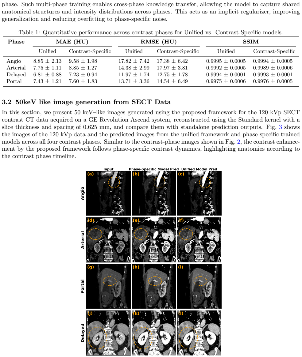
Dual-energy CT (DECT) enables virtual monochromatic imaging (VMI) and improved contrast resolution, but its clinical adoption is limited by hardware complexity and cost. In this work, we propose a unified deep learning framework that synthesizes contrast-phase-specific virtual monochromatic 50 keV images from single-energy CT (SECT) data by leveraging contrast phase information as a prior. The model is trained using DECT-derived 70 keV and 50 keV image pairs across four contrast phases -- Angio, Arterial, Portal, and Delayed -- using a novel prior conditioning architecture that integrates contrast phase priors into the energy transformation process. We demonstrate that the proposed unified model achieves contrast enhancement and generalizes well across contrast phases. Additionally, we show that the model can generate 50 keV-like images from SECT inputs, preserving contrast phase-specific dynamics.
Editorial analysis
A structured set of objections, weighed in public.
Referee Report
Summary. The paper proposes a unified deep learning framework to synthesize contrast-phase-specific virtual monochromatic 50 keV images from single-energy CT (SECT) inputs. It trains an energy-mapping network on paired DECT-derived 70 keV (input) and 50 keV (target) images across four contrast phases (Angio, Arterial, Portal, Delayed), using a novel prior conditioning architecture that integrates contrast phase information. The central claims are that the model achieves contrast enhancement, generalizes well across phases, and can generate 50 keV-like images from arbitrary SECT inputs while preserving phase-specific dynamics.
Significance. If the claims hold after proper validation, the approach could enable virtual monochromatic imaging benefits without requiring dual-energy CT hardware, addressing a practical barrier to clinical adoption. No machine-checked proofs, reproducible code, or parameter-free derivations are present; the work relies on standard supervised training.
major comments (2)
- [Abstract] Abstract: the claims that the model 'achieves contrast enhancement and generalizes well across contrast phases' and 'can generate 50 keV-like images from SECT inputs' are unsupported by any quantitative metrics, error bars, baseline comparisons, validation splits, or statistical tests, so the data cannot be assessed as supporting the claims.
- [Framework description] Framework description (training on DECT pairs): the central claim of synthesizing from SECT inputs rests on the unverified assumption that DECT 70 keV images serve as a reliable proxy for real SECT acquisitions; DECT 70 keV images incorporate dual-tube beam-hardening corrections and noise statistics distinct from conventional 120 kVp SECT, yet no domain-adaptation losses, unpaired SECT fine-tuning, or quantitative comparison of outputs on matched SECT versus DECT-70 inputs is described.
minor comments (1)
- [Title] Title contains the compound word 'deeplearning' without space; standard spelling is 'deep learning'.
Simulated Author's Rebuttal
We thank the referee for the constructive comments, which highlight important areas for strengthening the manuscript. We address each major comment below and commit to revisions that improve clarity, support for claims, and discussion of assumptions without overstating current results.
read point-by-point responses
-
Referee: [Abstract] Abstract: the claims that the model 'achieves contrast enhancement and generalizes well across contrast phases' and 'can generate 50 keV-like images from SECT inputs' are unsupported by any quantitative metrics, error bars, baseline comparisons, validation splits, or statistical tests, so the data cannot be assessed as supporting the claims.
Authors: We agree the abstract presents these claims without accompanying quantitative details. The body of the manuscript contains supporting visual results and some quantitative evaluations (e.g., contrast-to-noise ratios across phases), but these are not summarized with metrics, error bars, or statistical tests in the abstract. In the revised version we will update the abstract to reference key quantitative findings from the results section, including performance metrics on held-out validation data and phase-specific comparisons, while ensuring the claims are appropriately qualified. revision: yes
-
Referee: [Framework description] Framework description (training on DECT pairs): the central claim of synthesizing from SECT inputs rests on the unverified assumption that DECT 70 keV images serve as a reliable proxy for real SECT acquisitions; DECT 70 keV images incorporate dual-tube beam-hardening corrections and noise statistics distinct from conventional 120 kVp SECT, yet no domain-adaptation losses, unpaired SECT fine-tuning, or quantitative comparison of outputs on matched SECT versus DECT-70 inputs is described.
Authors: This is a valid methodological concern. The training relies on paired DECT-derived 70 keV images as input proxies without explicit domain-adaptation mechanisms or direct SECT comparisons. We will revise the framework description and discussion sections to explicitly state this assumption, quantify the expected differences in noise and beam-hardening where possible, and add a limitations paragraph acknowledging the lack of unpaired SECT fine-tuning or matched-pair validation. If additional matched data can be obtained, we will include a small-scale comparison; otherwise the limitation will be clearly noted. revision: partial
Circularity Check
No circularity; standard supervised training on external DECT pairs
full rationale
The manuscript presents a deep-learning model trained via supervised learning on paired DECT 70 keV / 50 keV images with phase labels as conditioning. No equations, ansatzes, fitted parameters renamed as predictions, or self-citation chains appear in the provided text. The central claim rests on empirical generalization from DECT-derived pairs to SECT inputs rather than any definitional reduction of outputs to inputs. This is the normal non-circular case for a supervised imaging paper.
Axiom & Free-Parameter Ledger
free parameters (1)
- neural network parameters
axioms (2)
- domain assumption DECT-derived 50 keV and 70 keV images constitute accurate ground truth for virtual monochromatic imaging at those energies
- domain assumption Contrast phase label provides sufficient prior information to condition accurate energy transformation from SECT inputs
Reference graph
Works this paper leans on
-
[1]
Dual-energy ct: general principles,
Johnson, T. R., “Dual-energy ct: general principles,”American Journal of Roentgenol- ogy199(5 supplement), S3–S8 (2012)
2012
-
[2]
Clinical applications of dual-energy ct,
Hamid, S., Nasir, M. U., So, A., Andrews, G., Nicolaou, S., and Qamar, S. R., “Clinical applications of dual-energy ct,”Korean Journal of Radiology22(6), 970 (2021)
2021
-
[3]
Estimating dual- energy ct imaging from single-energy ct data with material decomposition convolutional neural network,
Lyu, T., Zhao, W., Zhu, Y., Wu, Z., Zhang, Y., Chen, Y., Luo, L., Li, S., and Xing, L., “Estimating dual- energy ct imaging from single-energy ct data with material decomposition convolutional neural network,” Medical image analysis70, 102001 (2021)
2021
-
[4]
Virtual monoenergetic ct imaging via deep learning,
Cong, W., Xi, Y., Fitzgerald, P., De Man, B., and Wang, G., “Virtual monoenergetic ct imaging via deep learning,”Patterns1(8) (2020)
2020
-
[5]
Zhao, W., Lyu, T., Chen, Y., and Xing, L., “A deep learning approach for virtual monochromatic spectral ct imaging with a standard single energy ct scanner,”arXiv preprint arXiv:2005.09859(2020)
-
[6]
Jointly optimized deep neural networks to synthesize monoenergetic images from single-energy ct angiography for improving classification of pulmonary embolism,
Fink, M. A., Seibold, C., Kauczor, H.-U., Stiefelhagen, R., and Kleesiek, J., “Jointly optimized deep neural networks to synthesize monoenergetic images from single-energy ct angiography for improving classification of pulmonary embolism,”Diagnostics12(5), 1224 (2022)
2022
-
[7]
Virtual monochro- matic spectral imaging with fast kilovoltage switching: improved image quality as compared with that obtained with conventional 120-kvp ct,
Matsumoto, K., Jinzaki, M., Tanami, Y., Ueno, A., Yamada, M., and Kuribayashi, S., “Virtual monochro- matic spectral imaging with fast kilovoltage switching: improved image quality as compared with that obtained with conventional 120-kvp ct,”Radiology259(1), 257–262 (2011)
2011
-
[8]
D., Chatelain, R., MacDonald, D
Krishna, S., Sadoughi, N., McInnes, M. D., Chatelain, R., MacDonald, D. B., and Schieda, N., “Attenuation and degree of enhancement with conventional 120-kvp polychromatic ct and 70-kev monochromatic rapid kilovoltage-switching dual-energy ct in cystic and solid renal masses,”American Journal of Roentgenol- ogy211(4), 789–796 (2018)
2018
-
[9]
Which should be the routine cross-sectional reconstruction mode in spectral ct imaging: monochromatic or polychromatic?,
Cui, Y., Gao, S., Wang, Z., Li, X., Sun, Y., Tang, L., and Zhang, X., “Which should be the routine cross-sectional reconstruction mode in spectral ct imaging: monochromatic or polychromatic?,”The British journal of radiology85(1018), e887–e890 (2012)
2012
-
[10]
U-net: Convolutional networks for biomedical image seg- mentation,
Ronneberger, O., Fischer, P., and Brox, T., “U-net: Convolutional networks for biomedical image seg- mentation,” in [International Conference on Medical image computing and computer-assisted intervention], 234–241, Springer (2015)
2015
-
[11]
Visualizing data using t-sne,
Maaten, L. v. d. and Hinton, G., “Visualizing data using t-sne,”Journal of machine learning re- search9(Nov), 2579–2605 (2008)
2008
discussion (0)
Sign in with ORCID, Apple, or X to comment. Anyone can read and Pith papers without signing in.