Distortion-Corrected Diffusion MRI Using Rotated-View EPI and Joint Field-Map/Image Estimation with Gaussian Primitives
Pith reviewed 2026-07-01 02:38 UTC · model grok-4.3
The pith
Joint k-space estimation of B0 field and image using Gaussian primitives corrects EPI distortions more accurately than sequential methods.
A machine-rendered reading of the paper's core claim, the machinery that carries it, and where it could break.
Core claim
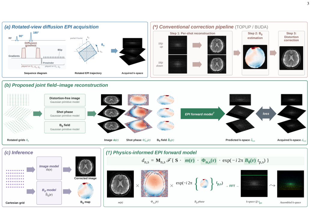
The central claim is that representing both the B0 inhomogeneity and the diffusion-weighted image as superpositions of Gaussian primitives embedded in a joint MRI physics forward model, combined with rotated-view acquisitions, permits direct estimation from k-space and yields superior distortion correction on in vivo brain data compared with sequential reconstruction-then-correction pipelines, especially at high b-value and high acceleration.
What carries the argument
Superposition of Gaussian primitives inside the MRI forward model for joint B0-field and image estimation from k-space.
If this is right
- Avoidance of reconstruction artifacts from parallel imaging at high acceleration because estimation occurs directly from k-space.
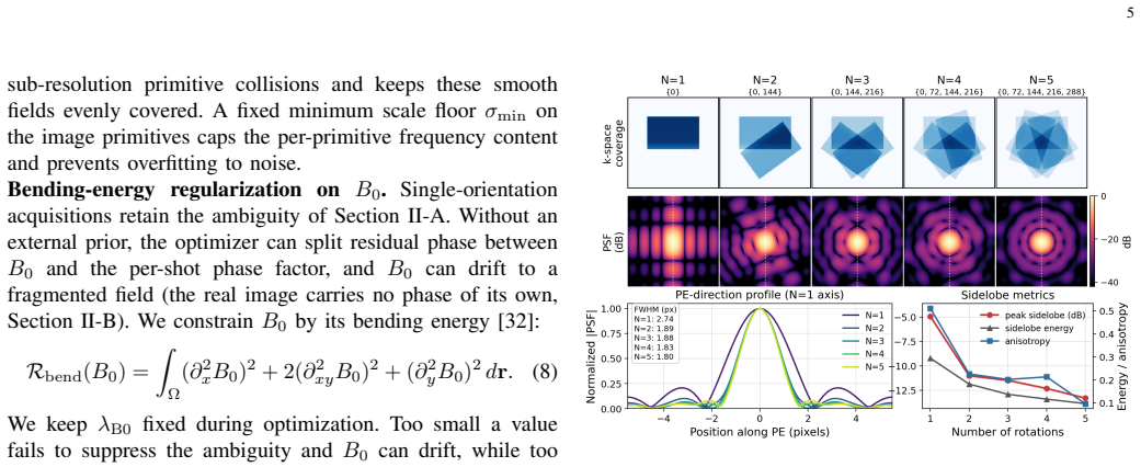
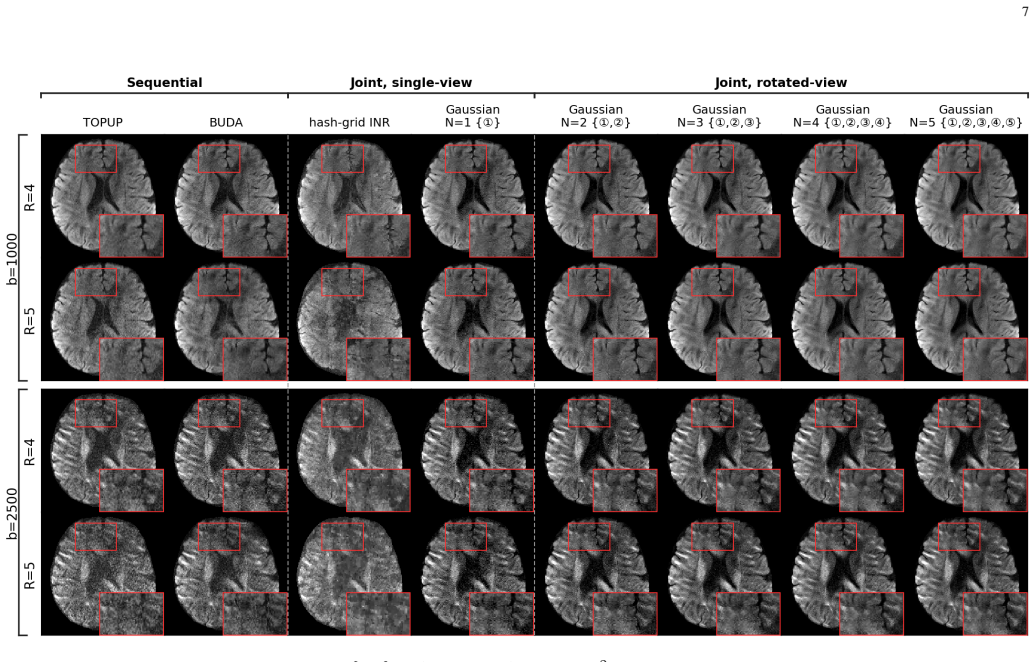
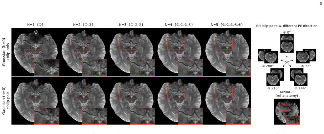
- Rotated views distribute distortions across multiple phase-encoding orientations, improving point-spread-function isotropy and strengthening constraints on B0 estimation.
- The diffusion-weighted image is constrained to be real and non-negative while per-shot phase factors absorb the image phase.
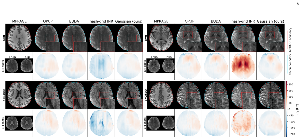
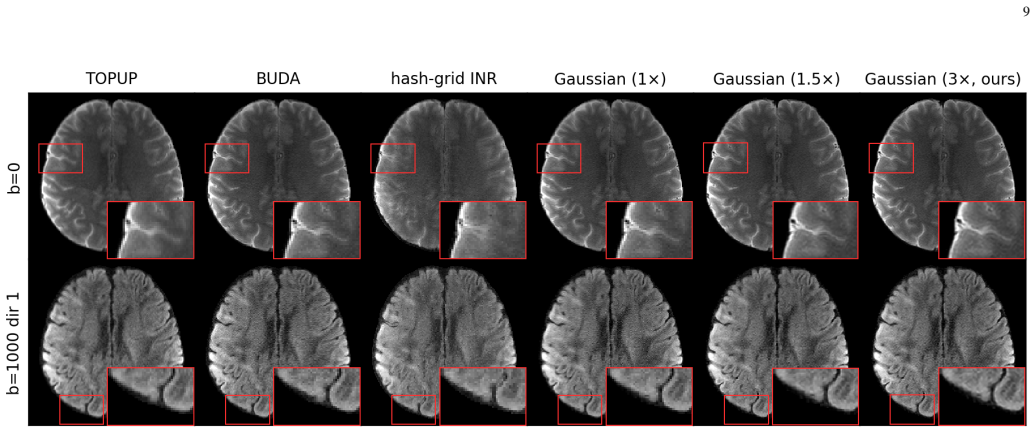
- Improved detail fidelity and noise suppression appear in visual comparisons on brain data.
Where Pith is reading between the lines
- The continuous parameterization could reduce reliance on post-acquisition interpolation or smoothing steps in multi-shot or high-acceleration protocols.
- If the Gaussian representation generalizes, the same joint model might apply to other contrasts such as functional MRI where B0-induced distortion also limits spatial specificity.
- Stronger k-space constraints from multiple rotated views may allow higher acceleration factors while preserving geometric accuracy, which would shorten scan times in clinical diffusion protocols.
Load-bearing premise
Both the B0 field and the diffusion-weighted image can be accurately represented as superpositions of Gaussian primitives inside the MRI forward model without introducing systematic bias at tissue boundaries or requiring post-hoc tuning of the number or placement of primitives.
What would settle it
On the same in vivo brain diffusion EPI datasets, if the proposed joint method fails to produce the closest brain-boundary agreement with the distortion-free structural reference or shows no larger improvement than sequential methods at high b-value and high acceleration, the central claim would be falsified.
Figures
read the original abstract
Echo Planar Imaging (EPI) is the standard acquisition technique for diffusion and functional neuroimaging, enabling rapid imaging but suffering from geometric distortions caused by B0 field inhomogeneities. Existing correction methods first reconstruct distorted images using parallel imaging, then estimate the B0 field and correct the distortion in the image domain. In this sequential process, reconstruction artifacts at high acceleration factors and low SNR at high diffusion b-values degrade B0 estimation and limit the overall correction quality. We propose a physics-informed framework that jointly estimates the B0 field and distortion-free image directly from k-space data, without depending on an intermediate parallel-imaging reconstruction for the correction. The image and the B0 field are each represented as a superposition of Gaussian primitives embedded within an MRI physics forward model. The explicit, continuous parameterization captures both smooth regions and tissue boundaries and supports rotated-view EPI acquisitions without interpolation. The diffusion-weighted image is modeled as real and non-negative, with the image phase absorbed into a per-shot phase factor. Rotated views distribute distortions across multiple phase-encoding orientations, improving point spread function isotropy and providing stronger constraints for B0 estimation. On in vivo brain diffusion EPI, the proposed method attains the closest brain-boundary agreement with a distortion-free structural reference, with the largest improvement over sequential methods at high b-value and high acceleration. Extensive visual comparisons further show improved detail fidelity and noise suppression.
Editorial analysis
A structured set of objections, weighed in public.
Referee Report
Summary. The paper proposes a joint physics-informed optimization framework for estimating the B0 field and distortion-free diffusion-weighted image directly from k-space data in rotated-view EPI. Both quantities are represented as superpositions of Gaussian primitives inside the MRI forward model; the image is constrained to be real and non-negative with phase absorbed into per-shot factors. The method is claimed to yield the closest brain-boundary agreement to a distortion-free structural reference on in-vivo brain data, with the largest gains over sequential parallel-imaging-plus-correction pipelines at high b-value and high acceleration.
Significance. If the empirical superiority is confirmed with quantitative, reproducible metrics and the Gaussian-primitive representation is shown not to introduce systematic boundary bias, the approach would offer a principled route to higher-fidelity distortion correction in accelerated diffusion MRI, improving alignment with structural references and downstream analyses.
major comments (3)
- [Abstract] Abstract: the central claim of 'closest brain-boundary agreement' and 'largest improvement' is presented without any quantitative boundary-distance metric, error bars, number of subjects or slices, or statistical comparison; these omissions prevent verification that the reported superiority is robust rather than anecdotal.
- [Abstract] Abstract (paragraph describing the parameterization): the boundary-agreement metric directly depends on the fidelity of the Gaussian-primitive representation at tissue interfaces, yet no ablation on primitive count, placement strategy, or edge-gradient fidelity test is described; if the finite superposition smooths or rings at boundaries, the metric may be biased toward the joint method by construction.
- [Abstract] Abstract: the non-negativity constraint and per-shot phase absorption are presented as enabling features, but no sensitivity analysis or comparison with/without these constraints is supplied, leaving open whether they are load-bearing for the reported gains at high b-value.
minor comments (1)
- [Abstract] Abstract: the phrase 'supports rotated-view EPI acquisitions without interpolation' would benefit from a brief statement of how the continuous parameterization achieves this.
Simulated Author's Rebuttal
We thank the referee for the constructive comments. We address each major comment point by point below, indicating planned revisions to the manuscript.
read point-by-point responses
-
Referee: [Abstract] Abstract: the central claim of 'closest brain-boundary agreement' and 'largest improvement' is presented without any quantitative boundary-distance metric, error bars, number of subjects or slices, or statistical comparison; these omissions prevent verification that the reported superiority is robust rather than anecdotal.
Authors: We agree that the abstract would benefit from quantitative support for the boundary-agreement claim. The full results section relies on visual comparisons across in-vivo datasets, but we will revise the abstract to report mean boundary-distance errors with standard deviations, the number of subjects and slices evaluated, and the outcomes of statistical comparisons against the sequential pipelines. revision: yes
-
Referee: [Abstract] Abstract (paragraph describing the parameterization): the boundary-agreement metric directly depends on the fidelity of the Gaussian-primitive representation at tissue interfaces, yet no ablation on primitive count, placement strategy, or edge-gradient fidelity test is described; if the finite superposition smooths or rings at boundaries, the metric may be biased toward the joint method by construction.
Authors: The potential for the finite Gaussian superposition to affect boundary fidelity is a legitimate concern. The primitives are optimized jointly within the forward model to capture both smooth field variations and sharp image edges, but we acknowledge the absence of an explicit ablation. We will add an ablation study varying primitive count and placement, together with quantitative edge-gradient fidelity comparisons against the structural reference, in the revised manuscript. revision: yes
-
Referee: [Abstract] Abstract: the non-negativity constraint and per-shot phase absorption are presented as enabling features, but no sensitivity analysis or comparison with/without these constraints is supplied, leaving open whether they are load-bearing for the reported gains at high b-value.
Authors: The non-negativity constraint and per-shot phase factors are physically motivated, yet we agree that their specific contribution at high b-value requires explicit verification. We will include a sensitivity analysis in the revision that compares reconstructions with and without these constraints on the high-b-value, high-acceleration data to quantify their effect on boundary agreement and detail fidelity. revision: yes
Circularity Check
No circularity: joint estimation and empirical boundary agreement are independent of model inputs by construction
full rationale
The paper introduces a joint k-space optimization of B0 and image, each parameterized as superpositions of Gaussian primitives inside an MRI forward model. The central claim (closest brain-boundary agreement with a distortion-free reference, largest gains at high b-value/high acceleration) is an external empirical comparison, not a quantity that reduces to a fitted parameter or self-citation by construction. No self-definitional step, fitted-input-called-prediction, or load-bearing self-citation chain appears; the Gaussian ansatz is an explicit modeling choice whose sufficiency is asserted but whose performance metric remains falsifiable against an independent structural reference. This is the common honest case of a self-contained method whose results do not collapse to its inputs.
Axiom & Free-Parameter Ledger
axioms (2)
- domain assumption The diffusion-weighted image is real and non-negative, with phase absorbed into a per-shot factor.
- domain assumption Gaussian primitives can represent both smooth regions and tissue boundaries without interpolation when embedded in the MRI physics forward model.
invented entities (1)
-
Gaussian primitives for joint image and B0 representation
no independent evidence
Reference graph
Works this paper leans on
-
[1]
Improving diffusion MRI using simultaneous multi-slice echo planar imaging,
K. Setsompopet al., “Improving diffusion MRI using simultaneous multi-slice echo planar imaging,”NeuroImage, vol. 63, no. 1, pp. 569– 580, 2012
2012
-
[2]
How to correct susceptibil- ity distortions in spin-echo echo-planar images: application to diffusion tensor imaging,
J. L. Andersson, S. Skare, and J. Ashburner, “How to correct susceptibil- ity distortions in spin-echo echo-planar images: application to diffusion tensor imaging,”NeuroImage, vol. 20, no. 2, pp. 870–888, 2003
2003
-
[3]
Advances in functional and structural MR image analysis and implementation as FSL,
S. M. Smithet al., “Advances in functional and structural MR image analysis and implementation as FSL,”NeuroImage, vol. 23, pp. S208– S219, 2004
2004
-
[4]
Correction for geometric distortion in echo planar images from B0 field variations,
P. Jezzard and R. S. Balaban, “Correction for geometric distortion in echo planar images from B0 field variations,”Magnetic Resonance in Medicine, vol. 34, no. 1, pp. 65–73, 1995
1995
-
[5]
Advances in diffusion MRI acquisition and processing in the Human Connectome Project,
S. N. Sotiropouloset al., “Advances in diffusion MRI acquisition and processing in the Human Connectome Project,”NeuroImage, vol. 80, pp. 125–143, 2013
2013
-
[6]
Efficient correction of inhomogeneous static magnetic field-induced distortion in Echo Planar Imaging,
D. Holland, J. M. Kuperman, and A. M. Dale, “Efficient correction of inhomogeneous static magnetic field-induced distortion in Echo Planar Imaging,”NeuroImage, vol. 50, no. 1, pp. 175–183, 2010
2010
-
[7]
DR-BUDDI (Diffeomorphic Registration for Blip-Up blip- Down Diffusion Imaging) method for correcting echo planar imaging distortions,
M. O. Irfanoglu, P. Modi, A. Nayak, E. B. Hutchinson, J. Sarlls, and C. Pierpaoli, “DR-BUDDI (Diffeomorphic Registration for Blip-Up blip- Down Diffusion Imaging) method for correcting echo planar imaging distortions,”NeuroImage, vol. 106, pp. 284–299, 2015
2015
-
[8]
Improved B0-distortion correction in diffusion MRI using interlaced q-space sam- pling and constrained reconstruction,
C. Bhushan, A. A. Joshi, R. M. Leahy, and J. P. Haldar, “Improved B0-distortion correction in diffusion MRI using interlaced q-space sam- pling and constrained reconstruction,”Magnetic Resonance in Medicine, vol. 72, no. 5, pp. 1218–1232, 2014
2014
-
[9]
Multi-shot sensitivity- encoded diffusion data recovery using structured low-rank matrix com- pletion (MUSSELS),
M. Mani, M. Jacob, D. Kelley, and V . Magnotta, “Multi-shot sensitivity- encoded diffusion data recovery using structured low-rank matrix com- pletion (MUSSELS),”Magnetic Resonance in Medicine, vol. 78, no. 2, pp. 494–507, 2017
2017
-
[10]
Advances in sensitivity encoding with arbitrary k-space trajectories,
K. P. Pruessmann, M. Weiger, P. B ¨ornert, and P. Boesiger, “Advances in sensitivity encoding with arbitrary k-space trajectories,”Magnetic Resonance in Medicine, vol. 46, no. 4, pp. 638–651, 2001
2001
-
[11]
An integrated approach to correction for off-resonance effects and subject movement in diffusion MR imaging,
J. L. Andersson and S. N. Sotiropoulos, “An integrated approach to correction for off-resonance effects and subject movement in diffusion MR imaging,”NeuroImage, vol. 125, pp. 1063–1078, 2016
2016
-
[12]
Fast, iterative image reconstruction for MRI in the presence of field inhomogeneities,
B. P. Sutton, D. C. Noll, and J. A. Fessler, “Fast, iterative image reconstruction for MRI in the presence of field inhomogeneities,”IEEE Transactions on Medical Imaging, vol. 22, no. 2, pp. 178–188, 2003
2003
-
[13]
Highly efficient MRI through multi-shot echo planar imaging,
C. Liao, X. Cao, J. Cho, Z. Zhang, K. Setsompop, and B. Bilgic, “Highly efficient MRI through multi-shot echo planar imaging,” inProc. SPIE Wavelets and Sparsity XVIII, vol. 11138, 2019, pp. 353–365
2019
-
[14]
Distortion-free, high-isotropic-resolution diffusion MRI with gSlider BUDA-EPI and multicoil dynamic B0 shimming,
C. Liaoet al., “Distortion-free, high-isotropic-resolution diffusion MRI with gSlider BUDA-EPI and multicoil dynamic B0 shimming,”Magnetic Resonance in Medicine, vol. 86, no. 2, pp. 791–803, 2021
2021
-
[15]
An unsupervised deep learning technique for susceptibility artifact correction in reversed phase-encoding EPI images,
S. T. M. Duong, S. L. Phung, A. Bouzerdoum, and M. M. Schira, “An unsupervised deep learning technique for susceptibility artifact correction in reversed phase-encoding EPI images,”Magnetic Resonance Imaging, vol. 71, pp. 1–10, 2020
2020
-
[16]
FD-Net: An unsupervised deep forward-distortion model for susceptibility artifact correction in EPI,
Z. Alkilani, T. C ¸ ukur, and E. U. Saritas, “FD-Net: An unsupervised deep forward-distortion model for susceptibility artifact correction in EPI,”Magnetic Resonance in Medicine, vol. 91, no. 1, pp. 280–296, 2024
2024
-
[17]
On hallucinations in tomographic image reconstruction,
S. Bhadra, V . A. Kelkar, F. J. Brooks, and M. A. Anastasio, “On hallucinations in tomographic image reconstruction,”IEEE Transactions on Medical Imaging, vol. 40, no. 11, pp. 3249–3260, 2021
2021
-
[18]
Motion correction with PROPELLER MRI: application to head motion and free-breathing cardiac imaging,
J. G. Pipe, “Motion correction with PROPELLER MRI: application to head motion and free-breathing cardiac imaging,”Magnetic Resonance in Medicine, vol. 42, no. 5, pp. 963–969, 1999
1999
-
[19]
Physics-informed implicit neural representations for joint B0 estimation and echo planar imaging,
W. Huanget al., “Physics-informed implicit neural representations for joint B0 estimation and echo planar imaging,” inProc. Medical Image Computing and Computer-Assisted Intervention (MICCAI), 2025
2025
-
[20]
Instant neural graphics primitives with a multiresolution hash encoding,
T. M ¨uller, A. Evans, C. Schied, and A. Keller, “Instant neural graphics primitives with a multiresolution hash encoding,”ACM transactions on graphics (TOG), vol. 41, no. 4, pp. 1–15, 2022
2022
-
[21]
NeRF: Representing scenes as neural radiance fields for view synthesis,
B. Mildenhall, P. P. Srinivasan, M. Tancik, J. T. Barron, R. Ramamoorthi, and R. Ng, “NeRF: Representing scenes as neural radiance fields for view synthesis,”Communications of the ACM, vol. 65, no. 1, pp. 99– 106, 2022
2022
-
[22]
NeRP: implicit neural representation learning with prior embedding for sparsely sampled image reconstruc- tion,
L. Shen, J. Pauly, and L. Xing, “NeRP: implicit neural representation learning with prior embedding for sparsely sampled image reconstruc- tion,”IEEE Transactions on Neural Networks and Learning Systems, vol. 35, no. 1, pp. 770–782, 2024
2024
-
[23]
Neural implicit k-space for binning-free non-Cartesian cardiac MR imaging,
W. Huang, H. B. Li, J. Pan, G. Cruz, D. Rueckert, and K. Hammernik, “Neural implicit k-space for binning-free non-Cartesian cardiac MR imaging,” inProc. Information Processing in Medical Imaging (IPMI), 2023, pp. 548–560
2023
-
[24]
ICoNIK: Generating respiratory-resolved abdominal MR reconstructions using neural implicit representations in k-space,
V . Spiekeret al., “ICoNIK: Generating respiratory-resolved abdominal MR reconstructions using neural implicit representations in k-space,” in Proc. Deep Generative Models Workshop (DGM4MICCAI), 2023, pp. 183–192
2023
-
[25]
3D Gaussian Splatting for real-time radiance field rendering,
B. Kerbl, G. Kopanas, T. Leimk ¨uhler, and G. Drettakis, “3D Gaussian Splatting for real-time radiance field rendering,”ACM Transactions on Graphics, vol. 42, no. 4, pp. 1–14, 2023
2023
-
[26]
Three-dimensional MRI reconstruction with 3D Gaussian representations: Tackling the under- sampling problem,
T. Peng, R. Zha, Z. Li, X. Liu, and Q. Zou, “Three-dimensional MRI reconstruction with 3D Gaussian representations: Tackling the under- sampling problem,”IEEE Transactions on Medical Imaging, vol. 45, pp. 1905–1917, 2025
1905
-
[27]
Fast undersampled dynamic MRI reconstruction using explicit representation learning with Gaussian splatting,
M. Terpstra and C. van den Berg, “Fast undersampled dynamic MRI reconstruction using explicit representation learning with Gaussian splatting,” inProc. ISMRM Workshop on Data Sampling and Image Reconstruction, Sedona, AZ, USA, 2026
2026
-
[28]
Back to basis: Spatially continuous MRI with adaptive Gaussians,
I. Singh, A. Dupuis, S. Kukran, C. Badve, and M. Griswold, “Back to basis: Spatially continuous MRI with adaptive Gaussians,” inProc. ISMRM Workshop on Data Sampling and Image Reconstruction, Sedona, AZ, USA, 2026
2026
-
[29]
Real diffusion-weighted MRI enabling true signal averaging and increased diffusion contrast,
C. Eichneret al., “Real diffusion-weighted MRI enabling true signal averaging and increased diffusion contrast,”NeuroImage, vol. 122, pp. 373–384, 2015
2015
-
[30]
Nonlinear phase correction for navigated diffusion imaging,
K. L. Miller and J. M. Pauly, “Nonlinear phase correction for navigated diffusion imaging,”Magnetic Resonance in Medicine, vol. 50, no. 2, pp. 343–353, 2003
2003
-
[31]
Fast poisson disk sampling in arbitrary dimensions,
R. Bridson, “Fast poisson disk sampling in arbitrary dimensions,” in Proc. ACM SIGGRAPH Sketches, 2007, pp. 22–es
2007
-
[32]
Nonrigid registration using free-form deformations: application to breast MR images,
D. Rueckert, L. I. Sonoda, C. Hayes, D. L. Hill, M. O. Leach, and D. J. Hawkes, “Nonrigid registration using free-form deformations: application to breast MR images,”IEEE Transactions on Medical Imaging, vol. 18, no. 8, pp. 712–721, 1999
1999
-
[33]
ESPIRiT—an eigenvalue approach to autocalibrating parallel MRI: where SENSE meets GRAPPA,
M. Ueckeret al., “ESPIRiT—an eigenvalue approach to autocalibrating parallel MRI: where SENSE meets GRAPPA,”Magnetic Resonance in Medicine, vol. 71, no. 3, pp. 990–1001, 2014
2014
-
[34]
Three-dimensional magnetization-prepared rapid gradient-echo imaging (3D MP RAGE),
J. P. Mugler III and J. R. Brookeman, “Three-dimensional magnetization-prepared rapid gradient-echo imaging (3D MP RAGE),” Magnetic Resonance in Medicine, vol. 15, no. 1, pp. 152–157, 1990
1990
-
[35]
Adam: A method for stochastic optimization,
D. P. Kingma and J. Ba, “Adam: A method for stochastic optimization,” inProc. International Conference on Learning Representations (ICLR), 2015
2015
-
[36]
An iterative image registration technique with an application to stereo vision,
B. D. Lucas and T. Kanade, “An iterative image registration technique with an application to stereo vision,” inProc. 7th International Joint Conference on Artificial Intelligence (IJCAI), 1981, pp. 674–679
1981
-
[37]
Synthmorph: learning contrast-invariant registration without acquired images,
M. Hoffmann, B. Billot, D. N. Greve, J. E. Iglesias, B. Fischl, and A. V . Dalca, “Synthmorph: learning contrast-invariant registration without acquired images,”IEEE Transactions on Medical Imaging, vol. 41, no. 3, pp. 543–558, 2022
2022
-
[38]
Synthstrip: skull-stripping for any brain image,
A. Hoopes, J. S. Mora, A. V . Dalca, B. Fischl, and M. Hoffmann, “Synthstrip: skull-stripping for any brain image,”NeuroImage, vol. 260, p. 119474, 2022
2022
-
[39]
Implicit neural representations with periodic activation functions,
V . Sitzmann, J. Martel, A. Bergman, D. Lindell, and G. Wetzstein, “Implicit neural representations with periodic activation functions,” Advances in Neural Information Processing Systems, vol. 33, pp. 7462– 7473, 2020
2020
discussion (0)
Sign in with ORCID, Apple, or X to comment. Anyone can read and Pith papers without signing in.