Delving Deep into Liver Focal Lesion Detection: A Preliminary Study
Pith reviewed 2026-05-24 16:58 UTC · model grok-4.3
The pith
A CNN framework detects liver lesions in 3D CT scans by chaining image processing, feature extraction, region proposal, registration, and classification.
A machine-rendered reading of the paper's core claim, the machinery that carries it, and where it could break.
Core claim
Because liver lesions vary widely in shape and the liver receives blood from two major vessels, automatic detection is required; the authors therefore introduce a CNN framework that performs image processing, feature extraction, region proposal, image registration, and classification recognition to locate lesions in CT volumes where two-dimensional methods cannot exploit spatial information.
What carries the argument
The liver cancer-detection framework with CNN, a staged pipeline that adapts convolution networks to three-dimensional CT data through sequential processing, extraction, proposal, alignment, and recognition steps.
If this is right
- Automatic lesion detection becomes possible for 3D medical volumes where existing 2D networks lose spatial context.
- The framework directly incorporates radiologists' clinical workflow steps into the detection process.
- Large existing annotated CT collections can be reused to train and evaluate the system without requiring new data.
- Doctors facing high scan volumes gain a tool that targets the specific difficulties of liver tumor appearance.
Where Pith is reading between the lines
- Registration between phases of contrast-enhanced CT could improve consistency across arterial and portal-venous images.
- The same staged approach might transfer to lesion detection in other abdominal organs imaged in 3D.
- Performance would likely depend on how well each stage is tuned to the specific noise and resolution properties of liver CT.
Load-bearing premise
That applying the listed sequence of standard CNN stages in order will overcome the recognition problems caused by variable lesion shapes in 3D liver CT images.
What would settle it
A head-to-head test on the same liver CT dataset in which the proposed multi-stage framework shows no improvement in detection accuracy or sensitivity over a straightforward 2D CNN baseline would falsify the claim that the new pipeline is needed.
Figures


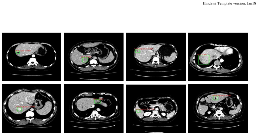
read the original abstract
Hepatocellular carcinoma (HCC) is the second most frequent cause of malignancy-related death and is one of the diseases with the highest incidence in the world. Because the liver is the only organ in the human body that is supplied by two major vessels: the hepatic artery and the portal vein, various types of malignant tumors can spread from other organs to the liver. And due to the liver masses' heterogeneous and diffusive shape, the tumor lesions are very difficult to be recognized, thus automatic lesion detection is necessary for the doctors with huge workloads. To assist doctors, this work uses the existing large-scale annotation medical image data to delve deep into liver lesion detection from multiple directions. To solve technical difficulties, such as the image-recognition task, traditional deep learning with convolution neural networks (CNNs) has been widely applied in recent years. However, this kind of neural network, such as Faster Regions with CNN features (R-CNN), cannot leverage the spatial information because it is applied in natural images (2D) rather than medical images (3D), such as computed tomography (CT) images. To address this issue, we propose a novel algorithm that is appropriate for liver CT imaging. Furthermore, according to radiologists' experience in clinical diagnosis and the characteristics of CT images of liver cancer, a liver cancer-detection framework with CNN, including image processing, feature extraction, region proposal, image registration, and classification recognition, was proposed to facilitate the effective detection of liver lesions.
Editorial analysis
A structured set of objections, weighed in public.
Referee Report
Summary. The manuscript proposes a CNN-based framework for automatic detection of liver focal lesions in 3D CT images. The framework comprises standard stages (image processing, feature extraction, region proposal, image registration, and classification) intended to address the difficulties posed by heterogeneous and diffusive lesion shapes; the work is positioned as a preliminary study leveraging existing annotated medical image data.
Significance. If a concrete 3D-adapted implementation were supplied together with reproducible experiments on public CT datasets and quantitative comparisons against 3D Faster R-CNN or U-Net baselines, the framework could contribute to computer-aided diagnosis of hepatocellular carcinoma. As written, however, the absence of any architecture details, training procedure, or results leaves the contribution at the level of an untested high-level outline.
major comments (3)
- [Abstract] Abstract: the central claim that the listed pipeline 'facilitates the effective detection of liver lesions' is unsupported because the manuscript contains no description of 3D-specific modifications (e.g., 3D convolutions, volumetric RPN, or 3D registration algorithm), no loss function, and no training protocol.
- [Abstract] Abstract / manuscript body: no experiments, datasets, metrics (Dice, sensitivity, false-positive rate), or baseline comparisons are reported, rendering the assertion that the framework solves the stated 3D lesion-detection problem unverifiable.
- [Abstract] Abstract: the statement that standard 2D Faster R-CNN 'cannot leverage the spatial information' in 3D CT is presented without reference to existing 3D extensions (e.g., 3D R-CNN or V-Net) or any quantitative motivation for the new proposal.
minor comments (2)
- [Title] The title uses 'Preliminary Study' yet the text supplies neither preliminary results nor a clear roadmap for future validation.
- [Abstract] Minor grammatical issues appear (e.g., 'the liver masses' heterogeneous and diffusive shape').
Simulated Author's Rebuttal
We thank the referee for their thorough review of our preliminary study on liver focal lesion detection. We acknowledge that the manuscript presents a high-level framework without detailed implementation or experimental validation, consistent with its 'preliminary' designation. We will revise the manuscript to better align claims with the content provided and to include references and future directions.
read point-by-point responses
-
Referee: [Abstract] Abstract: the central claim that the listed pipeline 'facilitates the effective detection of liver lesions' is unsupported because the manuscript contains no description of 3D-specific modifications (e.g., 3D convolutions, volumetric RPN, or 3D registration algorithm), no loss function, and no training protocol.
Authors: We agree with this assessment. The manuscript is intended as a preliminary outline of a framework inspired by clinical practice rather than a fully specified and trained model. We will revise the abstract to replace the claim with language indicating that the framework is proposed to address the detection challenges, and we will add a statement clarifying that specific 3D adaptations, loss functions, and training details are planned for future implementation. revision: yes
-
Referee: [Abstract] Abstract / manuscript body: no experiments, datasets, metrics (Dice, sensitivity, false-positive rate), or baseline comparisons are reported, rendering the assertion that the framework solves the stated 3D lesion-detection problem unverifiable.
Authors: As this is explicitly a preliminary study, the current version focuses on describing the proposed multi-component framework (image processing, feature extraction, region proposal, registration, classification) without empirical results. We will add a dedicated section on 'Limitations and Future Work' that specifies intended evaluation on public CT datasets (e.g., LiTS), using standard metrics such as sensitivity, Dice coefficient, and false positive rate, along with comparisons to 3D-adapted baselines like 3D U-Net or 3D Faster R-CNN. revision: yes
-
Referee: [Abstract] Abstract: the statement that standard 2D Faster R-CNN 'cannot leverage the spatial information' in 3D CT is presented without reference to existing 3D extensions (e.g., 3D R-CNN or V-Net) or any quantitative motivation for the new proposal.
Authors: We will incorporate references to 3D CNN extensions including 3D R-CNN and V-Net in the revised introduction. The motivation for our pipeline remains the need for explicit registration and multi-stage processing to handle the diffusive and heterogeneous nature of liver lesions in CT, which may complement pure 3D convolutional approaches; we will expand this discussion with additional clinical context. revision: yes
Circularity Check
No circularity; proposal is high-level description without derivations
full rationale
The paper's central claim is a high-level proposal of a CNN framework (image processing, feature extraction, region proposal, registration, classification) for 3D liver CT lesion detection. No equations, fitted parameters, self-citations, or derivation steps appear in the provided text. The content does not reduce any prediction or result to its inputs by construction, nor invoke uniqueness theorems or ansatzes from prior work. This is a standard non-circular preliminary proposal.
Axiom & Free-Parameter Ledger
Reference graph
Works this paper leans on
-
[1]
Cancer incidence and mortality worldwide: sources, methods and major patterns in GLOBOCAN 2012
Ferlay J, Soerjomataram I, Dikshit R, et al. Cancer incidence and mortality worldwide: sources, methods and major patterns in GLOBOCAN 2012. Int J Cancer;136(5): E359–E386, 2015
work page 2012
-
[2]
Sowell, Elizabeth R., et al. "Development of cortical and subcortical brain structures in childhood and adolescence: a structural MRI study." Developmental Medicine & Child Neurology 44.01: 4-16, 2002
work page 2002
-
[3]
The ischemic penumbra operationally defined by diffusion and perfusion MRI
Schlaug, G., et al. "The ischemic penumbra operationally defined by diffusion and perfusion MRI." Neurology 53.7: 1528-1528, 1999
work page 1999
-
[4]
Sharpe, James. "Optical projection tomography." Annu. Rev. Biomed. Eng. 6: 209- 228, 2004
work page 2004
-
[6]
Brain tumor segmentation with deep neural networks
Havaei, Mohammad, et al. "Brain tumor segmentation with deep neural networks." Medical image analysis 35 : 18-31, 2017
work page 2017
-
[7]
Histograms of oriented gradients for human detection
Dalal, Navneet, and Bill Triggs. "Histograms of oriented gradients for human detection." Computer Vision and Pattern Recognition, 2005. CVPR 2005. IEEE Computer Society Conference on. Vol. 1. IEEE, 2005. Hindawi Template version: Jan18 17
work page 2005
-
[8]
Multiresolution gray-scale and rotation invariant texture classification with local binary patterns
Ojala, Timo, Matti Pietikainen, and Topi Maenpaa. "Multiresolution gray-scale and rotation invariant texture classification with local binary patterns." IEEE Transactions on pattern analysis and machine intelligence 24.7: 971-987, 2002
work page 2002
-
[9]
SIFT: Predicting amino acid changes that affect protein function
Ng, Pauline C., and Steven Henikoff. "SIFT: Predicting amino acid changes that affect protein function." Nucleic acids research 31.13: 3812-3814, 2003
work page 2003
-
[11]
Imagenet classification with deep convolutional neural networks
Krizhevsky, Alex, Ilya Sutskever, and Geoffrey E. Hinton. "Imagenet classification with deep convolutional neural networks." Advances in neural information processing systems. 2012
work page 2012
-
[12]
Very Deep Convolutional Networks for Large-Scale Image Recognition
Simonyan, Karen, and Andrew Zisserman. "Very deep convolutional networks for large-scale image recognition." arXiv preprint arXiv:1409.1556(2014)
work page internal anchor Pith review Pith/arXiv arXiv 2014
-
[13]
Going deeper with convolutions
Szegedy, Christian, et al. "Going deeper with convolutions." Proceedings of the IEEE Conference on Computer Vision and Pattern Recognition. 2015
work page 2015
-
[14]
Deep Residual Learning for Image Recognition
He, Kaiming, et al. "Deep residual learning for image recognition." arXiv preprint arXiv:1512.03385 (2015)
work page internal anchor Pith review Pith/arXiv arXiv 2015
-
[15]
A LOGICAL CALCULUS OF THE IDEAS IMMANENT IN NERVOUS ACTIVITY
MCCULLOCH, WARREN S., and WALTER PITTS. "A LOGICAL CALCULUS OF THE IDEAS IMMANENT IN NERVOUS ACTIVITY."1943
work page 1943
-
[16]
Backpropagation applied to handwritten zip code recognition
LeCun, Yann, et al. "Backpropagation applied to handwritten zip code recognition." Neural computation 1.4: 541-551, 1989
work page 1989
-
[17]
Ravikumar, T. S., et al. "Intraoperative ultrasonography of liver: detection of occult liver tumors and treatment by cryosurgery." Cancer detection and prevention 18.2: 131-138, 1994
work page 1994
-
[18]
Pooler, B. Dustin, et al. "Prospective evaluation of reduced dose computed tomography for the detection of low-contrast liver lesions: direct comparison with concurrent standard dose imaging." European radiology 27.5: 2055-2066, 2017. Hindawi Template version: Jan18 18
work page 2055
-
[19]
Rich feature hierarchies for accurate object detection and semantic segmentation
Girshick, Ross, et al. "Rich feature hierarchies for accurate object detection and semantic segmentation." Proceedings of the IEEE conference on computer vision and pattern recognition. 2014
work page 2014
-
[20]
Faster R-CNN: towards real-time object detection with region proposal networks
Ren, Shaoqing, et al. "Faster R-CNN: towards real-time object detection with region proposal networks." IEEE transactions on pattern analysis and machine intelligence 39.6 : 1137-1149, 2017
work page 2017
-
[21]
V-net: Fully convolutional neural networks for volumetric medical image segmentation
Milletari, Fausto, Nassir Navab, and Seyed-Ahmad Ahmadi. "V-net: Fully convolutional neural networks for volumetric medical image segmentation." 3D Vision (3DV), 2016 Fourth International Conference on. IEEE, 2016
work page 2016
-
[22]
Efficient multi-scale 3D CNN with fully connected CRF for accurate brain lesion segmentation
Kamnitsas, Konstantinos, et al. "Efficient multi-scale 3D CNN with fully connected CRF for accurate brain lesion segmentation." Medical Image Analysis 36: 61-78, 2017
work page 2017
-
[23]
3d deeply supervised network for automatic liver segmentation from ct volumes
Dou, Qi, et al. "3d deeply supervised network for automatic liver segmentation from ct volumes." International Conference on Medical Image Computing and Computer- Assisted Intervention. Springer International Publishing, 2016
work page 2016
-
[24]
Deep convolutional neural networks and data augmentation for environmental sound classification
Salamon, Justin, and Juan Pablo Bello. "Deep convolutional neural networks and data augmentation for environmental sound classification." IEEE Signal Processing Letters 24.3: 279-283, 2017
work page 2017
-
[25]
Data augmentation for deep neural network acoustic modeling
Cui, Xiaodong, Vaibhava Goel, and Brian Kingsbury. "Data augmentation for deep neural network acoustic modeling." IEEE/ACM Transactions on Audio, Speech, and Language Processing 23.9: 1469-1477, 2015
work page 2015
-
[26]
Optical projection tomography as a tool for 3D microscopy and gene expression studies
[100] Sharpe, James, et al. "Optical projection tomography as a tool for 3D microscopy and gene expression studies." Science 296.5567 : 541-545, 2002
work page 2002
-
[27]
MXNet: A Flexible and Efficient Machine Learning Library for Heterogeneous Distributed Systems
Chen, Tianqi, et al. "Mxnet: A flexible and efficient machine learning library for heterogeneous distributed systems." arXiv preprint arXiv:1512.01274 (2015)
work page internal anchor Pith review Pith/arXiv arXiv 2015
-
[28]
Pan, Sinno Jialin, and Qiang Yang. "A survey on transfer learning." IEEE Transactions on knowledge and data engineering 22.10 : 1345-1359, 2010. Hindawi Template version: Jan18 19
work page 2010
-
[29]
Improving neural networks by preventing co-adaptation of feature detectors
Hinton, Geoffrey E., et al. "Improving neural networks by preventing co-adaptation of feature detectors." arXiv preprint arXiv:1207.0580 (2012)
work page internal anchor Pith review Pith/arXiv arXiv 2012
-
[30]
Dropout: a simple way to prevent neural networks from overfitting
Srivastava, Nitish, et al. "Dropout: a simple way to prevent neural networks from overfitting." Journal of Machine Learning Research 15.1: 1929-1958, 2014
work page 1929
-
[31]
Batch Normalization: Accelerating Deep Network Training by Reducing Internal Covariate Shift
Ioffe, Sergey, and Christian Szegedy. "Batch normalization: Accelerating deep network training by reducing internal covariate shift." arXiv preprint arXiv:1502.03167 (2015)
work page internal anchor Pith review Pith/arXiv arXiv 2015
-
[32]
Long J, Shelhamer E, Darrell T. Fully convolutional networks for semantic segmentation[C]//Proceedings of the IEEE Conference on Computer Vision and Pattern Recognition.: 3431-3440, 2015
work page 2015
-
[33]
Instance-aware semantic segmentation via multi-task network cascades
Dai, Jifeng, Kaiming He, and Jian Sun. "Instance-aware semantic segmentation via multi-task network cascades." Proceedings of the IEEE Conference on Computer Vision and Pattern Recognition. 2016
work page 2016
-
[34]
Ssd: Single shot multibox detector
Liu, Wei, et al. "Ssd: Single shot multibox detector." European conference on computer vision. Springer, Cham, 2016
work page 2016
-
[35]
You only look once: Unified, real-time object detection
Redmon, Joseph, et al. "You only look once: Unified, real-time object detection." Proceedings of the IEEE conference on computer vision and pattern recognition. 2016
work page 2016
-
[36]
YOLO9000: better, faster, stronger
Redmon, Joseph, and Ali Farhadi. "YOLO9000: better, faster, stronger." arXiv preprint (2017)
work page 2017
-
[37]
A-Fast-RCNN: Hard Positive Generation via Adversary for Object Detection
Wang, Xiaolong, Abhinav Shrivastava, and Abhinav Gupta. "A-fast-rcnn: Hard positive generation via adversary for object detection." arXiv preprint arXiv:1704.03414 2 (2017)
work page internal anchor Pith review Pith/arXiv arXiv 2017
-
[38]
Learning to refine object segments
Pinheiro, Pedro O., et al. "Learning to refine object segments." European Conference on Computer Vision. Springer, Cham, 2016
work page 2016
-
[39]
Feature pyramid networks for object detection
Lin, Tsung-Yi, et al. "Feature pyramid networks for object detection." CVPR. Vol. 1. No. 2. 2017. Hindawi Template version: Jan18 20
work page 2017
-
[40]
Learning to segment object candidates
Pinheiro, Pedro O., Ronan Collobert, and Piotr Dollár. "Learning to segment object candidates." Advances in Neural Information Processing Systems. 2015
work page 2015
-
[41]
A MultiPath Network for Object Detection
Zagoruyko, Sergey, et al. "A multipath network for object detection." arXiv preprint arXiv:1604.02135 (2016)
work page internal anchor Pith review Pith/arXiv arXiv 2016
discussion (0)
Sign in with ORCID, Apple, or X to comment. Anyone can read and Pith papers without signing in.