Deep Learning-Enabled Modality Transfer Between Independent Microscopes for High-Throughput Imaging
Pith reviewed 2026-05-24 02:50 UTC · model grok-4.3
The pith
A GAN trained on paired images from separate instruments transfers confocal-level quality to wide-field microscope captures.
A machine-rendered reading of the paper's core claim, the machinery that carries it, and where it could break.
Core claim
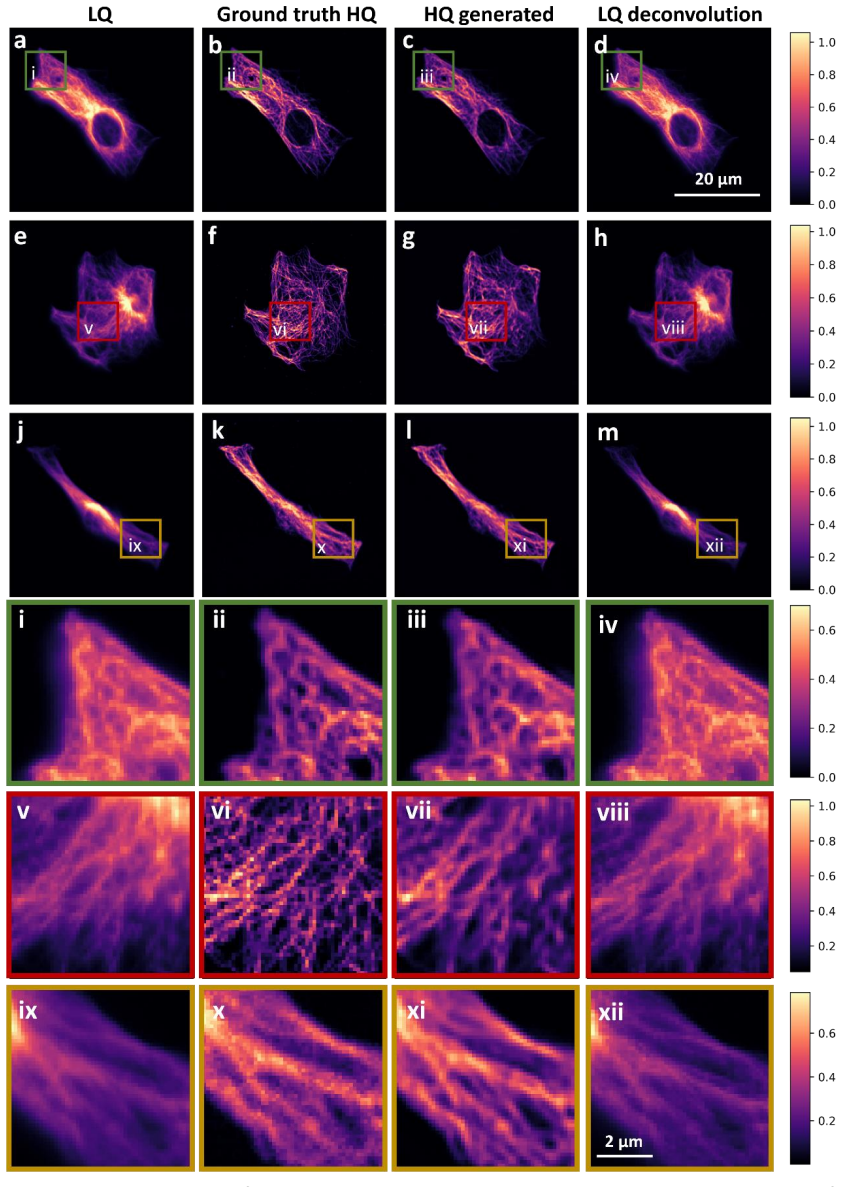
A GAN-based model trained on paired datasets acquired on physically separate wide-field and confocal microscopes can reliably transfer image quality, recovering key structural features so that the enhanced wide-field images reach median SSIM of 0.94 and PSNR of 31.87 versus the original values of 0.83 and 21.48.
What carries the argument
Generative adversarial network trained on paired wide-field and confocal images from independent microscopes, which learns a stable mapping from low- to high-quality representations.
If this is right
- High-throughput imaging can be performed on fast wide-field systems while high-quality structural information is recovered computationally.
- High-resolution confocal time can be reserved for targeted validation only, lowering total acquisition time.
- Key structural features are recovered with high accuracy, enabling scalable high-content workflows across independent instruments.
Where Pith is reading between the lines
- The same paired-training strategy could be tested on other modality pairs, such as wide-field versus super-resolution or light-sheet systems, provided alignment is feasible.
- If inference speed is sufficient, the transfer could be inserted into live acquisition pipelines to guide real-time decisions.
- Cross-lab standardization of image quality might become possible by training on shared paired reference datasets from different sites.
Load-bearing premise
The paired images from the two separate microscopes are accurately aligned and show identical biological structures rather than registration errors or instrument-specific artifacts.
What would settle it
Application of the trained model to new wide-field images yields no consistent improvement in SSIM or PSNR when compared against actual confocal images of the same fields.
Figures
read the original abstract
High-throughput biological imaging is often constrained by a trade-off between acquisition speed and image quality. Fast imaging modalities, such as wide-field fluorescence microscopy, enable large-scale data acquisition but suffer from reduced contrast and resolution, whereas high-resolution techniques, including confocal microscopy or single-molecule localization microscopy-based super-resolution techniques, provide superior image quality at the cost of throughput and instrument time. Here, we present a deep learning-based approach for modality transfer across independent microscopes, enabling the transformation of low-quality images acquired on fast systems into high-quality representations comparable to those obtained using advanced imaging platforms. To achieve this, we employ a generative adversarial network (GAN)-based model trained on paired datasets acquired on physically separate wide-field and confocal microscopes, demonstrating that image quality can be reliably transferred between independent instruments. Quantitative evaluation shows substantial improvement in structural similarity and signal fidelity, with median SSIM and PSNR of 0.94 and 31.87, respectively, compared to 0.83 and 21.48 for the original wide-field images. These results indicate that key structural features can be recovered with high accuracy. Importantly, this approach enables a workflow in which high-throughput imaging can be performed on fast, accessible microscopy systems while preserving the ability to computationally recover high-quality structural information. High-resolution microscopy can then be reserved for targeted validation, reducing acquisition time and improving overall experimental efficiency. Together, our results establish deep learning-enabled modality transfer as a practical strategy for bridging independent microscopy systems and supporting scalable, high-content imaging workflows.
Editorial analysis
A structured set of objections, weighed in public.
Referee Report
Summary. The manuscript presents a GAN-based model trained on paired image datasets acquired from physically separate wide-field and confocal microscopes. It claims that this enables reliable modality transfer, transforming low-quality wide-field images into high-quality representations with median SSIM of 0.94 and PSNR of 31.87 (versus 0.83 and 21.48 for the originals), supporting high-throughput workflows where high-resolution imaging is reserved for validation.
Significance. If the transfer function is shown to recover true structural features rather than instrument-specific or alignment artifacts, the result would be significant for scalable biological imaging by decoupling acquisition speed from final image quality. The direct empirical metrics on held-out pairs are a strength, but the absence of registration validation limits the strength of the central claim.
major comments (1)
- [Abstract / Methods] Abstract and Methods: The central claim requires that paired images from independent microscopes capture identical biological structures after alignment. No quantitative registration error metric (e.g., mean landmark displacement, Fourier-ring correlation, or residual translation/rotation statistics) is reported, and no ablation that removes registration steps before retraining is described. This leaves open the possibility that the reported SSIM/PSNR gains reflect compensation for geometric mismatches rather than modality transfer.
minor comments (2)
- [Abstract] The abstract does not specify the number of training/validation image pairs, the biological samples imaged, or the exact network architecture and loss terms used.
- [Results] Figure captions and results text should clarify whether the reported median metrics are computed on the full test set or on selected fields of view.
Simulated Author's Rebuttal
We thank the referee for highlighting the need for explicit registration validation to support the modality-transfer claim. We address this point directly below.
read point-by-point responses
-
Referee: [Abstract / Methods] Abstract and Methods: The central claim requires that paired images from independent microscopes capture identical biological structures after alignment. No quantitative registration error metric (e.g., mean landmark displacement, Fourier-ring correlation, or residual translation/rotation statistics) is reported, and no ablation that removes registration steps before retraining is described. This leaves open the possibility that the reported SSIM/PSNR gains reflect compensation for geometric mismatches rather than modality transfer.
Authors: We agree that quantitative registration metrics are required to rule out the possibility that SSIM/PSNR gains arise from correcting geometric mismatches. The manuscript describes rigid registration of the paired wide-field and confocal images but does not report error statistics. In the revised manuscript we will add these metrics (mean residual translation/rotation and landmark displacement on held-out pairs) to the Methods and Results sections. An ablation that removes the registration step before retraining is not feasible with the existing dataset, because unpaired structures would no longer correspond; we will instead expand the Methods to detail the registration procedure and its necessity for paired training, thereby clarifying that the reported improvements are measured after alignment. revision: partial
Circularity Check
No circularity; empirical metrics on held-out pairs
full rationale
The paper trains a standard GAN on paired wide-field/confocal images acquired from physically separate microscopes and reports median SSIM/PSNR directly computed on held-out test pairs. No equations, parameter fits, self-citations, or ansatzes are present that would reduce any reported quantity to its own inputs by construction. The central results are straightforward empirical measurements, not predictions forced by the training procedure itself.
Axiom & Free-Parameter Ledger
Lean theorems connected to this paper
-
IndisputableMonolith/Cost/FunctionalEquation.leanwashburn_uniqueness_aczel unclear?
unclearRelation between the paper passage and the cited Recognition theorem.
GAN architecture with U-NET generator, CNN discriminator, loss ℒ_G|D = α·MSE + β·SSIM + γ·BCE; median SSIM 0.9413, PSNR 31.87 on testing set
What do these tags mean?
- matches
- The paper's claim is directly supported by a theorem in the formal canon.
- supports
- The theorem supports part of the paper's argument, but the paper may add assumptions or extra steps.
- extends
- The paper goes beyond the formal theorem; the theorem is a base layer rather than the whole result.
- uses
- The paper appears to rely on the theorem as machinery.
- contradicts
- The paper's claim conflicts with a theorem or certificate in the canon.
- unclear
- Pith found a possible connection, but the passage is too broad, indirect, or ambiguous to say the theorem truly supports the claim.
Reference graph
Works this paper leans on
-
[1]
Dazzi, M., Rowland, E. M., Mohri, Z. & Weinberg, P . D. 3D confocal microscope imaging of macromolecule uptake in the intact brachiocephalic artery. Atherosclerosis 310, (2020)
work page 2020
-
[2]
White, S. L., Lam, A. T. & Buck, H. D. 3D Imaging for Cleared Tissues and Thicker Samples on Confocal and Light-Sheet Microscopes. in Methods in Molecular Biology vol. 2593 (2023)
work page 2023
-
[3]
Swaim, W. D. Overview of confocal microscopy. Methods Mol Biol 588, (2010)
work page 2010
-
[4]
Elliott, A. D. Confocal Microscopy: Principles and Modern Practices. Curr Protoc Cytom 92, (2020)
work page 2020
-
[5]
Hickey, S. M. et al. Fluorescence microscopy—an outline of hardware, biological handling, and fluorophore considerations. Cells vol. 11 Preprint at https://doi.org/10.3390/cells11010035 (2022)
-
[6]
Adamczyk, O., Baster, Z., Szczypior, M. & Rajfur, Z. Substrate stiffness mediates formation of novel cytoskeletal structures in fibroblasts during cell–microspheres interaction. Int J Mol Sci 22, (2021)
work page 2021
-
[7]
Haberkiewicz, O. et al. Decreased Expression of the Slc31a1 Gene and Cytoplasmic Relocalization of Membrane CTR1 Protein in Renal Epithelial Cells: A Potent Protective Mechanism against Copper Nephrotoxicity in a Mouse Model of Menkes Disease. Int J Mol Sci 23, (2022)
work page 2022
-
[8]
Szafranska, K. et al. From fixed-dried to wet-fixed to live - Comparative super-resolution microscopy of liver sinusoidal endothelial cell fenestrations. Nanophotonics 11, (2022)
work page 2022
-
[9]
Stemmer, A., Beck, M. & Fiolka, R. Widefield fluorescence microscopy with extended resolution. Histochemistry and Cell Biology vol. 130 Preprint at https://doi.org/10.1007/s00418-008-0506-8 (2008)
-
[10]
Combs, C. A. & Shroff, H. Fluorescence microscopy: A concise guide to current imaging methods. Curr Protoc Neurosci 2017, (2017)
work page 2017
-
[11]
Lan, L. et al. Generative Adversarial Networks and Its Applications in Biomedical Informatics. Frontiers in Public Health vol. 8 Preprint at https://doi.org/10.3389/fpubh.2020.00164 (2020)
-
[12]
Generative Adversarial Networks and Other Generative Models
Wenzel, M. Generative Adversarial Networks and Other Generative Models. in Neuromethods vol. 197 (2023)
work page 2023
-
[13]
Chen, R. et al. Single-frame deep-learning super-resolution microscopy for intracellular dynamics imaging. Nat Commun 14, (2023)
work page 2023
-
[14]
Melanthota, S. K. et al. Deep learning-based image processing in optical microscopy. Biophysical Reviews vol. 14 Preprint at https://doi.org/10.1007/s12551-022-00949-3 (2022)
-
[15]
Yang, T., Luo, Y ., Ji, W. & Yang, G. Advancing biological super-resolution microscopy through deep learning: a brief review. Biophysics Reports vol. 7 Preprint at https://doi.org/10.52601/bpr.2021.210019 (2021)
-
[16]
Wang, H. et al. Deep learning enables cross-modality super-resolution in fluorescence microscopy. Nat Methods 16, (2019)
work page 2019
-
[17]
Qiao, C. et al. Rationalized deep learning super-resolution microscopy for sustained live imaging of rapid subcellular processes. Nat Biotechnol 41, (2023)
work page 2023
-
[18]
Zhang, H. et al. High-throughput, high-resolution deep learning microscopy based on registration-free generative adversarial network. Biomed Opt Express 10, (2019)
work page 2019
-
[19]
Park, M., Lee, M. & Yu, S. HRGAN: A Generative Adversarial Network Producing Higher- Resolution Images than Training Sets. Sensors 22, (2022)
work page 2022
-
[20]
J., De León-Cuevas, A., González-Gutiérrez, A., Gorrostieta- Hurtado, E
Quintana-Quintana, O. J., De León-Cuevas, A., González-Gutiérrez, A., Gorrostieta- Hurtado, E. & Tovar-Arriaga, S. Dual U-Net-Based Conditional Generative Adversarial Network for Blood Vessel Segmentation with Reduced Cerebral MR Training Volumes. Micromachines (Basel) 13, (2022)
work page 2022
-
[21]
Alam, M. S. et al. Super-resolution enhancement method based on generative adversarial network for integral imaging microscopy. Sensors 21, (2021)
work page 2021
-
[22]
Huang, B. et al. Enhancing image resolution of confocal fluorescence microscopy with deep learning. PhotoniX 4, (2023)
work page 2023
- [23]
- [24]
-
[25]
Microscopy cell nuclei segmentation with enhanced U-Net
Long, F. Microscopy cell nuclei segmentation with enhanced U-Net. BMC Bioinformatics 21, (2020)
work page 2020
-
[26]
Z., Yakopcic, C., Hasan, M., Taha, T
Alom, M. Z., Yakopcic, C., Hasan, M., Taha, T. M. & Asari, V. K. Recurrent residual U-Net for medical image segmentation. Journal of Medical Imaging 6, (2019)
work page 2019
-
[27]
Khouy, M., Jabrane, Y ., Ameur, M. & Hajjam El Hassani, A. Medical Image Segmentation Using Automatic Optimized U-Net Architecture Based on Genetic Algorithm. J Pers Med 13, (2023)
work page 2023
-
[28]
Hirose, I. et al. U-Net-Based Segmentation of Microscopic Images of Colorants and Simplification of Labeling in the Learning Process. J Imaging 8, (2022)
work page 2022
-
[29]
Shihan, M. H., Novo, S. G., Le Marchand, S. J., Wang, Y . & Duncan, M. K. A simple method for quantitating confocal fluorescent images. Biochem Biophys Rep 25, (2021)
work page 2021
-
[30]
Schroeder, A. B. et al. The ImageJ ecosystem: Open-source software for image visualization, processing, and analysis. Protein Science 30, (2021)
work page 2021
-
[31]
Schindelin, J., Rueden, C. T., Hiner, M. C. & Eliceiri, K. W. The ImageJ ecosystem: An open platform for biomedical image analysis. Molecular Reproduction and Development vol. 82 Preprint at https://doi.org/10.1002/mrd.22489 (2015)
-
[32]
Schindelin, J. et al. Fiji: An open-source platform for biological-image analysis. Nature Methods vol. 9 Preprint at https://doi.org/10.1038/nmeth.2019 (2012)
-
[33]
Wang, Z., Bovik, A. C., Sheikh, H. R. & Simoncelli, E. P . Image quality assessment: From error visibility to structural similarity. IEEE Transactions on Image Processing 13, (2004)
work page 2004
-
[34]
Aguet, F., Van De Ville D. & Unser, M. Model-based 2.5-d deconvolution for extended depth of feld in brightfeld microscopy. IEEE Trans. Image Process 17, 1144–1153 (2008)
work page 2008
- [35]
-
[36]
Kirshner, H. , Aguet, F. , Sage, D. , & Unser. 3-D PSF fitting for fluorescence microscopy: implementation and localization application. J Microsc 249, 13–25 (2013)
work page 2013
-
[37]
Baster, Z. & Rajfur, Z. Batchdeconvolution: A Fiji plugin for increasing deconvolution workflow. Bio-Algorithms and Med-Systems 16, (2020)
work page 2020
-
[38]
Richardson, W. H. Bayesian-Based Iterative Method of Image Restoration*. J Opt Soc Am 62, (1972)
work page 1972
-
[39]
Lucy, L. B. An iterative technique for the rectification of observed distributions. Astron J 79, (1974)
work page 1974
-
[40]
Ounkomol, C., Seshamani, S., Maleckar, M. M., Collman, F. & Johnson, G. R. Label-free prediction of three-dimensional fluorescence images from transmitted-light microscopy. Nat Methods 15, (2018)
work page 2018
-
[41]
von Chamier, L. et al. Democratising deep learning for microscopy with ZeroCostDL4Mic. Nat Commun 12, (2021). Cross-system biological image quality enhancement based on the generative adversarial network as a foundation for establishing a multi-institute microscopy cooperative network Dominik Paneka, b, Carina Rzącaa, b, Maksymilian Szczypiorb,c, Joanna S...
work page 2021
-
[42]
Cells were passaged every 2-3 days when the confluence of cells reached about 75%
Cell culture and staining Mouse embryonic fibroblasts (MEF NIH/3T3 - ATCC CRL-1658 |) were culture in medium DMEM Low Glucose medium (L0066, Biowest, Nuaillé, France) supplemented with 10% Gibco FBS (10270106, Thermo Fisher Scientific, Waltham, MA, USA), and 100 I.U./mL Penicillin and 100 μg/mL Streptomycin Solution (L0022, Biowest, Nuaillé, France) at 37...
-
[43]
Calibrations were carried out for each microscope before each experiment
Wide-field fluorescence and confocal images gathering Two microscopes were used for this purpose: a laser scanning confocal (Zeiss LSM 710 set on Zeiss Axio Observer Z1 body, oil-immersion Plan-Apochromat 40x NA 1.4 objective, ZEN black version 8.10.484 software) and wide-field fluorescence microscopes (Zeiss Axio Observer Z1 , oil-immersion EC Plan-Neofl...
-
[44]
Data preprocessing Some of the images were slightly misaligned, i.e. were rotated relative to each other and /or shifted in different axes. The steps of the alignment algorithm were as follows: a. evaluation of the rotation angle and rotation of one image (rotation relative to each other of the images was performed using the angle measurement tool in Fiji...
discussion (0)
Sign in with ORCID, Apple, or X to comment. Anyone can read and Pith papers without signing in.