REVIEW 4 major objections 9 minor 61 references
4D VQ-GAN: Synthesising Medical Scans at Any Time Point for Personalised Disease Progression Modelling of Idiopathic Pulmonary Fibrosis
T0 review · 4 major / 9 minor · reviewed 2026-08-08 · deepseek-v4-flash
Pith's one-line read Given two CT scans of an IPF patient, 4D-VQ-GAN synthesises realistic CT volumes at any requested time point, and biomarkers from those synthetic scans predict survival nearly as well as biomarkers from real scans.
desk verdict The model combination is new and the survival check is a real external validation, but the paper has not yet shown that its generated scans beat a no-change baseline, so the progression claims need work before publication. read the letter →
The pith
A machine-rendered reading of the paper's core claim, the machinery that carries it, and where it could break.
The reading
What carries the argument
The central object is the 4D-VQ-GAN, a two-stage architecture that couples a 3D-VQ-GAN with a latent neural ODE. The 3D-VQ-GAN learns a discrete codebook of volumetric image patterns and reconstructs CT volumes from quantised embeddings; the neural ODE $\mathrm{d}h/\mathrm{d}t = f_\theta(h,t)$ then defines how those embeddings drift over time. A 3D-ConvGRU encoder compresses the observed embedding sequence into an initial state $h_{t_0}$, the ODE solver produces hidden states at arbitrary target times, and a convolutional projector converts the difference between successive hidden states into a difference map added to the previous embedding. This difference-plus-skip-connection design is what lets the model generate a continuous trajectory from only two observations.
What would settle it
A decisive test would be to take two scans from patients with documented different IPF subtypes or rates of decline and compare the model's generated third scans; if the synthetic future lungs are statistically indistinguishable across groups, the patient-specific progression claim fails. Quantitatively, the claim would be falsified if, on a held-out cohort stratified by subtype, the C-index of biomarkers from generated scans drops to chance (0.5) once subtype is accounted for.
Extended reading notes
Core claim
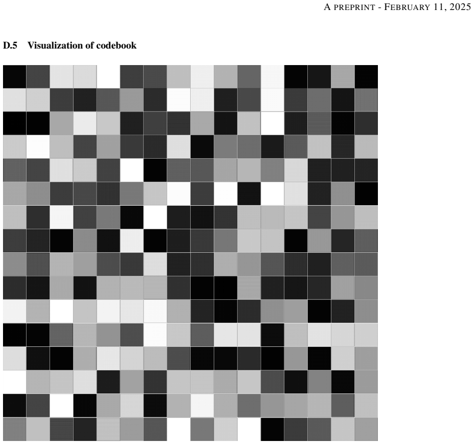
The central claim is that continuous disease progression in volumetric CT can be learned in a discrete latent space rather than modelled directly on voxels. A 3D-VQ-GAN first compresses each CT volume into a compact set of codebook indices whose entries correspond to local imaging patterns such as reticulation and ground-glass opacity. A second-stage temporal model, built around a neural ODE $f_\theta$, evolves the latent embedding over time; given two observed scans, the ODE solver is run from the encoded initial state to produce embeddings at any queried timestep, and skip connections between consecutive embeddings let the model learn only the difference between adjacent time points. The frozen 3D-VQ-GAN decoder then turns those embeddings back into CT volumes. The paper demonstrates interpolation and extrapolation on IPF patients and validates the generated scans through Cox survival analysis, reporting that biomarkers from generated scans achieve a C-index of 0.886 (cross-sectional) and 0.959 (longitudinal change) versus 0.929 and 1.0 for real scans.
Load-bearing premise
One fixed mathematical rule is assumed to describe how every patient's lungs change over time, and the paper itself says this is unlikely for a disease with distinct subtypes and progression patterns.
Editorial extensions
If this is right
- Given two CT scans, the model outputs synthetic scans at any requested interval and horizon, covering both interpolation and extrapolation.
- The best extrapolation configuration (ConvGRU encoder with embedding skip connections) reaches MSE 0.019, SSIM 0.489, and PSNR 17.816 on held-out patients.
- Survival-relevant texture is preserved: cross-sectional biomarkers from generated third scans give C-index 0.886 versus 0.929 for real scans, and longitudinal biomarkers give 0.959 versus 1.0.
- Synthetic longitudinal scans could be used to impute missing visits, augment sparse IPF datasets, and simulate treatment effects by comparing generated trajectories under different assumptions.
Reading between the lines
- Beyond the paper, the same two-stage recipe could transfer to other diseases with sparse longitudinal imaging, such as multiple sclerosis or osteoarthritis, because it only requires a learnable discrete codebook and a smooth latent trajectory.
- The paper's shared disease dynamic assumption is the obvious pressure point; conditioning the ODE on a per-patient latent code or subtype label would be a natural, untested upgrade that makes the personalised claim more literal.
- A sharp testable extension would be to feed generated scans into a separate segmentation or staging model and measure whether performance improves; if the synthetic volumes carry the same prognostic texture as real ones, they could function as data augmentation in rare-disease cohorts.
Editorial analysis
A structured set of objections, weighed in public.
Referee Report
Summary. This manuscript proposes 4D-VQ-GAN, a two-stage generative model for synthesizing longitudinal CT volumes in IPF patients. Stage 1 trains a 3D-VQ-GAN to compress CT volumes into a discrete codebook; Stage 2 trains a neural ODE on the quantized latent embeddings, conditioned on two observed scans, to generate volumes at arbitrary time points. The authors evaluate interpolation and extrapolation quality with MSE/SSIM/PSNR, perform an ablation over architectural variants, and validate clinical utility by deriving imaging biomarkers from generated scans and computing C-indices for survival prediction. They report C-indices of 0.886 (cross-sectional) and 0.959 (longitudinal) for generated scans, compared with 0.929 and 1.0 for real scans. The paper is clearly written, but the experimental validation is not sufficient to establish the central claim that the model captures disease progression in a personalized manner.
Significance. The paper addresses a relevant and difficult problem: generating longitudinal 3D medical images from sparse, irregular observations. The two-stage design (VQ-GAN compression + latent ODE) is sensible, and the use of a real longitudinal IPF cohort with survival outcomes is a strength. The codebook visualization and the external validation of the reconstruction stage are also commendable. However, as presented, the experimental validation does not establish the central claim that the model 'captures disease progression' or provides 'personalised' predictions. The absence of trivial baselines, the lack of statistical comparison for survival C-indices, and the untested assumption of a shared dynamics mean the quantitative results could be explained by simpler mechanisms such as copying the last observed scan or reflecting baseline severity. If the required baselines and analyses confirm the added value, the contribution would be significant for the medical imaging community.
major comments (4)
- [Section 4, Table 1] The ablation compares only variants of the proposed architecture (ConvGRU vs ODE-ConvGRU, skip connections, masked inputs), with no comparison against trivial baselines such as copying the last observed scan or its latent embedding, or linear interpolation in latent space. Since IPF changes between scans can be subtle, the reported extrapolation SSIM of 0.489 and PSNR of 17.816 may be close to a no-change baseline; without such comparisons, the quantitative results do not demonstrate that the model predicts progression beyond the information already present in the input scans.
- [Section 4, Survival outcome prediction] The claim that generated scans yield C-indices 'comparable' to real scans is not supported by appropriate statistical evidence: no confidence intervals, no significance tests, and the test set is small (44 patients). The longitudinal C-index of 1.0 for real scans suggests overfitting or an overly homogeneous test set, and the cross-sectional difference (0.886 vs 0.929) is not shown to be within sampling variability. Moreover, there is no comparison with biomarkers derived from the most recent real scan, so the high C-index of generated scans may reflect baseline disease severity rather than the model's predicted temporal change.
- [Section 2, Eq. (6); Section 5] The model uses a single deterministic neural ODE f_theta, which the authors acknowledge 'assumes a shared disease dynamic across all patients.' This assumption is not tested, yet the title and introduction claim 'personalised disease progression modelling.' To support this claim, the authors should demonstrate that generated trajectories vary meaningfully across patients (e.g., by testing whether patient-specific initial states lead to distinct predicted changes, or by comparing against a model with patient-specific dynamics).
- [Section 3, Evaluation Metrics] The quantitative evaluation relies on image-similarity metrics (MSE, SSIM, PSNR) that do not directly measure whether the generated changes correspond to pathological progression. The authors should include clinically meaningful metrics, such as changes in quantitative fibrosis extent or the imaging biomarkers already used in their survival analysis, to show that the generated temporal changes are in the right direction and of plausible magnitude.
minor comments (9)
- [Table 1] The variant names are confusing; for example, 'ODE encoder (ConvGRU)' and 'ODE encoder (ODE-ConvGRU)' could be more clearly distinguished, and the table should state which components are ablated in each row.
- [Section 4] The paper should report 95% confidence intervals for all C-indices (e.g., via bootstrap) and a test of the difference between generated and real biomarkers.
- [Appendix D.2, Eq. (3)] The GAN loss is written for a single discriminator, but the text mentions two discriminators D2d and D3d; the equation should include both terms explicitly.
- [Sections 2 and D.3] The 3D-ConvGRU is described as the 'encoder of the neural ODE solver', but it is actually the encoder that produces the initial latent state; rephrase to avoid confusion.
- [Section 3, Datasets] Provide more details on the test cohort: number of patients, number of scans per patient, and the distribution of time intervals between scans.
- [Appendix A] The phrase 'visual inspection by me' should be rewritten in a professional style, e.g., 'visual inspection by a trained researcher.'
- [Figure 3] Add annotations indicating which generated volumes correspond to which time points and which are interpolated versus extrapolated.
- [Appendix D.4, Table 3] The reconstruction errors on the external Southampton test set are considerably higher than on the internal test set; this deserves a comment in the main text.
- [Related Work] The paper would benefit from a comparison with at least one existing longitudinal image synthesis method, such as the latent linear mixed-effects model of Sauty et al. or TR-GAN, to contextualize the results.
Circularity Check
No significant circularity: the two-stage latent ODE model is trained on real CT volumes and validated against held-out survival outcomes, with no fitted parameter renamed as a prediction.
full rationale
The derivation chain is self-contained and externally validated. Stage 1 learns a discrete codebook by reconstructing real CT volumes; Stage 2 fits a neural ODE to the temporal trajectory of those codes; neither stage defines its target in terms of the evaluation. The survival analysis uses real, held-out survival outcomes: the top five code-frequency biomarkers are selected on training scans by Cox p-values, and the C-index is then computed on a separate test set for both real and generated scans, so no fitted parameter is renamed as a prediction. The only couplings—the same codebook is used for generation and biomarker extraction, and the deterministic ODE assumes a shared disease dynamic—are potential biases or modelling limitations, not reductions of the claimed result to its inputs. The reported absence of a 'no-change' baseline is a validation gap that affects how strongly the progression claim is supported, but it does not make the derivation circular. No self-citation chain or imported uniqueness theorem is load-bearing.
Assumptions & free parameters
free parameters (4)
- Codebook vocabulary size M =
256
- Compression rate r =
4
- Loss weights for 3D-VQ-GAN =
lambda_perc=4, lambda_rec=4, lambda_GAN=1
- Number of top biomarkers selected =
5
assumptions (3)
- domain assumption The latent embedding space of the 3D-VQ-GAN supports smooth, continuous disease trajectories that can be described by a single neural ODE fθ shared across all patients.
- domain assumption Normalized frequencies of codebook indices serve as meaningful prognostic imaging biomarkers.
- domain assumption Lung registration correctly aligns longitudinal scans so that residual differences reflect disease progression, not motion or positioning.
Cite this review
Pith. "Pith review of 4D VQ-GAN: Synthesising Medical Scans at Any Time Point for Personalised Disease Progression Modelling of Idiopathic Pulmonary Fibrosis." pith.science (2026). https://pith.science/paper/K3JYY5IF
@misc{pith2026250205713,
  author       = {Pith},
  title        = {Pith review of: 4D VQ-GAN: Synthesising Medical Scans at Any Time Point for Personalised Disease Progression Modelling of Idiopathic Pulmonary Fibrosis},
  year         = {2026},
  howpublished = {\url{https://pith.science/paper/K3JYY5IF}},
  note         = {Machine review of arXiv:2502.05713}
}
read the original abstract
Understanding the progression trajectories of diseases is crucial for early diagnosis and effective treatment planning. This is especially vital for life-threatening conditions such as Idiopathic Pulmonary Fibrosis (IPF), a chronic, progressive lung disease with a prognosis comparable to many cancers. Computed tomography (CT) imaging has been established as a reliable diagnostic tool for IPF. Accurately predicting future CT scans of early-stage IPF patients can aid in developing better treatment strategies, thereby improving survival outcomes. In this paper, we propose 4D Vector Quantised Generative Adversarial Networks (4D-VQ-GAN), a model capable of generating realistic CT volumes of IPF patients at any time point. The model is trained using a two-stage approach. In the first stage, a 3D-VQ-GAN is trained to reconstruct CT volumes. In the second stage, a Neural Ordinary Differential Equation (ODE) based temporal model is trained to capture the temporal dynamics of the quantised embeddings generated by the encoder in the first stage. We evaluate different configurations of our model for generating longitudinal CT scans and compare the results against ground truth data, both quantitatively and qualitatively. For validation, we conduct survival analysis using imaging biomarkers derived from generated CT scans and achieve a C-index comparable to that of biomarkers derived from the real CT scans. The survival analysis results demonstrate the potential clinical utility inherent to generated longitudinal CT scans, showing that they can reliably predict survival outcomes.
Figures


Figures from the paper (6 more)


Reference graph
Works this paper leans on
-
[1]
Delving deeper into convolutional networks for learning video representations
Nicolas Ballas et al. “Delving deeper into convolutional networks for learning video representations”. In: arXiv preprint arXiv:1511.06432 (2015)
arXiv 2015
-
[2]
Seeing what a GAN cannot generate
David Bau et al. “Seeing what a GAN cannot generate”. In: Proceedings of the IEEE/CVF International Conference on Computer Vision. 2019, pp. 4502–4511
work page 2019
-
[3]
Estimating or propagating gradients through stochastic neurons for conditional computation
Yoshua Bengio, Nicholas Léonard, and Aaron Courville. “Estimating or propagating gradients through stochastic neurons for conditional computation”. In: arXiv preprint arXiv:1308.3432 (2013)
arXiv 2013
-
[4]
Neural ordinary differential equations
Ricky TQ Chen et al. “Neural ordinary differential equations”. In: Advances in neural information processing systems. V ol. 31. 2018
work page 2018
-
[5]
Longitudinal self-supervision to disentangle inter- patient variability from disease progression
Raphaël Couronné, Paul Vernhet, and Stanley Durrleman. “Longitudinal self-supervision to disentangle inter- patient variability from disease progression”. In: International Conference on Medical image computing and computer-assisted intervention. Springer. 2021, pp. 231–241
work page 2021
-
[6]
Regression models and life-tables
David R Cox. “Regression models and life-tables”. In: Journal of the Royal Statistical Society: Series B (Methodological) 34.2 (1972), pp. 187–202
work page 1972
-
[7]
Generative adversarial networks: An overview
Antonia Creswell et al. “Generative adversarial networks: An overview”. In:IEEE signal processing magazine 35.1 (2018), pp. 53–65
work page 2018
-
[8]
Diffusion models in vision: A survey
Florinel-Alin Croitoru et al. “Diffusion models in vision: A survey”. In: IEEE Transactions on Pattern Analysis and Machine Intelligence 45.9 (2023), pp. 10850–10869
work page 2023
Show all 61 references
-
[9]
Estimating long-term multivariate progression from short-term data
Michael C Donohue et al. “Estimating long-term multivariate progression from short-term data”. In: Alzheimer’s & Dementia 10 (2014), S400–S410. 6 A PREPRINT - F EBRUARY 11, 2025
2014
-
[10]
Taming transformers for high-resolution image synthesis
Patrick Esser, Robin Rombach, and Bjorn Ommer. “Taming transformers for high-resolution image synthesis”. In: Proceedings of the IEEE/CVF Conference on Computer Vision and Pattern Recognition. 2021, pp. 12873–12883
2021
-
[11]
TR-Gan: multi-session future MRI prediction with temporal recurrent generative adversar- ial Network
Chen-Chen Fan et al. “TR-Gan: multi-session future MRI prediction with temporal recurrent generative adversar- ial Network”. In: IEEE Transactions on Medical Imaging 41.8 (2022), pp. 1925–1937
2022
-
[12]
TR-TransGAN: Temporal Recurrent Transformer Generative Adversarial Network for Longitudinal MRI Dataset Expansion
Chen-Chen Fan et al. “TR-TransGAN: Temporal Recurrent Transformer Generative Adversarial Network for Longitudinal MRI Dataset Expansion”. In: IEEE Transactions on Cognitive and Developmental Systems (2024), pp. 1–10
2024
-
[13]
GAN-based generation of realistic 3D data: A systematic review and taxonomy
André Ferreira et al. “GAN-based generation of realistic 3D data: A systematic review and taxonomy”. In: arXiv preprint arXiv:2207.01390 (2022)
2022 arXiv
-
[14]
GAN-based generation of realistic 3D volumetric data: A systematic review and taxonomy
André Ferreira et al. “GAN-based generation of realistic 3D volumetric data: A systematic review and taxonomy”. In: Medical Image Analysis (2024), p. 103100
2024
-
[15]
A fast operator for detection and precise location of distinct points, corners and centres of circular features
Wolfgang Förstner and Eberhard Gülch. “A fast operator for detection and precise location of distinct points, corners and centres of circular features”. In: Proc. ISPRS intercommission conference on fast processing of photogrammetric data. V ol. 6. 1987, pp. 281–305
1987
-
[16]
Long video generation with time-agnostic VQGAN and time-sensitive transformer
Songwei Ge et al. “Long video generation with time-agnostic VQGAN and time-sensitive transformer”. In: European Conference on Computer Vision. Springer. 2022, pp. 102–118
2022
-
[17]
GraphRegNet: Deep graph regularisation networks on sparse keypoints for dense registration of 3D lung CTs
Lasse Hansen and Mattias P Heinrich. “GraphRegNet: Deep graph regularisation networks on sparse keypoints for dense registration of 3D lung CTs”. In: IEEE Transactions on Medical Imaging 40.9 (2021), pp. 2246–2257
2021
-
[18]
Estimating large lung motion in COPD patients by symmetric regularised correspondence fields
Mattias P Heinrich, Heinz Handels, and Ivor JA Simpson. “Estimating large lung motion in COPD patients by symmetric regularised correspondence fields”. In: International Conference on Medical Image Computing and Computer-Assisted Intervention. Springer. 2015, pp. 338–345
2015
-
[19]
Learn2Reg: Comprehensive multi-task medical image registration challenge, dataset and evaluation in the era of deep learning
Alessa Hering et al. “Learn2Reg: Comprehensive multi-task medical image registration challenge, dataset and evaluation in the era of deep learning”. In: IEEE Transactions on Medical Imaging 42.3 (2022), pp. 697–712
2022
-
[20]
Denoising diffusion probabilistic models
Jonathan Ho, Ajay Jain, and Pieter Abbeel. “Denoising diffusion probabilistic models”. In: Advances in Neural Information Processing Systems. V ol. 33. 2020, pp. 6840–6851
2020
-
[21]
Imagen video: High definition video generation with diffusion models
Jonathan Ho et al. “Imagen video: High definition video generation with diffusion models”. In: arXiv preprint arXiv:2210.02303 (2022)
2022 arXiv
-
[22]
Automatic lung segmentation in routine imaging is primarily a data diversity problem, not a methodology problem
Johannes Hofmanninger et al. “Automatic lung segmentation in routine imaging is primarily a data diversity problem, not a methodology problem”. In: European Radiology Experimental 4.1 (2020), pp. 1–13
2020
-
[23]
Topological Neural Discrete Representation Learning à la Kohonen
Kazuki Irie, Róbert Csordás, and Jürgen Schmidhuber. “Topological Neural Discrete Representation Learning à la Kohonen”. In: arXiv preprint arXiv:2302.07950 (2023)
2023 arXiv
-
[24]
Brain β-amyloid load approaches a plateau
Clifford R Jack et al. “Brain β-amyloid load approaches a plateau”. In: Neurology 80.10 (2013), pp. 890–896
2013
-
[25]
A computational neurodegenerative disease progression score: method and results with the Alzheimer’s disease neuroimaging initiative cohort
Bruno M Jedynak et al. “A computational neurodegenerative disease progression score: method and results with the Alzheimer’s disease neuroimaging initiative cohort”. In: Neuroimage 63.3 (2012), pp. 1478–1486
2012
-
[26]
Simple video generation using neural ODEs
David Kanaa et al. “Simple video generation using neural ODEs”. In: arXiv preprint arXiv:2109.03292 (2021)
2021 arXiv
-
[27]
Diffusion models in medical imaging: A comprehensive survey
Amirhossein Kazerouni et al. “Diffusion models in medical imaging: A comprehensive survey”. In: Medical Image Analysis 88 (2023), p. 102846
2023
-
[28]
Medical Diffusion–Denoising Diffusion Probabilistic Models for 3D Medical Image Generation
Firas Khader et al. “Medical Diffusion–Denoising Diffusion Probabilistic Models for 3D Medical Image Generation”. In: arXiv preprint arXiv:2211.03364 (2022)
2022 arXiv
-
[29]
Transformers for CT reconstruction from monoplanar and biplanar radiographs
Firas Khader et al. “Transformers for CT reconstruction from monoplanar and biplanar radiographs”. In: International Workshop on Simulation and Synthesis in Medical Imaging. Springer. 2023, pp. 1–10
2023
-
[30]
Longitudinal brain MR image modeling using personal- ized memory for Alzheimer’s disease
Seong Tae Kim, Umut Küçükaslan, and Nassir Navab. “Longitudinal brain MR image modeling using personal- ized memory for Alzheimer’s disease”. In: IEEE Access 9 (2021), pp. 143212–143221
2021
-
[31]
Adam: A method for stochastic optimization
Diederik P Kingma and Jimmy Ba. “Adam: A method for stochastic optimization”. In: arXiv preprint arXiv:1412.6980 (2014)
2014 arXiv
-
[32]
Auto-encoding variational Bayes
Diederik P Kingma and Max Welling. “Auto-encoding variational Bayes”. In:arXiv preprint arXiv:1312.6114 (2013)
2013 arXiv
-
[33]
CCVS: Context-aware controllable video synthesis
Guillaume Le Moing, Jean Ponce, and Cordelia Schmid. “CCVS: Context-aware controllable video synthesis”. In: Advances in Neural Information Processing Systems. V ol. 34. 2021, pp. 14042–14055
2021
-
[34]
Hi-BEHRT: Hierarchical Transformer-based model for accurate prediction of clinical events using multimodal longitudinal electronic health records
Yikuan Li et al. “Hi-BEHRT: Hierarchical Transformer-based model for accurate prediction of clinical events using multimodal longitudinal electronic health records”. In: IEEE Journal of Biomedical and Health Informatics 27.2 (2022), pp. 1106–1117
2022
-
[35]
3D brain and heart volume generative models: A survey
Yanbin Liu et al. “3D brain and heart volume generative models: A survey”. In:ACM Computing Surveys 56.6 (2024), pp. 1–37. 7 A PREPRINT - F EBRUARY 11, 2025
2024
-
[36]
Disentangling normal aging from Alzheimer’s disease in structural magnetic resonance images
Marco Lorenzi et al. “Disentangling normal aging from Alzheimer’s disease in structural magnetic resonance images”. In: Neurobiology of Aging 36 (2015), S42–S52
2015
-
[37]
Decoupled weight decay regularization
Ilya Loshchilov and Frank Hutter. “Decoupled weight decay regularization”. In:arXiv preprint arXiv:1711.05101 (2017)
2017 arXiv
-
[38]
MC-RV AE: Multi-channel recurrent variational autoencoder for multimodal Alzheimer’s disease progression modelling
Gerard Martí-Juan et al. “MC-RV AE: Multi-channel recurrent variational autoencoder for multimodal Alzheimer’s disease progression modelling”. In: NeuroImage 268 (2023), p. 119892
2023
-
[39]
Learning imaging biomarker trajectories from noisy Alzheimer’s disease data using a Bayesian multilevel model
Neil P Oxtoby et al. “Learning imaging biomarker trajectories from noisy Alzheimer’s disease data using a Bayesian multilevel model”. In: Bayesian and grAphical Models for Biomedical Imaging: First International Workshop, BAMBI 2014, Cambridge, MA, USA, September 18, 2014, Rev...
2014
-
[40]
Vid-ODE: Continuous-time video generation with neural ordinary differential equation
Sunghyun Park et al. “Vid-ODE: Continuous-time video generation with neural ordinary differential equation”. In: Proceedings of the AAAI Conference on Artificial Intelligence. V ol. 35. 3. 2021, pp. 2412–2422
2021
-
[41]
Enhancing spatiotemporal disease progression models via latent diffusion and prior knowledge
Lemuel Puglisi, Daniel C Alexander, and Daniele Ravı‘. “Enhancing spatiotemporal disease progression models via latent diffusion and prior knowledge”. In: International Conference on Medical Image Computing and Computer-Assisted Intervention. Springer. 2024, pp. 173–183
2024
-
[42]
Degenerative adversarial neuroimage nets for brain scan simulations: Application in ageing and dementia
Daniele Ravi et al. “Degenerative adversarial neuroimage nets for brain scan simulations: Application in ageing and dementia”. In: Medical Image Analysis 75 (2022), p. 102257
2022
-
[43]
Generating diverse high-fidelity images with VQ-V AE-2
Ali Razavi, Aaron Van den Oord, and Oriol Vinyals. “Generating diverse high-fidelity images with VQ-V AE-2”. In: Advances in Neural Information Processing Systems. V ol. 32. 2019
2019
-
[44]
U-Net: Convolutional networks for biomedical image segmentation
Olaf Ronneberger, Philipp Fischer, and Thomas Brox. “U-Net: Convolutional networks for biomedical image segmentation”. In: International Conference on Medical Image Computing and Computer-Assisted Intervention. Springer. 2015, pp. 234–241
2015
-
[45]
Latent ordinary differential equations for irregularly- sampled time series
Yulia Rubanova, Ricky TQ Chen, and David K Duvenaud. “Latent ordinary differential equations for irregularly- sampled time series”. In: Advances in neural information processing systems. V ol. 32. 2019
2019
-
[46]
Progression models for imaging data with Longitudinal Variational Auto Encoders
Benoı‘t Sauty and Stanley Durrleman. “Progression models for imaging data with Longitudinal Variational Auto Encoders”. In: International Conference on Medical image computing and computer-assisted intervention. Springer. 2022, pp. 3–13
2022
-
[47]
InterfaceGAN: Interpreting the disentangled face representation learned by GANs
Yujun Shen et al. “InterfaceGAN: Interpreting the disentangled face representation learned by GANs”. In: IEEE Transactions on Pattern Analysis and Machine Intelligence44.4 (2020), pp. 2004–2018
2020
-
[48]
Very deep convolutional networks for large-scale image recognition
Karen Simonyan and Andrew Zisserman. “Very deep convolutional networks for large-scale image recognition”. In: arXiv preprint arXiv:1409.1556 (2014)
2014 arXiv
-
[49]
Medical image generation using generative adversarial networks: A review
Nripendra Kumar Singh and Khalid Raza. “Medical image generation using generative adversarial networks: A review”. In: Health informatics: A computational perspective in healthcare (2021), pp. 77–96
2021
-
[50]
A model of brain morphological changes related to aging and Alzheimer’s disease from cross-sectional assessments
Raphaël Sivera et al. “A model of brain morphological changes related to aging and Alzheimer’s disease from cross-sectional assessments”. In: NeuroImage 198 (2019), pp. 255–270
2019
-
[51]
Hierarchical amortized GAN for 3D high resolution medical image synthesis
Li Sun et al. “Hierarchical amortized GAN for 3D high resolution medical image synthesis”. In: IEEE Journal of Biomedical and Health Informatics 26.8 (2022), pp. 3966–3975
2022
-
[52]
Neuromorphologically-preserving volumetric data encoding using VQ-V AE
Petru-Daniel Tudosiu et al. “Neuromorphologically-preserving volumetric data encoding using VQ-V AE”. In: arXiv preprint arXiv:2002.05692 (2020)
2020 arXiv
-
[53]
Multi-scale GANs for memory-efficient generation of high resolution medical images
Hristina Uzunova et al. “Multi-scale GANs for memory-efficient generation of high resolution medical images”. In: International Conference on Medical Image Computing and Computer-Assisted Intervention. Springer. 2019, pp. 112–120
2019
-
[54]
Neural discrete representation learning
Aaron Van Den Oord, Oriol Vinyals, et al. “Neural discrete representation learning”. In: Advances in neural information processing systems. V ol. 30. 2017
2017
-
[55]
Amyloid β deposition, neurodegeneration, and cognitive decline in sporadic Alzheimer’s disease: a prospective cohort study
Victor L Villemagne et al. “Amyloid β deposition, neurodegeneration, and cognitive decline in sporadic Alzheimer’s disease: a prospective cohort study”. In: The Lancet Neurology 12.4 (2013), pp. 357–367
2013
-
[56]
MCVD-masked conditional video diffusion for prediction, generation, and interpolation
Vikram V oleti, Alexia Jolicoeur-Martineau, and Chris Pal. “MCVD-masked conditional video diffusion for prediction, generation, and interpolation”. In: Advances in Neural Information Processing Systems . V ol. 35. Curran Associates, Inc., 2022, pp. 23371–23385
2022
-
[57]
Modelling Latent Dynamics of StyleGAN using Neural ODEs
Weihao Xia, Yujiu Yang, and Jing-Hao Xue. “Modelling Latent Dynamics of StyleGAN using Neural ODEs”. In: arXiv preprint arXiv:2208.11197 (2022)
2022 arXiv
-
[58]
Videogpt: Video generation using VQ-V AE and transformers
Wilson Yan et al. “Videogpt: Video generation using VQ-V AE and transformers”. In: arXiv preprint arXiv:2104.10157 (2021)
2021 arXiv
-
[59]
SADM: Sequence-aware diffusion model for longitudinal medical image generation
Jee Seok Yoon et al. “SADM: Sequence-aware diffusion model for longitudinal medical image generation”. In: International Conference on Information Processing in Medical Imaging. Springer. 2023, pp. 388–400. 8 A PREPRINT - F EBRUARY 11, 2025
2023
-
[60]
The unreasonable effectiveness of deep features as a perceptual metric
Richard Zhang et al. “The unreasonable effectiveness of deep features as a perceptual metric”. In: Proceedings of the IEEE conference on computer vision and pattern recognition. 2018, pp. 586–595. A Pre-processing The registration process aligns corresponding structures across...
2018
-
[61]
VQ-GAN [10], a variant of VQ-V AE, incorporated a discriminator and perceptual loss, combining the strengths of both VQ-V AE and GAN to generate high-resolution images
utilized hierarchical multi-scale latent maps for large-scale image generation. VQ-GAN [10], a variant of VQ-V AE, incorporated a discriminator and perceptual loss, combining the strengths of both VQ-V AE and GAN to generate high-resolution images. Ge et al. [16] extended VQ-G...
2025
Reviewed August 8, 2026 · model on record in the stance chip above.
Discussion (0). Continue with ORCID to comment.