Recognition: 2 theorem links
· Lean TheoremPhysics-Informed Graph Neural Networks for Frequency-Aware Optical Aberration Correction
Pith reviewed 2026-05-17 00:40 UTC · model grok-4.3
The pith
A graph neural network models relationships between Zernike polynomials by azimuthal degree to correct large optical aberrations while aligning predictions with frequency-domain features.
A machine-rendered reading of the paper's core claim, the machinery that carries it, and where it could break.
Core claim
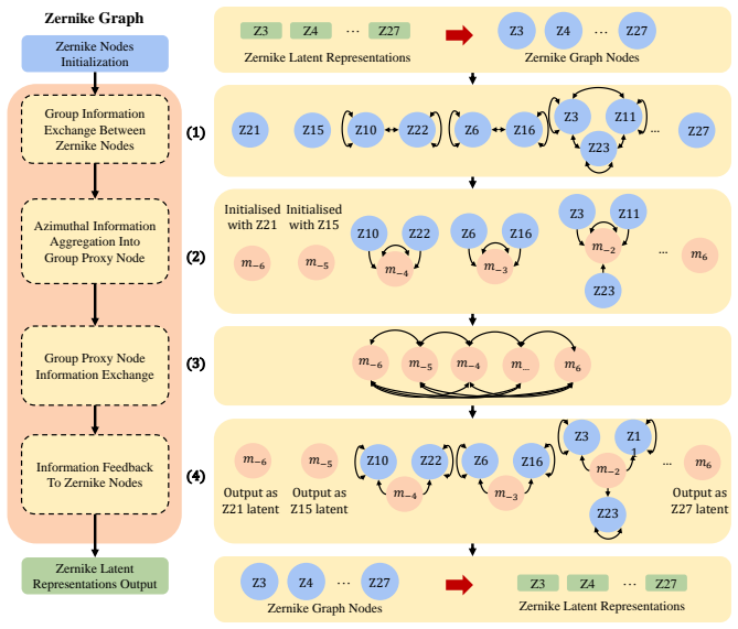
ZRNet jointly performs Zernike coefficient prediction and optical image restoration. The Zernike Graph module explicitly models physical relationships between Zernike polynomials based on their azimuthal degrees to ensure corrections align with fundamental optical principles. The Frequency-Aware Alignment loss enforces physical consistency by aligning Zernike predictions with image features in the Fourier domain.
What carries the argument
Zernike Graph module that connects Zernike polynomial terms according to azimuthal degrees, paired with Frequency-Aware Alignment loss that matches predictions to Fourier-domain image features.
If this is right
- The method achieves state-of-the-art results on both image restoration and Zernike coefficient prediction across diverse microscopy modalities and biological samples.
- Performance holds on experimental point-spread-function data collected from a physical microscope.
- The network remains effective under realistic sensor noise and generalizes past purely simulated conditions.
Where Pith is reading between the lines
- The same graph construction could be tested on other wavefront correction tasks such as adaptive optics in astronomy.
- Real-time versions might be paired with deformable mirrors for live-sample correction during imaging sessions.
- The frequency-alignment idea could extend to related inverse problems where coefficient estimates must stay consistent with measured intensities.
Load-bearing premise
That building a graph from azimuthal degrees between Zernike terms and adding the frequency alignment loss will force the network to follow optical physics rather than simply fitting training examples.
What would settle it
An ablation study on a dataset with known physical aberration patterns in which removing the graph module or the Frequency-Aware Alignment loss produces no measurable drop in restoration or coefficient accuracy would falsify the claim.
Figures


read the original abstract
Optical aberrations significantly degrade image quality in microscopy, particularly when imaging deeper into samples. These aberrations arise from distortions in the optical wavefront and can be mathematically represented using Zernike polynomials. Existing methods often address only mild aberrations on limited sample types and modalities, typically treating the problem as a black-box mapping without leveraging the underlying optical physics of wavefront distortions. We propose ZRNet, a physics-informed framework that jointly performs Zernike coefficient prediction and optical image Restoration. We contribute a Zernike Graph module that explicitly models physical relationships between Zernike polynomials based on their azimuthal degrees-ensuring that learned corrections align with fundamental optical principles. To further enforce physical consistency between image restoration and Zernike prediction, we introduce a Frequency-Aware Alignment (FAA) loss, which better aligns Zernike coefficient prediction and image features in the Fourier domain. Extensive experiments on CytoImageNet demonstrates that our approach achieves state-of-the-art performance in both image restoration and Zernike coefficient prediction across diverse microscopy modalities and biological samples with complex, large-amplitude aberrations. We further validate on experimental PSF data from a physical microscope and demonstrate robustness to realistic sensor noise, confirming generalisation beyond simulated conditions. Code is available at https://github.com/janetkok/ZRNet.
Editorial analysis
A structured set of objections, weighed in public.
Referee Report
Summary. The paper introduces ZRNet, a physics-informed graph neural network for joint Zernike coefficient prediction and optical image restoration in microscopy. It contributes a Zernike Graph module that connects polynomials according to azimuthal degree m to enforce optical relationships, plus a Frequency-Aware Alignment (FAA) loss that aligns features in the Fourier domain. The central claim is state-of-the-art performance on CytoImageNet for both restoration and coefficient prediction across modalities and large-amplitude aberrations, with additional validation on real experimental PSF data and robustness to sensor noise.
Significance. If the performance edge is shown to arise from the physics-informed components rather than added capacity, the work would provide a useful template for embedding wavefront physics into learned aberration correctors, with direct relevance to deep-tissue microscopy. The release of code and the inclusion of real PSF validation are concrete strengths that support reproducibility and generalization claims.
major comments (2)
- [Method and Experiments] The abstract and method description assert that the Zernike Graph (azimuthal-degree edges) plus FAA loss produce corrections that 'align with fundamental optical principles.' No ablation replaces the graph with a standard message-passing layer of matched capacity, nor is wavefront error reported on held-out experimental PSFs; without these controls the physics-informed framing remains untested against the alternative that the modules simply increase expressivity on the simulated training distribution.
- [Experiments] Table and figure captions reference SOTA results on CytoImageNet, yet the manuscript does not report the exact data splits, the full set of baselines (including non-graph CNNs and physics-agnostic GNNs), or statistical significance of the reported gains; these omissions make it impossible to assess whether the claimed superiority is robust or sensitive to post-hoc choices.
minor comments (2)
- [Method] Notation for the Zernike Graph adjacency matrix should be introduced once with a clear equation rather than described only in prose.
- [Method] The FAA loss is defined in the Fourier domain; a short derivation or reference showing why this particular alignment term is preferred over direct coefficient MSE would improve clarity.
Simulated Author's Rebuttal
We thank the referee for the thoughtful and constructive comments. We address each major point below and describe the revisions that will be incorporated into the next version of the manuscript.
read point-by-point responses
-
Referee: [Method and Experiments] The abstract and method description assert that the Zernike Graph (azimuthal-degree edges) plus FAA loss produce corrections that 'align with fundamental optical principles.' No ablation replaces the graph with a standard message-passing layer of matched capacity, nor is wavefront error reported on held-out experimental PSFs; without these controls the physics-informed framing remains untested against the alternative that the modules simply increase expressivity on the simulated training distribution.
Authors: We agree that isolating the contribution of the physics-informed design is important. In the revised manuscript we will add an ablation that replaces the Zernike Graph module with a standard message-passing GNN layer of matched capacity and report the resulting performance on both coefficient prediction and image restoration. We will also report wavefront error (Zernike coefficient RMSE) on the held-out experimental PSF data to provide direct evidence that the learned corrections generalize beyond the simulated distribution. revision: yes
-
Referee: [Experiments] Table and figure captions reference SOTA results on CytoImageNet, yet the manuscript does not report the exact data splits, the full set of baselines (including non-graph CNNs and physics-agnostic GNNs), or statistical significance of the reported gains; these omissions make it impossible to assess whether the claimed superiority is robust or sensitive to post-hoc choices.
Authors: We acknowledge these omissions limit the ability to fully evaluate the claims. The revised manuscript will explicitly document the train/validation/test splits used for CytoImageNet, expand the baseline set to include non-graph CNNs and physics-agnostic GNN variants, and report statistical significance (e.g., paired statistical tests with p-values) for the observed performance differences. revision: yes
Circularity Check
No circularity: architecture and loss are independent design choices validated on external data
full rationale
The paper defines the Zernike Graph from the known azimuthal order m of Zernike polynomials and introduces the FAA loss as a Fourier-domain alignment term; neither step reduces any claimed prediction or coefficient to a fitted input by construction. The central claims rest on end-to-end training plus experimental validation on CytoImageNet and real PSF measurements, not on self-referential equations or load-bearing self-citations. No derivation chain collapses to tautology.
Axiom & Free-Parameter Ledger
axioms (1)
- domain assumption Zernike polynomials provide a complete and accurate basis for representing optical aberrations in microscopy
Lean theorems connected to this paper
-
IndisputableMonolith/Foundation/AlexanderDuality.leanalexander_duality_circle_linking unclear?
unclearRelation between the paper passage and the cited Recognition theorem.
Frequency-Aware Alignment (FAA) loss... in the Fourier domain
What do these tags mean?
- matches
- The paper's claim is directly supported by a theorem in the formal canon.
- supports
- The theorem supports part of the paper's argument, but the paper may add assumptions or extra steps.
- extends
- The paper goes beyond the formal theorem; the theorem is a base layer rather than the whole result.
- uses
- The paper appears to rely on the theorem as machinery.
- contradicts
- The paper's claim conflicts with a theorem or certificate in the canon.
- unclear
- Pith found a possible connection, but the passage is too broad, indirect, or ambiguous to say the theorem truly supports the claim.
Reference graph
Works this paper leans on
-
[1]
K. M. Hampson, R. Turcotte, D. T. Miller, K. Kurokawa, J. R. Males, N. Ji, M. J. Booth, Adaptive optics for high-resolution imaging, Nature Reviews Methods Primers 1 (1) (2021) 68
work page 2021
-
[2]
M. Schwertner, M. J. Booth, M. A. Neil, T. Wilson, Measurement of specimen- induced aberrations of biological samples using phase stepping interferometry, Journal of microscopy 213 (1) (2004) 11–19
work page 2004
-
[3]
J. W. Hardy, Adaptive optics for astronomical telescopes, V ol. 16, Oxford Univer- sity Press on Demand, 1998
work page 1998
-
[4]
B. C. Platt, R. Shack, History and principles of shack-hartmann wavefront sensing (2001)
work page 2001
-
[5]
X. Tao, B. Fernandez, O. Azucena, M. Fu, D. Garcia, Y . Zuo, D. C. Chen, J. Kubby, Adaptive optics confocal microscopy using direct wavefront sensing, Optics letters 36 (7) (2011) 1062–1064
work page 2011
-
[6]
M. A. V orontsov, G. W. Carhart, M. Cohen, G. Cauwenberghs, Adaptive op- tics based on analog parallel stochastic optimization: analysis and experimental demonstration, JOSA A 17 (8) (2000) 1440–1453
work page 2000
- [7]
-
[8]
P. Yang, M. Ao, Y . Liu, B. Xu, W. Jiang, Intracavity transverse modes controlled by a genetic algorithm based on zernike mode coefficients, Optics express 15 (25) (2007) 17051–17062
work page 2007
-
[9]
R. W. Gerchberg, A practical algorithm for the determination of plane from image and diffraction pictures, Optik 35 (2) (1972) 237–246
work page 1972
-
[10]
A. J. Janssen, Extended nijboer–zernike approach for the computation of optical point-spread functions, JOSA A 19 (5) (2002) 849–857
work page 2002
-
[11]
J. Liu, P. Wang, X. Zhang, Y . He, X. Zhou, H. Ye, Y . Li, S. Xu, S. Chen, D. Fan, Deep learning based atmospheric turbulence compensation for orbital angular momentum beam distortion and communication, Optics express 27 (12) (2019) 16671–16688
work page 2019
-
[12]
H. Guo, Y . Xu, Q. Li, S. Du, D. He, Q. Wang, Y . Huang, Improved machine learning approach for wavefront sensing, Sensors 19 (16) (2019) 3533
work page 2019
-
[13]
K. Wang, M. Zhang, J. Tang, L. Wang, L. Hu, X. Wu, W. Li, J. Di, G. Liu, J. Zhao, Deep learning wavefront sensing and aberration correction in atmospheric turbulence, PhotoniX 2 (1) (2021) 1–11
work page 2021
-
[14]
Y . Nishizaki, M. Valdivia, R. Horisaki, K. Kitaguchi, M. Saito, J. Tanida, E. Vera, Deep learning wavefront sensing, Optics express 27 (1) (2019) 240–251
work page 2019
-
[15]
Y . Xu, D. He, Q. Wang, H. Guo, Q. Li, Z. Xie, Y . Huang, An improved method of measuring wavefront aberration based on image with machine learning in free space optical communication, Sensors 19 (17) (2019) 3665
work page 2019
-
[16]
Q. Xin, G. Ju, C. Zhang, S. Xu, Object-independent image-based wavefront sensing approach using phase diversity images and deep learning, Optics express 27 (18) (2019) 26102–26119
work page 2019
- [17]
-
[18]
M. R. Rai, C. Li, H. T. Ghashghaei, A. Greenbaum, Deep learning-based adaptive optics for light sheet fluorescence microscopy, Biomedical Optics Express 14 (6) (2023) 2905–2919
work page 2023
-
[19]
D. Fish, A. Brinicombe, E. Pike, J. Walker, Blind deconvolution by means of the richardson–lucy algorithm, JOSA A 12 (1) (1995) 58–65
work page 1995
-
[20]
A. Shajkofci, M. Liebling, Semi-blind spatially-variant deconvolution in optical microscopy with local point spread function estimation by use of convolutional neu- ral networks, in: 2018 25th IEEE International Conference on Image Processing (ICIP), IEEE, 2018, pp. 3818–3822
work page 2018
-
[21]
R. K. Tyson, B. W. Frazier, Principles of adaptive optics, CRC press, 2022
work page 2022
-
[22]
M. Guo, Y . Wu, C. M. Hobson, Y . Su, S. Qian, E. Krueger, R. Christensen, G. Kroeschell, J. Bui, M. Chaw, et al., Deep learning-based aberration com- pensation improves contrast and resolution in fluorescence microscopy, Nature Communications 16 (1) (2025) 313
work page 2025
-
[23]
A. P. Krishnan, C. Belthangady, C. Nyby, M. Lange, B. Yang, L. A. Royer, Optical aberration correction via phase diversity and deep learning, BioRxiv (2020) 2020– 04
work page 2020
- [24]
-
[25]
C. Qiao, H. Chen, R. Wang, T. Jiang, Y . Wang, D. Li, Deep learning-based optical aberration estimation enables offline digital adaptive optics and super-resolution imaging, Photonics Research 12 (3) (2024) 474–484
work page 2024
-
[26]
M. Schwertner, M. Booth, T. Wilson, Simulation of specimen-induced aberrations for objects with spherical and cylindrical symmetry, Journal of microscopy 215 (3) (2004) 271–280. 29
work page 2004
-
[27]
K. Wang, W. Sun, C. T. Richie, B. K. Harvey, E. Betzig, N. Ji, Direct wavefront sensing for high-resolution in vivo imaging in scattering tissue, Nature communi- cations 6 (1) (2015) 7276
work page 2015
-
[28]
M. J. Booth, Adaptive optical microscopy: the ongoing quest for a perfect image, Light: Science & Applications 3 (4) (2014) e165–e165
work page 2014
- [29]
- [30]
-
[31]
Z. Wang, X. Cun, J. Bao, W. Zhou, J. Liu, H. Li, Uformer: A general u-shaped transformer for image restoration, in: Proceedings of the IEEE/CVF conference on computer vision and pattern recognition, 2022, pp. 17683–17693
work page 2022
-
[32]
S. W. Zamir, A. Arora, S. Khan, M. Hayat, F. S. Khan, M.-H. Yang, Restormer: Efficient transformer for high-resolution image restoration, in: Proceedings of the IEEE/CVF conference on computer vision and pattern recognition, 2022, pp. 5728–5739
work page 2022
-
[33]
S. W. Zamir, A. Arora, S. Khan, M. Hayat, F. S. Khan, M.-H. Yang, L. Shao, Multi- stage progressive image restoration, in: Proceedings of the IEEE/CVF conference on computer vision and pattern recognition, 2021, pp. 14821–14831
work page 2021
- [34]
-
[35]
B. Xia, Y . Zhang, S. Wang, Y . Wang, X. Wu, Y . Tian, W. Yang, L. Van Gool, Diffir: Efficient diffusion model for image restoration, in: Proceedings of the IEEE/CVF International Conference on Computer Vision, 2023, pp. 13095–13105. 30
work page 2023
-
[36]
Z. Lu, Y . Liu, M. Jin, X. Luo, H. Yue, Z. Wang, S. Zuo, Y . Zeng, J. Fan, Y . Pang, et al., Virtual-scanning light-field microscopy for robust snapshot high-resolution volumetric imaging, Nature Methods 20 (5) (2023) 735–746
work page 2023
-
[37]
X. Qin, Z. Wang, Y . Bai, X. Xie, H. Jia, Ffa-net: Feature fusion attention network for single image dehazing, in: Proceedings of the AAAI conference on artificial intelligence, V ol. 34, 2020, pp. 11908–11915
work page 2020
-
[38]
Y . Song, Z. He, H. Qian, X. Du, Vision transformers for single image dehazing, IEEE Transactions on Image Processing 32 (2023) 1927–1941
work page 2023
-
[39]
W. Yinglong, H. Bin, Casdyf-net: Image dehazing via cascaded dynamic filters, arXiv preprint arXiv:2409.08510 (2024)
-
[40]
B. Lim, S. Son, H. Kim, S. Nah, K. Mu Lee, Enhanced deep residual networks for single image super-resolution, in: Proceedings of the IEEE conference on computer vision and pattern recognition workshops, 2017, pp. 136–144
work page 2017
-
[41]
X. Wang, L. Xie, C. Dong, Y . Shan, Real-esrgan: Training real-world blind super- resolution with pure synthetic data, in: Proceedings of the IEEE/CVF international conference on computer vision, 2021, pp. 1905–1914
work page 2021
- [42]
-
[43]
J. Su, B. Xu, H. Yin, A survey of deep learning approaches to image restoration, Neurocomputing 487 (2022) 46–65
work page 2022
-
[44]
X. Mao, C. Shen, Y .-B. Yang, Image restoration using very deep convolutional encoder-decoder networks with symmetric skip connections, Advances in neural information processing systems 29 (2016)
work page 2016
- [45]
-
[46]
D. Wang, J.-S. Pan, J.-H. Tang, Single image deraining using residual channel attention networks, Journal of Computer Science and Technology 38 (2) (2023) 439–454
work page 2023
- [47]
-
[48]
K. Purohit, M. Suin, A. Rajagopalan, V . N. Boddeti, Spatially-adaptive image restoration using distortion-guided networks, in: Proceedings of the IEEE/CVF international conference on computer vision, 2021, pp. 2309–2319
work page 2021
- [49]
-
[50]
Residual Non-local Attention Networks for Image Restoration
Y . Zhang, K. Li, K. Li, B. Zhong, Y . Fu, Residual non-local attention networks for image restoration, arXiv preprint arXiv:1903.10082 (2019)
work page internal anchor Pith review Pith/arXiv arXiv 1903
-
[51]
S. W. Zamir, A. Arora, S. Khan, M. Hayat, F. S. Khan, M.-H. Yang, L. Shao, Learn- ing enriched features for real image restoration and enhancement, in: Computer Vision–ECCV 2020: 16th European Conference, Glasgow, UK, August 23–28, 2020, Proceedings, Part XXV 16, Springer, 2020, pp. 492–511
work page 2020
-
[52]
J. Liu, Q. Wang, H. Fan, Y . Wang, Y . Tang, L. Qu, Residual denoising diffusion models, in: Proceedings of the IEEE/CVF Conference on Computer Vision and Pattern Recognition, 2024, pp. 2773–2783
work page 2024
- [53]
-
[54]
J. W. Goodman, Introduction to Fourier Optics, Roberts and Company Publishers, 2005
work page 2005
-
[55]
R. J. Noll, Zernike polynomials and atmospheric turbulence, JOsA 66 (3) (1976) 207–211. 32
work page 1976
-
[56]
D. Saha, U. Schmidt, Q. Zhang, A. Barbotin, Q. Hu, N. Ji, M. J. Booth, M. Weigert, E. W. Myers, Practical sensorless aberration estimation for 3d microscopy with deep learning, Optics express 28 (20) (2020) 29044–29053
work page 2020
-
[57]
Y . E. Kok, A. Bentley, A. J. Parkes, M. G. Somekh, A. J. Wright, M. P. Pound, Prac- tical aberration correction using deep transfer learning with limited experimental data, Optics Express 33 (6) (2025) 14431–14444
work page 2025
-
[58]
P. Velickovic, G. Cucurull, A. Casanova, A. Romero, P. Lio, Y . Bengio, et al., Graph attention networks, stat 1050 (20) (2017) 10–48550
work page 2017
-
[59]
B. Deng, S. Song, A. P. French, D. Schluppeck, M. P. Pound, Advancing saliency ranking with human fixations: Dataset models and benchmarks, in: Proceedings of the IEEE/CVF Conference on Computer Vision and Pattern Recognition, 2024, pp. 28348–28357
work page 2024
-
[60]
E. Williams, J. Moore, S. W. Li, G. Rustici, A. Tarkowska, A. Chessel, S. Leo, B. Antal, R. K. Ferguson, U. Sarkans, et al., Image data resource: a bioimage data integration and publication platform, Nature methods 14 (8) (2017) 775–781
work page 2017
- [61]
-
[62]
Q. Huynh-Thu, M. Ghanbari, Scope of validity of psnr in image/video quality assessment, Electronics letters 44 (13) (2008) 800–801
work page 2008
-
[63]
Z. Wang, A. C. Bovik, H. R. Sheikh, E. P. Simoncelli, Image quality assessment: from error visibility to structural similarity, IEEE transactions on image processing 13 (4) (2004) 600–612
work page 2004
- [64]
-
[65]
URLhttps://github.com/MrYxJ/calculate-flops.pytorch 33
xiaoju ye, calflops: a flops and params calculate tool for neural networks in pytorch framework (2023). URLhttps://github.com/MrYxJ/calculate-flops.pytorch 33
work page 2023
-
[66]
A. Maréchal, Study of the combined effects of diffraction and geometrical aber- rations on the image of a luminous point, Rev. Opt. Theor. Instrum 26 (1947) 257
work page 1947
- [67]
-
[68]
A. Ghasemabadi, M. K. Janjua, M. Salameh, C. Zhou, F. Sun, D. Niu, Cascad- edgaze: Efficiency in global context extraction for image restoration, arXiv preprint arXiv:2401.15235 (2024)
-
[69]
Z. Liu, Y . Lin, Y . Cao, H. Hu, Y . Wei, Z. Zhang, S. Lin, B. Guo, Swin transformer: Hierarchical vision transformer using shifted windows, in: Proceedings of the IEEE/CVF international conference on computer vision, 2021, pp. 10012–10022. 34
work page 2021
discussion (0)
Sign in with ORCID, Apple, or X to comment. Anyone can read and Pith papers without signing in.