Recognition: unknown
Quantifying the Spatiotemporal Dynamics of Engineered Cardiac Microbundles
Pith reviewed 2026-05-10 17:01 UTC · model grok-4.3
The pith
A computational pipeline applied to 670 cardiac microbundles shows continuous variation in contractile phenotypes instead of discrete clusters by condition.
A machine-rendered reading of the paper's core claim, the machinery that carries it, and where it could break.
Core claim
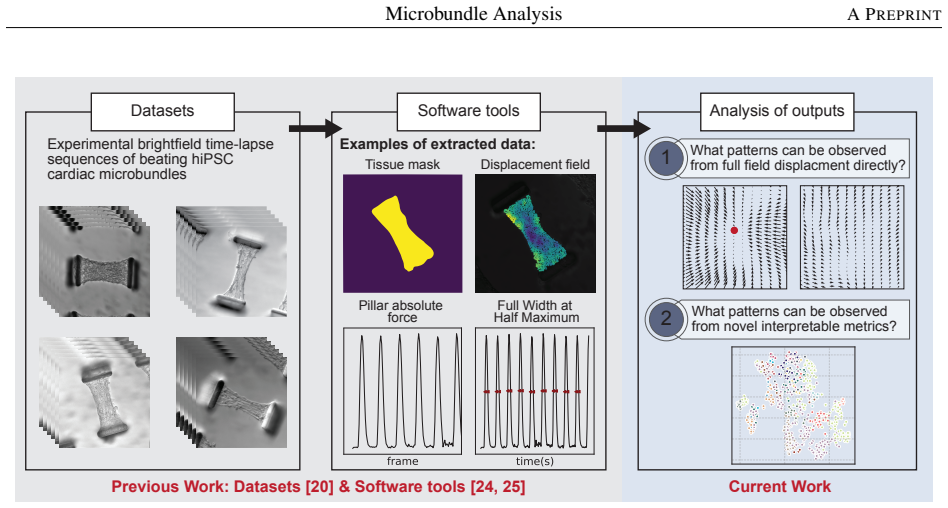
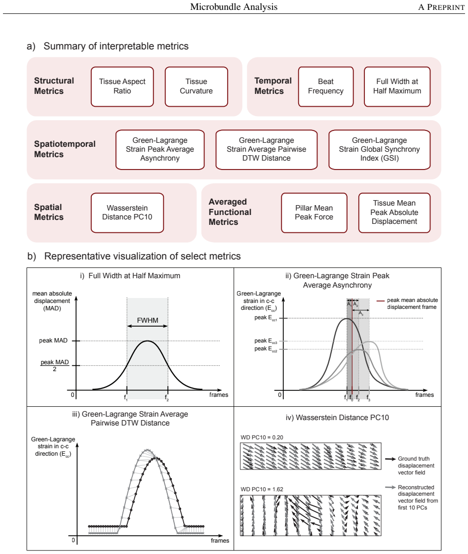
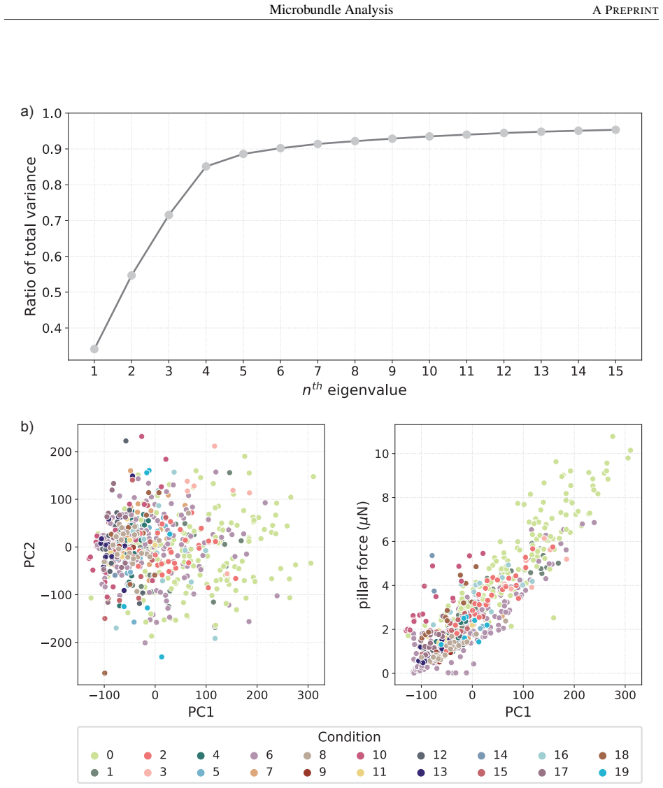
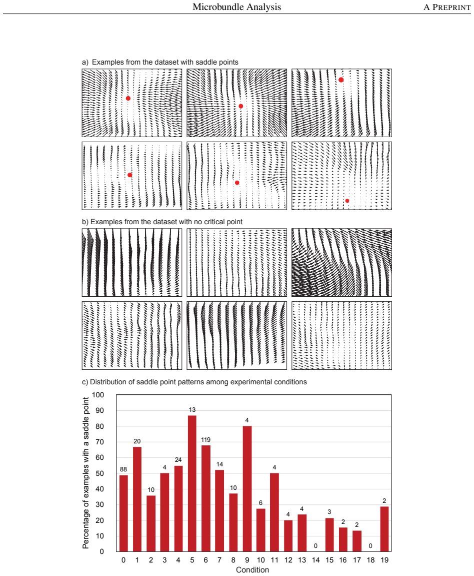
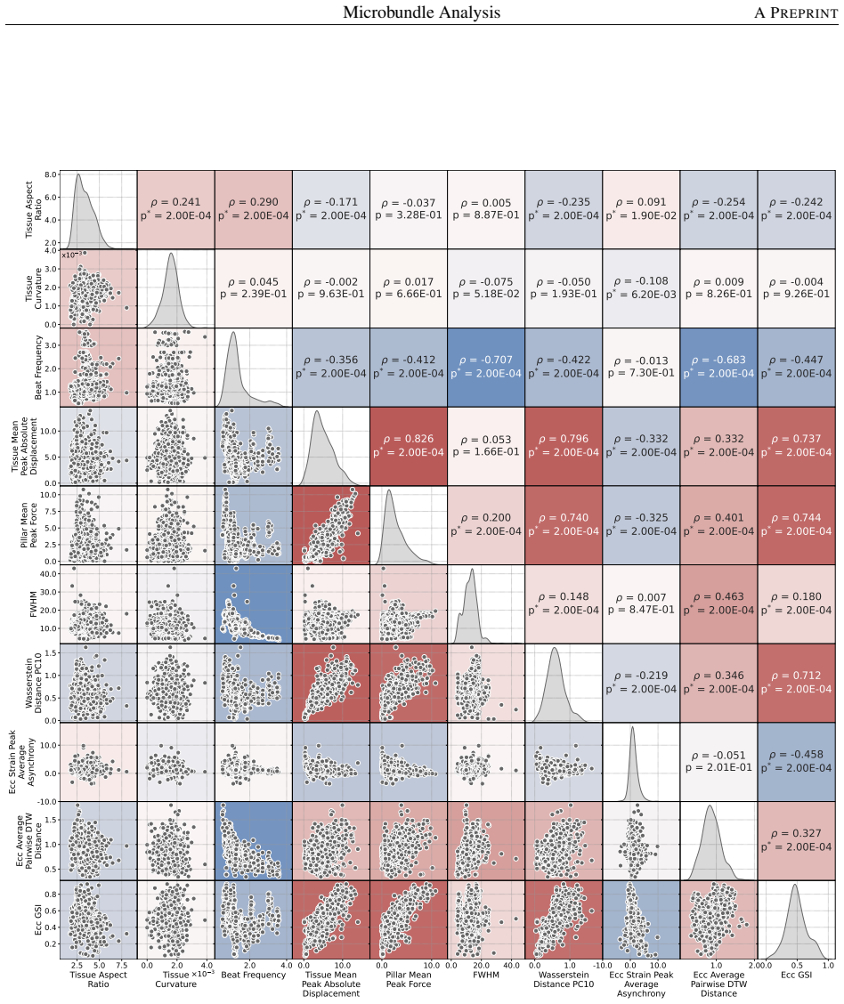
We present an open, scalable computational pipeline for quantifying spatiotemporal contractile dynamics in microscopy videos of human induced pluripotent stem cell-derived cardiac microbundles. Building on open-source tools, we define a suite of 16 interpretable metrics that capture tissue deformation, synchrony, and heterogeneity through full-field displacement tracking, strain reconstruction, and topology-based analysis. Applied to 670 samples across 20 conditions, this reveals continuous phenotypic variation with intra-condition variability often exceeding inter-condition differences, a dominant global isotropic contraction mode, and saddle-type patterns in roughly half the samples.
What carries the argument
The integrated workflow of full-field displacement tracking, strain reconstruction, spatial registration, dimensionality reduction, and topology-based vector-field analysis that produces 16 structural, functional, and spatiotemporal metrics.
If this is right
- The pipeline enables reproducible and scalable analysis of dynamic tissue mechanics across laboratories.
- Redundancy analysis identifies a reduced core set of 10 metrics that retain most informational content while minimizing multicollinearity.
- Contraction in these microbundles is dominated by a global isotropic mode.
- Localized saddle-type deformation patterns occur in approximately half of the samples.
Where Pith is reading between the lines
- Adoption of this pipeline could improve consistency in comparing results from different cardiac tissue engineering experiments.
- The observed continuous variation suggests that focusing on average behaviors per condition may overlook important individual sample differences.
- Similar analysis applied to other dynamic biological tissues might uncover analogous patterns of phenotypic continuity.
Load-bearing premise
The 16 metrics derived from displacement tracking and strain reconstruction accurately represent biologically meaningful contractile dynamics without significant artifacts introduced by imaging conditions or post-processing choices.
What would settle it
Reprocessing the 670 videos with an alternative displacement tracking method or a different set of metrics that results in clear discrete clusters corresponding to the 20 experimental conditions would indicate that the continuous variation finding depends on the specific analysis choices.
Figures


read the original abstract
Brightfield time-lapse imaging is widely used in cardiac tissue engineering, yet the absence of standardized, interpretable analytical frameworks limits reproducibility and cross-platform comparison. We present an open, scalable computational pipeline for quantifying spatiotemporal contractile dynamics in microscopy videos of human induced pluripotent stem cell-derived cardiac microbundles. Building on our open-source tools "MicroBundleCompute" and "MicroBundlePillarTrack," we define a suite of 16 interpretable structural, functional, and spatiotemporal metrics that capture tissue deformation, synchrony, and heterogeneity. The framework integrates full-field displacement tracking, strain reconstruction, spatial registration, dimensionality reduction, and topology-based vector-field analysis within a unified workflow. Applied to a dataset of 670 cardiac microbundles spanning 20 experimental conditions, the pipeline reveals continuous variation in contractile phenotypes rather than discrete condition-specific clustering, with intra-condition variability often exceeding inter-condition differences. Redundancy analysis identifies a reduced core set of 10 metrics that retain most informational content while minimizing multicollinearity. Analysis of denoised displacement fields shows that contraction is dominated by a global isotropic mode, with localized saddle-type deformation patterns present in approximately half of the samples. All software and workflows are released openly to enable reproducible, scalable analysis of dynamic tissue mechanics.
Editorial analysis
A structured set of objections, weighed in public.
Referee Report
Summary. The manuscript introduces an open-source computational pipeline for quantifying spatiotemporal contractile dynamics from brightfield time-lapse videos of hiPSC-derived cardiac microbundles. Building on MicroBundleCompute and MicroBundlePillarTrack, it defines 16 interpretable metrics for tissue deformation, synchrony, and heterogeneity, integrates full-field displacement tracking, strain reconstruction, spatial registration, dimensionality reduction, and topology-based vector-field analysis, and applies the workflow to a dataset of 670 microbundles spanning 20 experimental conditions. The analysis reports continuous phenotypic variation rather than discrete condition-specific clusters, with intra-condition variability often exceeding inter-condition differences; it further identifies a reduced core set of 10 metrics via redundancy analysis and shows that contraction is dominated by a global isotropic mode with localized saddle-type patterns in approximately half the samples. All software and workflows are released openly.
Significance. If the underlying tracking and metrics prove reliable, the work provides a much-needed standardized, reproducible framework for analyzing dynamic mechanics in cardiac tissue engineering, which should improve cross-study comparability and reduce reliance on ad-hoc image analysis. The open release of code and workflows is a clear strength that directly supports reproducibility and adoption. The reported finding of continuous rather than discrete phenotypes, if substantiated, could shift how variability is interpreted in experimental design for engineered cardiac tissues.
major comments (2)
- [Methods] Methods (pipeline description): The full-field displacement tracking (MicroBundlePillarTrack) and strain reconstruction steps lack any reported quantitative validation against synthetic ground-truth displacements or simultaneous fluorescent reference imaging under realistic brightfield conditions (low contrast, pillar boundary effects). Because the central claim of continuous phenotypic variation across 670 samples rests on these metrics faithfully capturing true contractile mechanics without systematic artifacts, the absence of such benchmarks directly undermines confidence in the intra- versus inter-condition variability results.
- [Results] Results (analysis of 670 samples): The claim that phenotypes exhibit continuous variation rather than discrete condition-specific clustering, and that intra-condition variability often exceeds inter-condition differences, is presented without explicit description of the dimensionality reduction technique, clustering algorithm, or statistical tests used to support these conclusions. In addition, no error bars, confidence intervals, or robustness checks on the 16 metrics are reported, making it impossible to assess whether the continuous-variation result is robust to metric selection or registration choices.
minor comments (2)
- [Abstract] Abstract and Results: The statement that contraction is 'dominated by a global isotropic mode' would benefit from a brief quantitative definition (e.g., the fraction of variance explained by the isotropic component or the topology metric threshold) to allow readers to evaluate the claim without consulting the full methods.
- [Methods] Figure captions and Methods: Ensure that all 16 metrics are explicitly named and their formulas or computation steps are cross-referenced to the open-source code repository so that independent replication is straightforward.
Simulated Author's Rebuttal
We thank the referee for their constructive and detailed review of our manuscript. We address each major comment point by point below, indicating where revisions will be made to improve clarity and rigor.
read point-by-point responses
-
Referee: [Methods] Methods (pipeline description): The full-field displacement tracking (MicroBundlePillarTrack) and strain reconstruction steps lack any reported quantitative validation against synthetic ground-truth displacements or simultaneous fluorescent reference imaging under realistic brightfield conditions (low contrast, pillar boundary effects). Because the central claim of continuous phenotypic variation across 670 samples rests on these metrics faithfully capturing true contractile mechanics without systematic artifacts, the absence of such benchmarks directly undermines confidence in the intra- versus inter-condition variability results.
Authors: We acknowledge the referee's concern regarding validation of the core tracking and strain steps. MicroBundlePillarTrack was introduced and quantitatively validated in our prior work, including comparisons to synthetic ground-truth displacements and cross-validation against fluorescent bead imaging under comparable brightfield conditions with pillar boundaries. The current manuscript applies this established pipeline to a new large-scale dataset rather than re-deriving the validation. To directly address the comment, we will add a dedicated Methods subsection that summarizes the prior validation results (with citations), reports any additional consistency checks performed on the 670-sample dataset (e.g., manual annotation agreement on a subset), and discusses potential brightfield-specific artifacts along with how the metrics are designed to mitigate them. This will strengthen confidence in the variability findings. revision: yes
-
Referee: [Results] Results (analysis of 670 samples): The claim that phenotypes exhibit continuous variation rather than discrete condition-specific clustering, and that intra-condition variability often exceeds inter-condition differences, is presented without explicit description of the dimensionality reduction technique, clustering algorithm, or statistical tests used to support these conclusions. In addition, no error bars, confidence intervals, or robustness checks on the 16 metrics are reported, making it impossible to assess whether the continuous-variation result is robust to metric selection or registration choices.
Authors: We agree that the description of the analytical pipeline for the 670-sample results can be made more explicit and transparent. The manuscript references dimensionality reduction and the observation of continuous phenotypic variation without discrete clusters, but does not fully detail the specific methods (e.g., PCA followed by UMAP embedding, quantitative assessment of clustering via silhouette scores on attempted k-means partitions, and variance-component analysis for intra- versus inter-condition comparisons). We will revise the Results section to include a clear, step-by-step account of these techniques with parameters. We will also add error bars or confidence intervals to metric distributions and include a supplementary robustness analysis varying metric subsets and registration parameters. These additions will allow readers to better evaluate the continuous-variation conclusion. revision: yes
Circularity Check
No circularity in pipeline description or empirical claims
full rationale
The paper describes an open-source computational pipeline (MicroBundleCompute, MicroBundlePillarTrack) that defines 16 metrics from displacement tracking and strain analysis, then applies them to 670 experimental samples. No mathematical derivations, first-principles predictions, or equations are presented that reduce by construction to fitted parameters or self-referential inputs. Self-references are to openly released code, which constitutes independent, reproducible support per the guidelines. The central observation of continuous phenotypic variation is a direct empirical result from data processing, not a tautological output. The work is self-contained against external benchmarks via the released software and workflows.
Axiom & Free-Parameter Ledger
Reference graph
Works this paper leans on
-
[1]
The tissue engineering revolution: from bench research to clinical reality
De Chiara F, Ferret-Miñana A, Fernández-Costa JM, Ramón-Azcón J. The tissue engineering revolution: from bench research to clinical reality. Biomedicines. 2024;12(2):453
2024
-
[2]
Tissue Engineering and Regenerative Medicine: Perspectives and Challenges
Hoang VT, Nguyen QT, Phan TTK, Pham TH, Dinh NTH, Anh LPH, et al. Tissue Engineering and Regenerative Medicine: Perspectives and Challenges. MedComm. 2025;6(5):e70192
2025
-
[3]
Robust cardiomyocyte differentiation from human pluripotent stem cells via temporal modulation of canonical Wnt signaling
Lian X, Hsiao C, Wilson G, Zhu K, Hazeltine LB, Azarin SM, et al. Robust cardiomyocyte differentiation from human pluripotent stem cells via temporal modulation of canonical Wnt signaling. Proceedings of the National Academy of Sciences. 2012;109(27):E1848-57
2012
-
[4]
Chemically defined generation of human cardiomyocytes
Burridge PW, Matsa E, Shukla P, Lin ZC, Churko JM, Ebert AD, et al. Chemically defined generation of human cardiomyocytes. Nature methods. 2014;11(8):855-60
2014
-
[5]
Biomaterials for tissue engineering applications and current updates in the field: a comprehensive review
Eldeeb AE, Salah S, Elkasabgy NA. Biomaterials for tissue engineering applications and current updates in the field: a comprehensive review. Aaps Pharmscitech. 2022;23(7):267
2022
-
[6]
Cardiac Tissue Engineering: A Journey from Scaffold Fabrication to In Vitro Characterization
Ketabat F, Alcorn J, Kelly ME, Badea I, Chen X. Cardiac Tissue Engineering: A Journey from Scaffold Fabrication to In Vitro Characterization. Small Science. 2024;4(9):2400079
2024
-
[7]
Scaffold-free cell-based tissue engineering therapies: advances, shortfalls and forecast
De Pieri A, Rochev Y , Zeugolis DI. Scaffold-free cell-based tissue engineering therapies: advances, shortfalls and forecast. NPJ Regenerative medicine. 2021;6(1):18
2021
-
[8]
Challenges and opportunities for the next generation of cardiovascular tissue engineering
Cho S, Discher DE, Leong KW, Vunjak-Novakovic G, Wu JC. Challenges and opportunities for the next generation of cardiovascular tissue engineering. Nature Methods. 2022;19(9):1064-71
2022
-
[9]
Microengineered platforms for characterizing the contractile function of in vitro cardiac models
Dou W, Malhi M, Zhao Q, Wang L, Huang Z, Law J, et al. Microengineered platforms for characterizing the contractile function of in vitro cardiac models. Microsystems & Nanoengineering. 2022;8(1):26
2022
-
[10]
The imaging tsunami: computational opportunities and challenges
Ouyang W, Zimmer C. The imaging tsunami: computational opportunities and challenges. Current Opinion in Systems Biology. 2017;4:105-13
2017
-
[11]
Highlights on advancing frontiers in tissue engineering
Ashammakhi N, GhavamiNejad A, Tutar R, Fricker A, Roy I, Chatzistavrou X, et al. Highlights on advancing frontiers in tissue engineering. Tissue Engineering Part B: Reviews. 2022;28(3):633-64
2022
-
[12]
A review on machine learning approaches in cardiac tissue engineering
Kalkunte N, Cisneros J, Castillo E, Zoldan J. A review on machine learning approaches in cardiac tissue engineering. Frontiers in Biomaterials Science. 2024;3:1358508
2024
-
[13]
Opportunities and challenges in cardiac tissue engineering from an analysis of two decades of advances
Zhuang RZ, Lock R, Liu B, Vunjak-Novakovic G. Opportunities and challenges in cardiac tissue engineering from an analysis of two decades of advances. Nature Biomedical Engineering. 2022;6(4):327-38
2022
-
[14]
Induced pluripotent stem cell-derived cardiomyocyte in vitro models: benchmarking progress and ongoing challenges
Ewoldt JK, DePalma SJ, Jewett ME, Karakan MÇ, Lin YM, Mir Hashemian P, et al. Induced pluripotent stem cell-derived cardiomyocyte in vitro models: benchmarking progress and ongoing challenges. Nature methods. 2025;22(1):24-40
2025
-
[15]
Current challenges in imaging the mechanical properties of tissue engineered grafts
Luo L, Okur KE, Bagnaninchi PO, El Haj AJ. Current challenges in imaging the mechanical properties of tissue engineered grafts. Frontiers in Biomaterials Science. 2024;3:1323763
2024
-
[16]
Advances in spatial transcriptomics and related data analysis strategies
Du J, Yang YC, An ZJ, Zhang MH, Fu XH, Huang ZF, et al. Advances in spatial transcriptomics and related data analysis strategies. Journal of translational medicine. 2023;21(1):330
2023
-
[17]
A practical guide to spatial transcriptomics: lessons from over 1000 samples
Grases D, Porta-Pardo E. A practical guide to spatial transcriptomics: lessons from over 1000 samples. Trends in Biotechnology. 2025
2025
-
[18]
Cardiac tissue engineering: state of the art
Hirt MN, Hansen A, Eschenhagen T. Cardiac tissue engineering: state of the art. Circulation research. 2014;114(2):354-67
2014
-
[19]
REMBI: Recommended Metadata for Biological Images—enabling reuse of microscopy data in biology
Sarkans U, Chiu W, Collinson L, Darrow MC, Ellenberg J, Grunwald D, et al. REMBI: Recommended Metadata for Biological Images—enabling reuse of microscopy data in biology. Nature methods. 2021;18(12):1418-22
2021
-
[20]
FibroTUG platforms: Time-lapse microscopy dataset of engineered cardiac microbundles; 2024
Kobeissi H, Gao X, DePalma SJ, Ewoldt JK, Wang MC, Das SL, et al.. FibroTUG platforms: Time-lapse microscopy dataset of engineered cardiac microbundles; 2024. Available from:10.5061/dryad.3r2280gqd
-
[21]
Strain gauge platforms: Time-lapse microscopy dataset of engineered cardiac microbundles; 2024
Kobeissi H, Gao X, DePalma SJ, Ewoldt JK, Wang MC, Das SL, et al.. Strain gauge platforms: Time-lapse microscopy dataset of engineered cardiac microbundles; 2024. Available from:10.5061/dryad.sqv9s4nbg
-
[22]
EMPIAR: the electron microscopy public image archive
Iudin A, Korir PK, Somasundharam S, Weyand S, Cattavitello C, Fonseca N, et al. EMPIAR: the electron microscopy public image archive. Nucleic Acids Research. 2023;51(D1):D1503-11
2023
-
[23]
SarcGraph - 2D Cardiac Muscle Bundle
Mohammadzadeh S, Tsan Yc, Kobeissi H, Lejeune E, Helms A. SarcGraph - 2D Cardiac Muscle Bundle. Harvard Dataverse; 2025. Available from:https://doi.org/10.7910/DVN/GHMKWJ. 32 Microbundle AnalysisA PREPRINT
-
[24]
MicroBundleCompute: Auto- mated segmentation, tracking, and analysis of subdomain deformation in cardiac microbundles
Kobeissi H, Jilberto J, Karakan MÇ, Gao X, DePalma SJ, Das SL, et al. MicroBundleCompute: Auto- mated segmentation, tracking, and analysis of subdomain deformation in cardiac microbundles. Plos one. 2024;19(3):e0298863. PMID: 38530829
2024
-
[25]
MicroBundlePillarTrack: A Python package for automated segmentation, tracking, and analysis of pillar deflection in cardiac microbundles
Kobeissi H, Gao X, DePalma SJ, Ewoldt JK, Wang MC, Das SL, et al. MicroBundlePillarTrack: A Python package for automated segmentation, tracking, and analysis of pillar deflection in cardiac microbundles. Microp- ublication Biology. 2024;2024. PMID: 39114859
2024
-
[26]
Automated video-based analysis of contractility and calcium flux in human-induced pluripotent stem cell-derived cardiomyocytes cultured over different spatial scales
Huebsch N, Loskill P, Mandegar MA, Marks NC, Sheehan AS, Ma Z, et al. Automated video-based analysis of contractility and calcium flux in human-induced pluripotent stem cell-derived cardiomyocytes cultured over different spatial scales. Tissue Engineering Part C: Methods. 2015;21(5):467-79
2015
-
[27]
MUSCLEMOTION: a versatile open software tool to quantify cardiomyocyte and cardiac muscle contraction in vitro and in vivo
Sala L, Van Meer BJ, Tertoolen LG, Bakkers J, Bellin M, Davis RP, et al. MUSCLEMOTION: a versatile open software tool to quantify cardiomyocyte and cardiac muscle contraction in vitro and in vivo. Circulation research. 2018;122(3):e5-e16
2018
-
[28]
Engineering of human cardiac muscle electromechanically matured to an adult-like phenotype
Ronaldson-Bouchard K, Yeager K, Teles D, Chen T, Ma S, Song L, et al. Engineering of human cardiac muscle electromechanically matured to an adult-like phenotype. Nature protocols. 2019;14(10):2781-817
2019
-
[29]
SarcTrack: an adaptable software tool for efficient large-scale analysis of sarcomere function in hiPSC-cardiomyocytes
Toepfer CN, Sharma A, Cicconet M, Garfinkel AC, Mücke M, Neyazi M, et al. SarcTrack: an adaptable software tool for efficient large-scale analysis of sarcomere function in hiPSC-cardiomyocytes. Circulation research. 2019;124(8):1172-83
2019
-
[30]
CalTrack: high-throughput automated calcium transient analysis in cardiomyocytes
Psaras Y , Margara F, Cicconet M, Sparrow AJ, Repetti GG, Schmid M, et al. CalTrack: high-throughput automated calcium transient analysis in cardiomyocytes. Circulation research. 2021;129(2):326-41
2021
-
[31]
Physiologic biomechanics enhance reproducible contractile development in a stem cell derived cardiac muscle platform
Tsan YC, DePalma SJ, Zhao YT, Capilnasiu A, Wu YW, Elder B, et al. Physiologic biomechanics enhance reproducible contractile development in a stem cell derived cardiac muscle platform. Nature Communications. 2021;12(1):6167
2021
-
[32]
milliPillar: a platform for the generation and real-time assessment of human engineered cardiac tissues
Tamargo MA, Nash TR, Fleischer S, Kim Y , Vila OF, Yeager K, et al. milliPillar: a platform for the generation and real-time assessment of human engineered cardiac tissues. ACS biomaterials science & engineering. 2021;7(11):5215-29
2021
-
[33]
Automated assessment of human engineered heart tissues using deep learning and template matching for segmentation and tracking
Rivera-Arbeláez JM, Keekstra D, Cofiño-Fabres C, Boonen T, Dostanic M, Ten Den SA, et al. Automated assessment of human engineered heart tissues using deep learning and template matching for segmentation and tracking. Bioengineering & Translational Medicine. 2023;8(3):e10513
2023
-
[34]
FORCETRACKER: A versatile tool for standardized assessment of tissue contractile properties in 3D Heart-on-Chip platforms
Rivera-Arbeláez JM, Dostani´c M, Windt LM, Stein JM, Cofiño-Fabres C, Boonen T, et al. FORCETRACKER: A versatile tool for standardized assessment of tissue contractile properties in 3D Heart-on-Chip platforms. PloS one. 2025;20(2):e0314985
2025
-
[35]
Quantifying HiPSC-CM structural organization at scale with deep learning- enhanced SarcGraph
Mohammadzadeh S, Lejeune E. Quantifying HiPSC-CM structural organization at scale with deep learning- enhanced SarcGraph. PLOS Computational Biology. 2025;21(10):e1013436
2025
-
[36]
SarcGraph for High-Throughput Regional Analysis of Sarcomere Organization and Contractile Function in 2D Cardiac Muscle Bundles
Mohammadzadeh S, Tsan YC, Renberg A, Kobeissi H, Helms A, Lejeune E. SarcGraph for High-Throughput Regional Analysis of Sarcomere Organization and Contractile Function in 2D Cardiac Muscle Bundles. arXiv preprint arXiv:251111913. 2025
2025
-
[37]
Matrix architecture and mechanics regulate myofibril organization, costamere assembly, and contractility in engineered myocardial microtissues
DePalma SJ, Jilberto J, Stis AE, Huang DD, Lo J, Davidson CD, et al. Matrix architecture and mechanics regulate myofibril organization, costamere assembly, and contractility in engineered myocardial microtissues. Advanced Science. 2024;11(47):2309740. PMID: 39558513
2024
-
[38]
The FAIR Guiding Principles for scientific data management and stewardship
Wilkinson MD, Dumontier M, Aalbersberg IJ, Appleton G, Axton M, Baak A, et al. The FAIR Guiding Principles for scientific data management and stewardship. Scientific data. 2016;3(1):1-9
2016
-
[39]
Open science
Bertram MG, Sundin J, Roche DG, Sánchez-Tójar A, Thoré ES, Brodin T. Open science. Current biology. 2023;33(15):R792-7
2023
-
[40]
Open science by design: Realizing a vision for 21st century research
National Academies of Sciences and Medicine and Global Affairs and Board on Research Data and Committee on Toward an Open Science Enterprise. Open science by design: Realizing a vision for 21st century research. The National Academies Press. 2018
2018
-
[41]
Building a FAIR image data ecosystem for microscopy communities
Kemmer I, Keppler A, Serrano-Solano B, Rybina A, Özdemir B, Bischof J, et al. Building a FAIR image data ecosystem for microscopy communities. Histochemistry and Cell Biology. 2023;160(3):199-209
2023
-
[42]
MITI minimum information guidelines for highly multiplexed tissue images
Schapiro D, Yapp C, Sokolov A, Reynolds SM, Chen Y A, Sudar D, et al. MITI minimum information guidelines for highly multiplexed tissue images. Nature methods. 2022;19(3):262-7
2022
-
[43]
FAIR high content screening in bioimaging
Hosseini R, Vlasveld M, Willemse J, van de Water B, Le Dévédec SE, Wolstencroft KJ. FAIR high content screening in bioimaging. Scientific Data. 2023;10(1):462. 33 Microbundle AnalysisA PREPRINT
2023
-
[44]
Myofibroblast activation in syn- thetic fibrous matrices composed of dextran vinyl sulfone
Davidson CD, Jayco DKP, Matera DL, DePalma SJ, Hiraki HL, Wang WY , et al. Myofibroblast activation in syn- thetic fibrous matrices composed of dextran vinyl sulfone. Acta biomaterialia. 2020;105:78-86. PMID:31945504
2020
-
[45]
Microenvironmental determinants of organized iPSC- cardiomyocyte tissues on synthetic fibrous matrices
DePalma SJ, Davidson CD, Stis AE, Helms AS, Baker BM. Microenvironmental determinants of organized iPSC- cardiomyocyte tissues on synthetic fibrous matrices. Biomaterials science. 2021;9(1):93-107. PMID:33325920
2021
-
[46]
A microfabricated platform to measure and manipulate the mechanics of engineered cardiac microtissues
Boudou T, Legant WR, Mu A, Borochin MA, Thavandiran N, Radisic M, et al. A microfabricated platform to measure and manipulate the mechanics of engineered cardiac microtissues. Tissue Engineering Part A. 2012;18(9-10):910-9
2012
-
[47]
Hypertrophic cardiomyopathy– associated mutations drive stromal activation via EGFR-mediated paracrine signaling
Ewoldt JK, Wang MC, McLellan MA, Cloonan PE, Chopra A, Gorham J, et al. Hypertrophic cardiomyopathy– associated mutations drive stromal activation via EGFR-mediated paracrine signaling. Science Advances. 2024;10(42):eadi6927
2024
-
[48]
A platform for generation of chamber- specific cardiac tissues and disease modeling
Zhao Y , Rafatian N, Feric NT, Cox BJ, Aschar-Sobbi R, Wang EY , et al. A platform for generation of chamber- specific cardiac tissues and disease modeling. Cell. 2019;176(4):913-27
2019
-
[49]
Geometry and length control of 3D engineered heart tissues using direct laser writing
Karakan MÇ, Ewoldt JK, Segarra AJ, Sundaram S, Wang MC, White AE, et al. Geometry and length control of 3D engineered heart tissues using direct laser writing. Lab on a Chip. 2024;24(6):1685-701
2024
-
[50]
Good features to track
Shi J, et al. Good features to track. In: 1994 Proceedings of IEEE conference on computer vision and pattern recognition. IEEE; 1994. p. 593-600
1994
-
[51]
An Iterative Image Registration Technique with an Application to Stereo Vision
Lucas BD, Kanade T. An Iterative Image Registration Technique with an Application to Stereo Vision. In: Proceedings of the 7th International Joint Conference on Artificial Intelligence - V olume 2. IJCAI’81. San Francisco, CA, USA: Morgan Kaufmann Publishers Inc.; 1981. p. 674–679
1981
-
[52]
Pyramidal implementation of the affine lucas kanade feature tracker description of the algorithm
Bouguet JY , et al. Pyramidal implementation of the affine lucas kanade feature tracker description of the algorithm. Intel corporation. 2001;5(1-10):4
2001
-
[53]
Light-driven biological actuators to probe the rheology of 3D microtissues
Méry A, Ruppel A, Revilloud J, Balland M, Cappello G, Boudou T. Light-driven biological actuators to probe the rheology of 3D microtissues. Nature Communications. 2023;14(1):717
2023
-
[54]
Dense optical flow software to quantify cellular contractility
Scalzo S, Afonso MQ, da Fonseca NJ, Jesus IC, Alves AP, Mendonça CA, et al. Dense optical flow software to quantify cellular contractility. Cell Reports Methods. 2021;1(4)
2021
-
[55]
Deformation gradients for continuum mechanical analysis of atomistic simulations
Zimmerman JA, Bammann DJ, Gao H. Deformation gradients for continuum mechanical analysis of atomistic simulations. International Journal of Solids and Structures. 2009;46(2):238-53
2009
-
[56]
Estimation of the deformation gradient tensor by particle tracking near a free boundary with quantified error
Benkley T, Li C, Kolinski J. Estimation of the deformation gradient tensor by particle tracking near a free boundary with quantified error. Experimental Mechanics. 2023;63(7):1255-70
2023
-
[57]
SciPy 1.0: fundamental algorithms for scientific computing in Python
Virtanen P, Gommers R, Oliphant TE, Haberland M, Reddy T, Cournapeau D, et al. SciPy 1.0: fundamental algorithms for scientific computing in Python. Nature methods. 2020;17(3):261-72
2020
-
[58]
scipy.interpolate.RBFInterpolator — SciPy v1.13.1 Manual; 2024
SciPy Developers. scipy.interpolate.RBFInterpolator — SciPy v1.13.1 Manual; 2024. Accessed on 2025- 11-19. Available from: https://docs.scipy.org/doc/scipy-1.13.1/reference/generated/scipy. interpolate.RBFInterpolator.html#scipy.interpolate.RBFInterpolator
2024
-
[59]
On lines and planes of closest fit to systems of points in space
Pearson K. On lines and planes of closest fit to systems of points in space. The London, Edinburgh, and Dublin philosophical magazine and journal of science. 1901;2(11):559-72
1901
-
[60]
Relations between two sets of variates
Hotelling H. Relations between two sets of variates. In: Breakthroughs in statistics: methodology and distribution. Springer; 1992. p. 162-90
1992
-
[61]
Principal component analysis of dynamic relative displacement fields estimated from MR images
Abney TM, Feng Y , Pless R, Okamoto RJ, Genin GM, Bayly PV . Principal component analysis of dynamic relative displacement fields estimated from MR images. PLoS One. 2011;6(7):e22063
2011
-
[62]
Computation of full-field strains using principal component analysis
Grama S, Subramanian S. Computation of full-field strains using principal component analysis. Experimental Mechanics. 2014;54(6):913-33
2014
-
[63]
Improving three-dimensional mechanical imaging of breast lesions with principal component analysis
Tyagi M, Wang Y , Hall TJ, Barbone PE, Oberai AA. Improving three-dimensional mechanical imaging of breast lesions with principal component analysis. Medical physics. 2017;44(8):4194-203
2017
-
[64]
Data-driven cardiovascular flow modelling: examples and opportunities
Arzani A, Dawson ST. Data-driven cardiovascular flow modelling: examples and opportunities. Journal of the Royal Society Interface. 2021;18(175):20200802
2021
-
[65]
MPCA: Multilinear principal component analysis of tensor objects
Lu H, Plataniotis KN, Venetsanopoulos AN. MPCA: Multilinear principal component analysis of tensor objects. IEEE transactions on Neural Networks. 2008;19(1):18-39
2008
-
[66]
A survey of multilinear subspace learning for tensor data
Lu H, Plataniotis KN, Venetsanopoulos AN. A survey of multilinear subspace learning for tensor data. Pattern Recognition. 2011;44(7):1540-51. 34 Microbundle AnalysisA PREPRINT
2011
-
[67]
Scikit-learn: Machine Learning in Python
Pedregosa F, Varoquaux G, Gramfort A, Michel V , Thirion B, Grisel O, et al. Scikit-learn: Machine Learning in Python. Journal of Machine Learning Research. 2011;12:2825-30
2011
-
[68]
In: Berrar DP, Dubitzky W, Granzow M, editors
Wall ME, Rechtsteiner A, Rocha LM. In: Berrar DP, Dubitzky W, Granzow M, editors. Singular Value Decomposition and Principal Component Analysis. Boston, MA: Springer US; 2003. p. 91-109. Available from: https://doi.org/10.1007/0-306-47815-3_5
-
[69]
Flow field analysis of a turbulent boundary layer over a riblet surface
Lee SJ, Lee SH. Flow field analysis of a turbulent boundary layer over a riblet surface. Experiments in fluids. 2001;30(2):153-66
2001
-
[70]
Analysis and interpretation of instantaneous turbulent velocity fields
Adrian RJ, Christensen KT, Liu ZC. Analysis and interpretation of instantaneous turbulent velocity fields. Experiments in fluids. 2000;29(3):275-90
2000
-
[71]
Representation and display of vector field topology in fluid flow data sets
Helman J, Hesselink L. Representation and display of vector field topology in fluid flow data sets. Computer. 1989;22(08):27-36
1989
-
[72]
Visualizing vector field topology in fluid flows
Helman JL, Hesselink L. Visualizing vector field topology in fluid flows. IEEE Computer Graphics and Applications. 1991;11(3):36-46
1991
-
[73]
Critical points in flow patterns
Perry AE, Fairlie B. Critical points in flow patterns. In: Advances in geophysics. vol. 18. Elsevier; 1975. p. 299-315
1975
-
[74]
A description of eddying motions and flow patterns using critical-point concepts
Perry AE, Chong MS. A description of eddying motions and flow patterns using critical-point concepts. Annual Review of Fluid Mechanics. 1987;19:125-55
1987
-
[75]
Vector field analysis for oriented patterns
Shu CF, Jain RC. Vector field analysis for oriented patterns. IEEE Transactions on Pattern Analysis and Machine Intelligence. 1994;16(9):946-50
1994
-
[76]
Finding and classifying critical points of 2D vector fields: a cell-oriented approach using group theory
Effenberger F, Weiskopf D. Finding and classifying critical points of 2D vector fields: a cell-oriented approach using group theory. Computing and Visualization in Science. 2010;13(8):377-96
2010
-
[77]
Detection and analysis of spatiotemporal patterns in brain activity
Townsend RG, Gong P. Detection and analysis of spatiotemporal patterns in brain activity. PLoS computational biology. 2018;14(12):e1006643
2018
-
[78]
Wave-like dopamine dynamics as a mechanism for spatiotemporal credit assignment
Hamid AA, Frank MJ, Moore CI. Wave-like dopamine dynamics as a mechanism for spatiotemporal credit assignment. Cell. 2021;184(10):2733-49
2021
-
[79]
Interacting spiral wave patterns underlie complex brain dynamics and are related to cognitive processing
Xu Y , Long X, Feng J, Gong P. Interacting spiral wave patterns underlie complex brain dynamics and are related to cognitive processing. Nature human behaviour. 2023;7(7):1196-215
2023
-
[80]
Mapping transcriptomic vector fields of single cells
Qiu X, Zhang Y , Martin-Rufino JD, Weng C, Hosseinzadeh S, Yang D, et al. Mapping transcriptomic vector fields of single cells. Cell. 2022;185(4):690-711
2022
discussion (0)
Sign in with ORCID, Apple, or X to comment. Anyone can read and Pith papers without signing in.