Recognition: unknown
A 3D SAM-Based Progressive Prompting Framework for Multi-Task Segmentation of Radiotherapy-induced Normal Tissue Injuries in Limited-Data Settings
Pith reviewed 2026-05-10 14:00 UTC · model grok-4.3
The pith
A 3D SAM framework progressively adds text, dose-guided box, and click prompts to segment radiotherapy-induced tissue injuries accurately despite limited labels.
A machine-rendered reading of the paper's core claim, the machinery that carries it, and where it could break.
Core claim
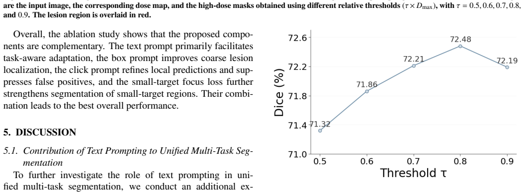
The 3D SAM-based progressive prompting framework, which incorporates text prompts for task-aware adaptation, dose-guided box prompts for coarse localization, and click prompts for iterative refinement together with a small-target focus loss, enables reliable multi-task segmentation of radiotherapy-induced normal tissue injuries across ORN, CE, and CRN in limited-data settings and outperforms prior state-of-the-art methods.
What carries the argument
The 3D SAM-based progressive prompting framework that chains text prompts, dose-guided box prompts, and click prompts with a small-target focus loss to handle task heterogeneity and small lesions.
If this is right
- The method supports simultaneous segmentation of three different injury types within one model rather than requiring separate networks.
- Dose information integrated via box prompts improves coarse localization before fine refinement begins.
- The small-target focus loss reduces errors on sparse or small lesions that standard losses often miss.
- Progressive addition of prompts allows the same backbone to adapt to varying lesion characteristics without full retraining.
Where Pith is reading between the lines
- The framework could lower the annotation burden for other rare or heterogeneous medical segmentation problems by reusing a single 3D SAM model with prompt layers.
- Interactive click refinement opens a path toward clinician-in-the-loop tools that start from automated outputs and require only minimal corrections.
- Because the approach explicitly uses radiation dose maps, it may integrate directly into existing radiotherapy planning software for longitudinal injury tracking.
Load-bearing premise
The curated dataset adequately represents real-world clinical variability and the prompting strategy plus small-target loss will generalize without overfitting to the limited annotations or imaging protocols used in the study.
What would settle it
An independent test set of head-and-neck radiotherapy injury scans, drawn from a different scanner or patient population, on which the method fails to match or exceed the segmentation accuracy of current leading approaches would disprove the generalization claim.
Figures


read the original abstract
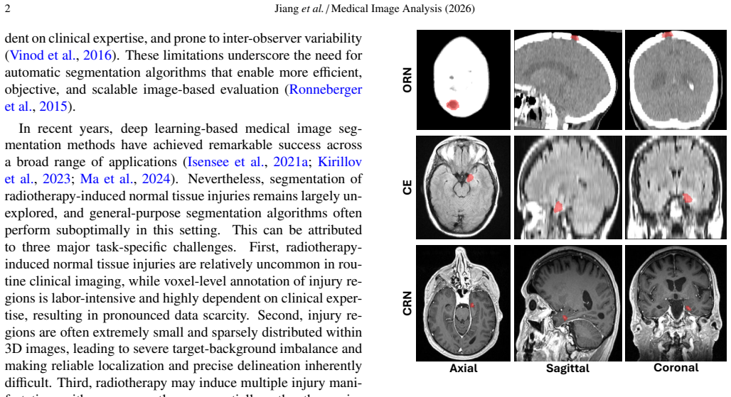
Radiotherapy-induced normal tissue injury is a clinically important complication, and accurate segmentation of injury regions from medical images could facilitate disease assessment, treatment planning, and longitudinal monitoring. However, automatic segmentation of these lesions remains largely unexplored because of limited voxel-level annotations and substantial heterogeneity across injury types, lesion size, and imaging modality. To address this gap, we curate a dedicated head-and-neck radiotherapy-induced normal tissue injury dataset covering three manifestations: osteoradionecrosis (ORN), cerebral edema (CE), and cerebral radiation necrosis (CRN). We further propose a 3D SAM-based progressive prompting framework for multi-task segmentation in limited-data settings. The framework progressively incorporates three complementary prompts: text prompts for task-aware adaptation, dose-guided box prompts for coarse localization, and click prompts for iterative refinement. A small-target focus loss is introduced to improve local prediction and boundary delineation for small and sparse lesions. Experiments on ORN, CE, and CRN demonstrate that the proposed method achieves reliable segmentation performance across diverse injury types and outperforms state-of-the-art methods.
Editorial analysis
A structured set of objections, weighed in public.
Referee Report
Summary. The manuscript curates a dedicated head-and-neck dataset covering osteoradionecrosis (ORN), cerebral edema (CE), and cerebral radiation necrosis (CRN) and introduces a 3D SAM-based progressive prompting framework that sequentially applies text prompts for task-aware adaptation, dose-guided box prompts for coarse localization, and click prompts for iterative refinement, together with a small-target focus loss, for multi-task segmentation of radiotherapy-induced injuries under limited annotations. Experiments on the curated collection are stated to demonstrate reliable performance across injury types and outperformance relative to state-of-the-art methods.
Significance. If the empirical results prove robust under proper validation, the work supplies a timely engineering solution for segmenting rare, heterogeneous lesions where voxel-level labels are scarce, potentially aiding clinical assessment, treatment planning, and longitudinal monitoring in radiotherapy. The release of a multi-injury dataset constitutes a concrete resource contribution.
major comments (2)
- [Abstract and Experiments] The central empirical claim of reliable multi-task segmentation and SOTA outperformance rests on internal experiments whose quantitative support (dataset sizes, cross-validation folds, statistical tests, exact metrics) is not supplied in the abstract or method overview, preventing verification of the headline result or assessment of post-hoc tuning risk.
- [Dataset Curation and Evaluation] The broader claim of utility in limited-data clinical settings is load-bearing on dataset representativeness and generalization; however, the evaluation uses a single self-curated collection with no external validation cohort, multi-center data, or prospective test set, leaving open the possibility that observed gains are idiosyncratic to the collection site's lesion-size distribution, imaging protocols, or annotation patterns.
minor comments (1)
- [Abstract] The abstract would be strengthened by inclusion of at least one key quantitative result (e.g., mean Dice or surface distance) to substantiate the outperformance statement.
Simulated Author's Rebuttal
We thank the referee for the constructive feedback on the presentation of results and the scope of our evaluation. We have revised the manuscript to improve clarity and transparency while preserving the core contributions of the curated dataset and the progressive prompting framework.
read point-by-point responses
-
Referee: [Abstract and Experiments] The central empirical claim of reliable multi-task segmentation and SOTA outperformance rests on internal experiments whose quantitative support (dataset sizes, cross-validation folds, statistical tests, exact metrics) is not supplied in the abstract or method overview, preventing verification of the headline result or assessment of post-hoc tuning risk.
Authors: We agree that the abstract and method overview would benefit from explicit quantitative summaries to enable immediate verification. The full experimental details—including per-task case counts, the cross-validation strategy, statistical significance testing, and exact metric values—are reported in the Experiments section. To address the concern directly, we will revise the abstract to include key summary statistics (dataset sizes and primary performance metrics) and add a concise paragraph in the method overview describing the validation protocol and any hyperparameter tuning procedures. This change improves accessibility without altering the reported results. revision: yes
-
Referee: [Dataset Curation and Evaluation] The broader claim of utility in limited-data clinical settings is load-bearing on dataset representativeness and generalization; however, the evaluation uses a single self-curated collection with no external validation cohort, multi-center data, or prospective test set, leaving open the possibility that observed gains are idiosyncratic to the collection site's lesion-size distribution, imaging protocols, or annotation patterns.
Authors: We acknowledge that external validation would provide stronger evidence of generalizability. The dataset is the first dedicated collection spanning ORN, CE, and CRN, assembled from a single center due to the rarity of these annotated cases. Internal validation via cross-validation and stratified splits was used to assess performance across injury types. In the revised manuscript we will add an explicit limitations subsection that discusses potential site-specific factors (lesion distribution, protocols, annotation style) and emphasizes that public release of the dataset is intended to enable community-driven external validation and multi-center studies. We cannot introduce new external data in this revision. revision: partial
- Absence of an external validation cohort or multi-center data, which cannot be supplied without additional data collection outside the current study scope.
Circularity Check
No circularity: empirical engineering framework with no load-bearing derivations or self-referential predictions
full rationale
The paper presents a 3D SAM-based progressive prompting framework for multi-task segmentation, including text prompts, dose-guided box prompts, click prompts, and a small-target focus loss. It curates a new head-and-neck dataset for ORN, CE, and CRN and reports experimental outperformance on that data. No equations, first-principles derivations, or predictions are claimed that reduce to the inputs by construction. There are no self-citations invoked as uniqueness theorems or ansatzes that bear the central result. The claims rest on empirical evaluation rather than any chain that collapses to fitted parameters or renamed inputs. This is a standard engineering contribution evaluated on a self-curated collection; the absence of mathematical reduction means the derivation chain is self-contained and non-circular.
Axiom & Free-Parameter Ledger
Reference graph
Works this paper leans on
-
[1]
A novel focal tversky loss function with improved attention u-net for lesion segmentation
A novel focal tversky loss function with improved attention u-net for lesion segmentation. arXiv preprint arXiv:1810.07842 . Alsentzer, E., Murphy, J., Boag, W., Weng, W.H., Jin, D., Naumann, T., McDer- mott, M.,
-
[2]
arXiv preprint arXiv:1904.03323
Publicly available clinical bert embeddings. arXiv preprint arXiv:1904.03323 . Barua, S., Elhalawani, H., V olpe, S., et al.,
-
[3]
Frontiers in Oncology 11, 689468
Computed tomography ra- diomics kinetics as early imaging correlates of osteoradionecrosis in oropha- ryngeal cancer patients. Frontiers in Oncology 11, 689468. doi:10.3389/ fonc.2021.689468. Bentzen, S.M., Constine, L.S., Deasy, J.O., Eisbruch, A., Jackson, A., Marks, L.B., Ten Haken, R.K., Yorke, E.D.,
-
[4]
Xcoop: Explainable prompt learning for computer-aided diagnosis via concept-guided context optimization, in: Medical Image Computing and Computer-Assisted Intervention (MICCAI), pp. 773–783. doi:10.1007/978-3-031-72390-2_72. Chen, C., Cui, J., Ma, X., Qian, X., Song, K., Zuo, W., Yan, Y ., Jin, M., Guo, Y ., Yin, Y ., et al.,
-
[5]
Computers in Biology and Medicine 181, 108984
Ma-sam: Modality-agnostic sam adaptation for 3d medical image segmentation. Computers in Biology and Medicine 181, 108984. doi:10.1016/j.compbiomed.2024.108984. Cubero, I., Esteban, P., et al.,
-
[6]
arXiv preprint arXiv:2404.14750
Grounded knowledge-enhanced medical vision- language pre-training for chest x-ray. arXiv preprint arXiv:2404.14750 . Denner, S., Bujotzek, M., Bounias, D., Zimmerer, D., Stock, R., J"ager, P.F., Maier-Hein, K.,
-
[7]
arXiv preprint arXiv:2408.15802
Visual prompt engineering for medical vision lan- guage models in radiology. arXiv preprint arXiv:2408.15802 . Dona Lemus, O.M., Cao, M., Cai, B., Cummings, M., Zheng, D.,
-
[8]
doi:10.3390/cancers16061206. Dosovitskiy, A., Beyer, L., Kolesnikov, A., Weissenborn, D., Zhai, X., Un- terthiner, T., Dehghani, M., Minderer, M., Heigold, G., Gelly, S., Uszkoreit, J., Houlsby, N.,
-
[9]
Clinical evaluation of deep learning and atlas- based auto-segmentation for critical organs at risk in radiation therapy. Jour- nal of Medical Radiation Sciences 70, 15–25. doi:10.1002/jmrs.618. Guo, Z., Xie, Y ., Zhang, Y ., Wang, S., Xia, T., Wang, F., Cheng, J., Chen, H., Li, X.,
-
[10]
Computers in Biology and Medicine 172, 108286
Clicksam: Fine-tuning segment anything model using click prompts for ultrasound image segmentation. Computers in Biology and Medicine 172, 108286. doi:10.1016/j.compbiomed.2024.108286. Hatamizadeh, A., Tang, Y ., Nath, V ., Yang, D., Myronenko, A., Landman, B., Roth, H., Xu, D., 2022a. Unetr: Transformers for 3d medical image segmen- tation, in: Proceedin...
-
[11]
Segment anything. arXiv preprint arXiv:2304.02643 . Lagedamon, V ., Leni, P.E., Gschwind, R.,
work page internal anchor Pith review arXiv
-
[12]
Can- cer/Radiothérapie 28, 402–414
Deep learning applied to dose prediction in external radiation therapy: A narrative review. Can- cer/Radiothérapie 28, 402–414. doi:10.1016/j.canrad.2024.03.005. Lai, H., Yao, Q., Jiang, Z., Wang, R., He, Z., Tao, X., Zhou, S.K.,
-
[13]
Computerized Medical Imaging and Graphics 99, 102092
A contrastive consistency semi-supervised left atrium segmentation model. Computerized Medical Imaging and Graphics 99, 102092. doi:10.1016/j.compmedimag.2022. 102092. Liu, Z., Lin, Y ., Cao, Y ., Hu, H., Wei, Y ., Zhang, Z., Lin, S., Guo, B.,
-
[14]
Multiscale Vision Transformers , isbn =
Swin transformer: Hierarchical vision transformer using shifted windows, in: Proceedings of the IEEE/CVF International Conference on Computer Vi- sion (ICCV), pp. 10012–10022. doi:10.1109/ICCV48922.2021.00986. Ma, J., He, Y ., Li, F., Han, L., You, C., Wang, B., et al.,
-
[15]
V-net: Fully convolutional neural networks for volumetric medical image segmentation, in: 2016 Fourth International Conference on 3D Vision (3DV), IEEE. pp. 565–571. doi:10.1109/3DV.2016.79. Myronenko, A.,
-
[16]
arXiv preprint arXiv:1810.11654
3d mri brain tumor segmentation using autoencoder regularization. arXiv preprint arXiv:1810.11654 . Pandey, S., Singh, P.R., Tian, J.,
-
[17]
Comparison of machine-learning and deep- learning methods for the prediction of osteoradionecrosis resulting from head and neck cancer radiation therapy. International Journal of Radia- tion Oncology*Biology*Physics 117, 367–375. doi:10.1016/j.ijrobp. 2023.03.008. Ronneberger, O., Fischer, P., Brox, T.,
-
[18]
Journal of Medical Imaging and Radiation Oncology 60, 393–406
A review of interventions to reduce inter-observer variability in volume delineation in radiation oncology. Journal of Medical Imaging and Radiation Oncology 60, 393–406. V orontsov, E., Molchanov, P., Gazda, M., Beckham, C., Kautz, J., Kadoury, S., 14 Jianget al./Medical Image Analysis (2026)
2026
-
[19]
Medical Image Analysis 82, 102624
Towards annotation-efficient segmentation via image-to-image trans- lation. Medical Image Analysis 82, 102624. doi:10.1016/j.media.2022. 102624. Wang, H., Guo, S., Ye, J., Deng, Z., Cheng, J., Li, T., Chen, J., Su, Y ., Huang, Z., Shen, Y ., et al.,
-
[20]
Journal of Applied Clinical Medical Physics 26, e14553
Clinical target volume (ctv) automatic delineation using deep learning network for cervical cancer radiotherapy: A study with external validation. Journal of Applied Clinical Medical Physics 26, e14553. doi:10.1002/acm2.14553. Yang, Y ., Li, X., Wu, Z., et al.,
-
[21]
Zhang, L., Jindal, B., Alaa, A., Weinreb, R., Wilson, D., Segal, E., Zou, J., Xie, P.,
doi:10.1186/s12880-025-01660-x. Zhang, L., Jindal, B., Alaa, A., Weinreb, R., Wilson, D., Segal, E., Zou, J., Xie, P.,
discussion (0)
Sign in with ORCID, Apple, or X to comment. Anyone can read and Pith papers without signing in.