Recognition: unknown
VOLT: Volumetric Wide-Field Microscopy via 3D-Native Probabilistic Transport
Pith reviewed 2026-05-10 02:53 UTC · model grok-4.3
The pith
A probabilistic transport framework reconstructs 3D wide-field microscopy volumes with better resolution and credibility estimates.
A machine-rendered reading of the paper's core claim, the machinery that carries it, and where it could break.
Core claim
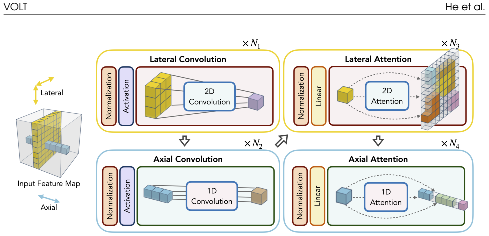
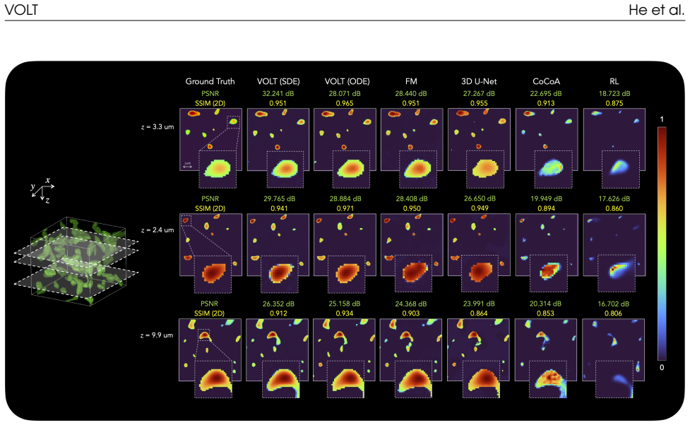
VOLT combines a transport-based formulation that maps degraded measurements to clean volumes via stochastic interpolants with a 3D-native anisotropic network that separates lateral and axial processing. This design operates directly in voxel space and achieves improved scalability to large volumes without relying on slice-wise approximations. Both stochastic (SDE) and deterministic (ODE) variants are developed, and validation on simulated datasets shows significant improvements in reconstruction quality in both lateral and axial directions while providing voxel-wise credibility estimates.
What carries the argument
The transport-based formulation using stochastic interpolants paired with a 3D-native anisotropic network that separates lateral and axial processing.
If this is right
- Improved reconstruction quality in lateral and axial directions for 3D volumes.
- Scalable operation on large high-dimensional volumes without slice-wise approximations.
- Voxel-wise credibility estimates for assessing reconstruction reliability.
- Both stochastic and deterministic reconstruction variants available in one framework.
Where Pith is reading between the lines
- This approach might allow biologists to obtain clearer 3D images of living samples where blur is hard to avoid.
- The credibility maps could be used to guide further processing or analysis by highlighting uncertain regions.
- If the method generalizes to real data, it could replace or complement traditional deconvolution techniques in microscopy workflows.
Load-bearing premise
The assumption that the transport-based formulation with stochastic interpolants accurately captures the physical out-of-focus blur in wide-field microscopy.
What would settle it
An experiment comparing VOLT reconstructions to ground-truth clean volumes on real wide-field microscopy datasets, checking for actual improvements in axial resolution and correlation of credibility estimates with errors.
Figures


read the original abstract
Three-dimensional (3D) wide-field fluorescence microscopy is a widely used modality for volumetric imaging, but suffers from characteristic out-of-focus blur. Existing reconstruction methods either struggle to operate on high-dimensional volumes or fail to provide credibility characterization of the reconstruction. In this work, we introduce Volumetric Transport (VOLT), a 3D-native probabilistic framework for wide-field fluorescence microscopy reconstruction. VOLT combines a transport-based formulation that maps degraded measurements to clean volumes via stochastic interpolants with a 3D-native anisotropic network that separates lateral and axial processing. This design operates directly in voxel space and achieves improved scalability to large volumes without relying on slice-wise approximations. We develop both stochastic (SDE) and deterministic (ODE) variants within the same framework. We validate VOLT on simulated wide-field microscopy datasets. Our results show that VOLT significantly improves reconstruction quality in both lateral and axial directions while providing voxel-wise credibility estimates.
Editorial analysis
A structured set of objections, weighed in public.
Referee Report
Summary. The manuscript introduces VOLT, a 3D-native probabilistic framework for wide-field fluorescence microscopy reconstruction. It combines a transport-based formulation that maps degraded measurements to clean volumes via stochastic interpolants with a 3D-native anisotropic network separating lateral and axial processing. Both SDE and ODE variants are developed, operating directly in voxel space for improved scalability, and the method is validated on simulated datasets claiming significant improvements in lateral and axial reconstruction quality along with voxel-wise credibility estimates.
Significance. If the stochastic-interpolant transport accurately captures the physical PSF convolution and noise degradation process and the anisotropic network scales without artifacts, this could advance volumetric imaging by enabling direct 3D probabilistic reconstruction with uncertainty quantification, avoiding slice-wise approximations common in prior work. The framework's design for large volumes and credibility estimates addresses practical needs in biological microscopy.
major comments (2)
- [§5] §5 (Validation): Results are reported exclusively on simulated wide-field microscopy phantoms; no real experimental volumes, PSF-calibration experiments, or quantitative metrics (e.g., PSNR, SSIM with baselines and error bars) are provided, which is load-bearing for the central claim that the transport map inverts the true physical degradation process rather than simulation-specific statistics.
- [§4] §4 (Method and Network Design): The assertion that the 3D-native anisotropic network scales effectively to large volumes (>512^3) without slice-wise artifacts lacks supporting memory/time scaling curves or ablation studies on real-scale stacks, undermining the scalability advantage over existing methods.
minor comments (2)
- [Abstract] Abstract: The claim of 'significantly improves reconstruction quality' would be strengthened by referencing specific quantitative results or tables from the experiments section.
- [§3] Notation: The distinction between the SDE and ODE variants could be clarified earlier with explicit equations showing how the stochastic interpolants are adapted in each case.
Simulated Author's Rebuttal
We thank the referee for the constructive comments on our manuscript. We address each major point below and indicate planned revisions to strengthen the work.
read point-by-point responses
-
Referee: [§5] §5 (Validation): Results are reported exclusively on simulated wide-field microscopy phantoms; no real experimental volumes, PSF-calibration experiments, or quantitative metrics (e.g., PSNR, SSIM with baselines and error bars) are provided, which is load-bearing for the central claim that the transport map inverts the true physical degradation process rather than simulation-specific statistics.
Authors: We acknowledge that the validation relies on simulated phantoms, which were chosen to provide controlled ground-truth evaluation of the transport map's inversion of the PSF convolution and noise model. To directly address the concern, the revised manuscript will include quantitative metrics such as PSNR and SSIM with baseline comparisons and error bars computed over multiple realizations. We will also expand the discussion to clarify the design of the phantoms to match physical degradation statistics and note the value of future real-data experiments with PSF calibration. revision: yes
-
Referee: [§4] §4 (Method and Network Design): The assertion that the 3D-native anisotropic network scales effectively to large volumes (>512^3) without slice-wise artifacts lacks supporting memory/time scaling curves or ablation studies on real-scale stacks, undermining the scalability advantage over existing methods.
Authors: We agree that empirical scaling evidence is needed to support the scalability claims. In the revision we will add memory and runtime scaling curves for volumes exceeding 512^3, together with ablation studies on large stacks that quantify the absence of slice-wise artifacts and compare against slice-wise baselines. revision: yes
Circularity Check
No significant circularity in derivation chain
full rationale
The paper introduces VOLT as a new 3D-native probabilistic framework that maps degraded wide-field measurements to clean volumes using stochastic interpolants within a transport formulation, paired with an anisotropic network for lateral/axial separation. No equations or steps in the provided abstract or description reduce a claimed prediction or result to a fitted parameter or self-referential definition by construction. No load-bearing self-citations, uniqueness theorems from prior author work, or ansatzes smuggled via citation are present. Validation claims rest on simulated data performance rather than tautological re-derivation of inputs. The derivation chain is self-contained against external benchmarks.
Axiom & Free-Parameter Ledger
Reference graph
Works this paper leans on
-
[1]
Imaging 3D cell cultures with optical microscopy,
H.-C. Hsieh, Q. Han, D. Brenes, K. W. Bishop, R. Wang, Y. Wang, C. Poudel, A. K. Glaser, B. S. Freedman, J. C. Vaughanet al., “Imaging 3D cell cultures with optical microscopy,”Nature Methods, vol. 22, no. 6, pp. 1167–1190, 2025
2025
-
[2]
Advanced3Dimaging and organoid bioprintingfor biomedical research andtherapeutic applications,
S.Maharjan, C. Ma, B.Singh, H.Kang, G.Orive, J. Yao, andY.S. Zhang, “Advanced3Dimaging and organoid bioprintingfor biomedical research andtherapeutic applications,”Advanced Drug Delivery Reviews, vol. 208, p. 115237, 2024
2024
-
[3]
J. B. Pawley,Handbook of Biological Confocal Microscopy, 3rd ed. New York: Springer, 2006
2006
-
[4]
Deep tissue two-photon microscopy,
F. Helmchen and W. Denk, “Deep tissue two-photon microscopy,”Nature Methods, vol. 2, no. 12, pp. 932–940, 2005
2005
-
[5]
Mertz,Introduction to Optical Microscopy, 2nd ed
J. Mertz,Introduction to Optical Microscopy, 2nd ed. Cambridge: Cambridge University Press, 2019
2019
-
[6]
R. C. Gonzalez and R. E. Woods,Digital Image Processing, 4th ed. New York: Pearson, 2018
2018
-
[7]
Bayesianestimationofregularizationandpointspread function parameters for Wiener–Hunt deconvolution,
F.Orieux,J.-F.Giovannelli,andT.Rodet,“Bayesianestimationofregularizationandpointspread function parameters for Wiener–Hunt deconvolution,”Journal of the Optical Society of America A, vol. 27, no. 7, pp. 1593–1607, 2010
2010
-
[8]
Bayesian-based iterative method of image restoration,
W. H. Richardson, “Bayesian-based iterative method of image restoration,”Journal of the Optical Society of America, vol. 62, no. 1, pp. 55–59, 1972
1972
-
[9]
Aniterativetechniquefortherectificationofobserveddistributions,
L.B.Lucy,“Aniterativetechniquefortherectificationofobserveddistributions,”TheAstronomical Journal, vol. 79, p. 745, 1974
1974
-
[10]
Content-awareimagerestoration: pushingthelimitsoffluorescence microscopy,
M. Weigert, U. Schmidt, T. Boothe, A. Müller, A. Dibrov, A. Jain, B. Wilhelm, D. Schmidt, C.Broaddus,S.Culleyetal.,“Content-awareimagerestoration: pushingthelimitsoffluorescence microscopy,”Nature Methods, vol. 15, no. 12, pp. 1090–1097, 2018
2018
-
[11]
Efficient and accurate inversion of multiple scattering with deep learning,
Y. Sun, Z. Xia, and U. S. Kamilov, “Efficient and accurate inversion of multiple scattering with deep learning,”Optics Express, vol. 26, no. 11, pp. 14678–14688, 2018. 13 VOLT He et al
2018
-
[12]
Deep-3D microscope: 3D volumetric microscopy of thick scattering samples using a wide-field microscope and machine learning,
B. Li, S. Tan, J. Dong, X. Lian, Y. Zhang, X. Ji, and A. Veeraraghavan, “Deep-3D microscope: 3D volumetric microscopy of thick scattering samples using a wide-field microscope and machine learning,”Biomedical Optics Express, vol. 13, no. 1, pp. 284–299, 2022
2022
-
[13]
Recovery of continuous 3D refractive index mapsfromdiscreteintensity-onlymeasurementsusingneuralfields,
R. Liu, Y. Sun, J. Zhu, L. Tian, and U. S. Kamilov, “Recovery of continuous 3D refractive index mapsfromdiscreteintensity-onlymeasurementsusingneuralfields,”NatureMachineIntelligence, vol. 4, no. 9, pp. 781–791, 2022
2022
-
[14]
Coordinate-based neural representations for compu- tational adaptive optics in widefield microscopy,
I. Kang, Q. Zhang, S. X. Yu, and N. Ji, “Coordinate-based neural representations for compu- tational adaptive optics in widefield microscopy,”Nature Machine Intelligence, vol. 6, no. 6, pp. 714–725, 2024
2024
-
[15]
Quantifying generative model uncertainty in posterior sampling methods for computational imaging,
C. Ekmekci and M. Cetin, “Quantifying generative model uncertainty in posterior sampling methods for computational imaging,” inNeurIPS 2023 Workshop on Deep Learning and Inverse Problems, 2023
2023
-
[16]
Conformalizedgenerativebayesianimaging: anuncertaintyquantificationframeworkfor computational imaging,
——,“Conformalizedgenerativebayesianimaging: anuncertaintyquantificationframeworkfor computational imaging,”IEEE Transactions on Computational Imaging, vol. 12, pp. 216–229, 2025
2025
-
[17]
DiffuseIR: Diffusion models for isotropic reconstruction of 3D microscopic images,
M. Pan, Y. Gan, F. Zhou, J. Liu, Y. Zhang, A. Wang, S. Zhang, and D. Li, “DiffuseIR: Diffusion models for isotropic reconstruction of 3D microscopic images,” inMedical Image Computing and Computer Assisted Intervention – MICCAI 2023. Springer, 2023, pp. 323–332
2023
-
[18]
Mi- croscopyimagereconstructionwithphysics-informeddenoisingdiffusionprobabilisticmodel,
R. Li, G. della Maggiora, J. Andilla, P. Loza-Alvarez, M. Lakadamyali, and D. K. Bhatt, “Mi- croscopyimagereconstructionwithphysics-informeddenoisingdiffusionprobabilisticmodel,” Communications Engineering, vol. 3, p. 186, 2024
2024
-
[19]
Score-based gener- ative modeling through stochastic differential equations,
Y. Song, J. Sohl-Dickstein, D. P. Kingma, A. Kumar, S. Ermon, and B. Poole, “Score-based gener- ative modeling through stochastic differential equations,” inInternational Conference on Learning Representations, 2021
2021
-
[20]
Denoising diffusion probabilistic models,
J. Ho, A. Jain, and P. Abbeel, “Denoising diffusion probabilistic models,”Advances in Neural Information Processing Systems, vol. 33, pp. 6840–6851, 2020
2020
-
[21]
Flow matching for generative modeling,
Y. Lipman, R. T. Q. Chen, H. Ben-Hamu, M. Nickel, and M. Le, “Flow matching for generative modeling,” inInternational Conference on Learning Representations, 2023
2023
-
[22]
Flowstraightandfast: Learningtogenerateandtransferdatawith rectified flow,
X.Liu,C.Gong,andQ.Liu,“Flowstraightandfast: Learningtogenerateandtransferdatawith rectified flow,” inThe Eleventh International Conference on Learning Representations (ICLR), 2023
2023
-
[23]
Deep learning optical- sectioning method,
X. Zhang, Y. Chen, K. Ning, C. Zhou, Y. Han, H. Gong, and J. Yuan, “Deep learning optical- sectioning method,”Optics Express, vol. 26, no. 23, pp. 30762–30772, 2018
2018
-
[24]
Q.Li,C.Lou,Y.Cheng,B.Gong,X.Chen,H.Chen,B.Li,J.Wang,Y.Wang,S.Yang,Y.Tang,and L.Dai,“ComputationalTIRFenablesopticalsectioningbeyondtheevanescentfieldforwidefield fluorescence microscopy,” 2026. [Online]. Available: https://arxiv.org/abs/2511.06853
-
[25]
High-resolutionimagesynthesis with latent diffusion models,
R.Rombach,A.Blattmann,D.Lorenz,P.Esser,andB.Ommer,“High-resolutionimagesynthesis with latent diffusion models,” inProceedings of the IEEE/CVF Conference on Computer Vision and Pattern Recognition (CVPR), 2022, pp. 10684–10695. 14 VOLT He et al
2022
-
[26]
PSI3D: Plug-and-play 3D stochastic inference with slice-wise latent diffusion prior,
W. Guo, J. Yu, Y. Wang, J. U. Kang, and Y. Sun, “PSI3D: Plug-and-play 3D stochastic inference with slice-wise latent diffusion prior,” in2025 59th Asilomar Conference on Signals, Systems, and Computers, 2025, pp. 484–488
2025
-
[27]
Videodiffusionmodels,
J.Ho,T.Salimans,A.Gritsenko,W.Chan,M.Norouzi,andD.J.Fleet,“Videodiffusionmodels,” inAdvances in Neural Information Processing Systems (NeurIPS), vol. 35, 2022
2022
-
[28]
Make-A-Video: Text-to-video generation without text- video data,
U. Singer, A. Polyak, T. Hayes, X. Yin, J. An, S. Zhang, Q. Hu, H. Yang, O. Ashual, O. Gafni, D. Parikh, S. Gupta, and Y. Taigman, “Make-A-Video: Text-to-video generation without text- video data,” inThe Eleventh International Conference on Learning Representations (ICLR), 2023
2023
-
[29]
ModelScope Text-to-Video Technical Report
J.Wang,H.Yuan,D.Chen,Y.Zhang,X.Wang,andS.Zhang,“ModelScopetext-to-videotechnical report,”arXiv preprint arXiv:2308.06571, 2023
work page internal anchor Pith review arXiv 2023
-
[30]
Stochasticinterpolants: Aunifyingframework for flows and diffusions,
M.Albergo,N.M.Boffi,andE.Vanden-Eijnden,“Stochasticinterpolants: Aunifyingframework for flows and diffusions,”Journal of Machine Learning Research, vol. 26, no. 209, pp. 1–80, 2025
2025
-
[31]
Buildingnormalizingflowswithstochasticinterpolants,
M.S.AlbergoandE.Vanden-Eijnden,“Buildingnormalizingflowswithstochasticinterpolants,” inThe Eleventh International Conference on Learning Representations
-
[32]
Diffusion posterior sampling for general noisy inverse problems,
H. Chung, J. Kim, M. T. McCann, M. L. Klasky, and J. C. Ye, “Diffusion posterior sampling for general noisy inverse problems,” inInternational Conference on Learning Representations, 2023
2023
-
[33]
FlowDPS:Flow-drivenposteriorsamplingforinverseproblems,
J.Kim,B.S.Kim,andJ.C.Ye,“FlowDPS:Flow-drivenposteriorsamplingforinverseproblems,” inProceedingsoftheIEEE/CVFInternationalConferenceonComputerVision,2025,pp.12328–12337
2025
-
[34]
Practical sensorless aberration estimation for 3D microscopy with deep learning,
D. Saha, U. Schmidt, Q. Zhang, A. Barbotin, Q. Hu, N. Ji, M. J. Booth, M. Weigert, and E. W. Myers, “Practical sensorless aberration estimation for 3D microscopy with deep learning,”Opt. Express, vol. 28, no. 20, pp. 29044–29053, Sep 2020. [Online]. Available: https://opg.optica.org/oe/abstract.cfm?URI=oe-28-20-29044
2020
-
[35]
3D deconvolution microscopy,
D. S. C. Biggs, “3D deconvolution microscopy,”Current Protocols in Cytometry, vol. 52, no. 1, pp. 12.19.1–12.19.20, 2010
2010
-
[36]
Three-dimensional imaging by de- convolution microscopy,
J. G. McNally, T. Karpova, J. Cooper, and J.-A. Conchello, “Three-dimensional imaging by de- convolution microscopy,”Methods, vol. 19, no. 3, pp. 373–385, 1999
1999
-
[37]
Richardson–Lucy algorithm with total variation regularization for 3D confocal microscope deconvolution,
N. Dey, L. Blanc-Féraud, C. Zimmer, P. Roux, Z. Kam, J.-C. Olivo-Marin, and J. Zerubia, “Richardson–Lucy algorithm with total variation regularization for 3D confocal microscope deconvolution,”Microscopy Research and Technique, vol. 69, no. 4, pp. 260–266, 2006
2006
-
[38]
Deep convolutional neural network for inverseproblemsinimaging,
K. H. Jin, M. T. McCann, E. Froustey, and M. Unser, “Deep convolutional neural network for inverseproblemsinimaging,”IEEETransactionsonImageProcessing,vol.26,no.9,pp.4509–4522, 2017
2017
-
[39]
Deep equilibrium architectures for inverse problems in imaging,
D. Gilton, G. Ongie, and R. Willett, “Deep equilibrium architectures for inverse problems in imaging,”IEEE Transactions on Computational Imaging, vol. 7, pp. 1123–1133, 2021
2021
-
[40]
Accurate and versatile 3D segmentation of plant tissues at cellular resolution,
A. Wolny, L. Cerrone, A. Vijayan, R. Tofanelli, A. V. Barro, M. Louveaux, C. Wenzl, S. Strauss, D. Wilson-Sánchezet al., “Accurate and versatile 3D segmentation of plant tissues at cellular resolution,”eLife, vol. 9, p. e57613, 2020. 15 VOLT He et al
2020
-
[41]
NeRF: Representing scenes as neural radiance fields for view synthesis,
B. Mildenhall, P. P. Srinivasan, M. Tancik, J. T. Barron, R. Ramamoorthi, and R. Ng, “NeRF: Representing scenes as neural radiance fields for view synthesis,”Communications of the ACM, vol. 65, no. 1, pp. 99–106, 2021
2021
-
[42]
Principled probabilistic imag- ing using diffusion models as plug-and-play priors,
Z. Wu, Y. Sun, Y. Chen, B. Zhang, Y. Yue, and K. L. Bouman, “Principled probabilistic imag- ing using diffusion models as plug-and-play priors,”Advances in Neural Information Processing Systems, vol. 37, pp. 118389–118427, 2024
2024
-
[43]
Inversionbydirectiteration: Analternativetodenoisingdiffusion for image restoration,
M.DelbracioandP.Milanfar,“Inversionbydirectiteration: Analternativetodenoisingdiffusion for image restoration,”Transactions on Machine Learning Research, 2023, featured Certification, OutstandingCertification.[Online].Available: https://openreview.net/forum?id=VmyFF5lL3F
2023
-
[44]
Denoisingdiffusion restorationmodels,
B.Kawar, M. Elad, S. Ermon, andJ.Song, “Denoisingdiffusion restorationmodels,” inAdvances in Neural Information Processing Systems, vol. 35, 2022, pp. 23593–23606
2022
-
[45]
Back to Basics: Let Denoising Generative Models Denoise
T. Li and K. He, “Back to basics: Let denoising generative models denoise,”arXiv preprint arXiv:2511.13720, 2025
work page internal anchor Pith review arXiv 2025
-
[46]
Image quality assessment: from error visibilitytostructuralsimilarity,
Z. Wang, A. C. Bovik, H. R. Sheikh, and E. P. Simoncelli, “Image quality assessment: from error visibilitytostructuralsimilarity,”IEEETransactionsonImageProcessing,vol.13,no.4,pp.600–612, 2004
2004
-
[47]
K. Perlin, “Improving noise,” inProceedings of the 29th Annual Conference on Computer Graphics andInteractiveTechniques,ser.SIGGRAPH’02. NewYork,NY,USA:AssociationforComputing Machinery, 2002, p. 681–682. [Online]. Available: https://doi.org/10.1145/566570.566636
-
[48]
FlashAttention: Fast and memory-efficient exact attention with IO-awareness,
T. Dao, D. Y. Fu, S. Ermon, A. Rudra, and C. Ré, “FlashAttention: Fast and memory-efficient exact attention with IO-awareness,” inAdvances in Neural Information Processing Systems, 2022
2022
-
[49]
Diffusion models beat GANs on image synthesis,
P. Dhariwal and A. Nichol, “Diffusion models beat GANs on image synthesis,” inAdvances in Neural Information Processing Systems, vol. 34, 2021, pp. 8780–8794
2021
-
[50]
Multiscale structural similarity for image quality assessment,
Z. Wang, E. P. Simoncelli, and A. C. Bovik, “Multiscale structural similarity for image quality assessment,” inThe Thrity-Seventh Asilomar Conference on Signals, Systems & Computers, 2003, vol. 2. Ieee, 2003, pp. 1398–1402
2003
-
[51]
The unreasonable effectiveness of deepfeaturesasaperceptualmetric,
R. Zhang, P. Isola, A. A. Efros, E. Shechtman, and O. Wang, “The unreasonable effectiveness of deepfeaturesasaperceptualmetric,”inProceedingsoftheIEEEConferenceonComputerVisionand Pattern Recognition, 2018, pp. 586–595
2018
-
[52]
Pattern recognition and machine learning
C. M. Bishop, “Pattern recognition and machine learning.” New York: Springer, 2006. 16
2006
discussion (0)
Sign in with ORCID, Apple, or X to comment. Anyone can read and Pith papers without signing in.