Recognition: unknown
Computation of frequency- and time-domain Jacobians in optical tomography with Monte Carlo simulations
Pith reviewed 2026-05-07 10:02 UTC · model grok-4.3
The pith
Monte Carlo simulations now compute sensitivity profiles for frequency- and time-domain optical tomography.
A machine-rendered reading of the paper's core claim, the machinery that carries it, and where it could break.
Core claim
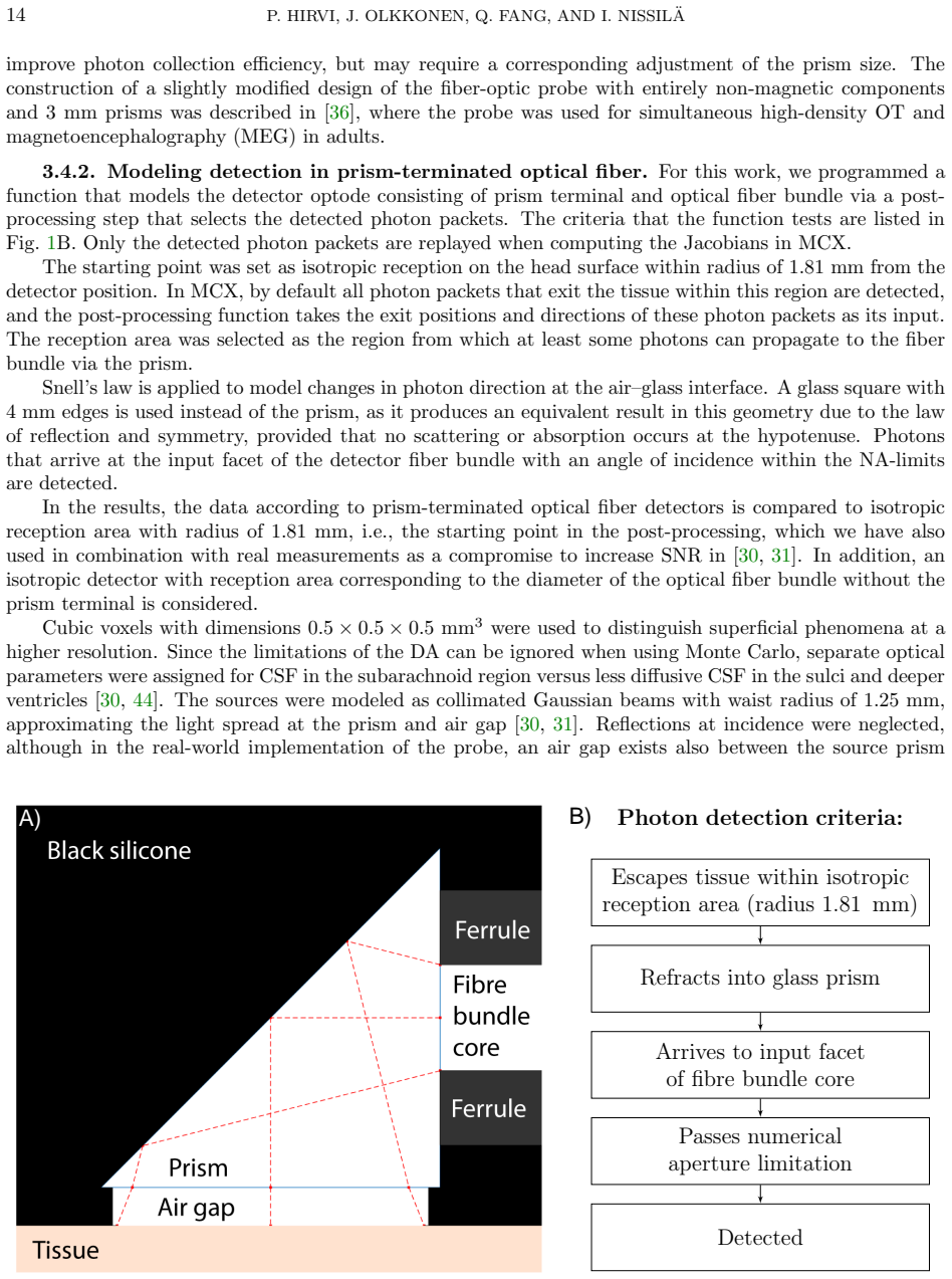
The central discovery is a complete theoretical framework that derives frequency- and time-domain Jacobians within the perturbation Monte Carlo approach, implemented as software in the MCX simulator. This includes extensions for split voxels on curved surfaces and a detector model applied in post-processing for prism-terminated fibers. Validation against finite-element diffusion approximation solutions in neonatal head models reveals that the two methods agree closely only in high-scattering regimes.
What carries the argument
Perturbation Monte Carlo framework for absorption and scattering Jacobians in frequency- and time-domain, combined with post-processing detector modeling.
Load-bearing premise
The perturbation Monte Carlo framework accurately derives the Jacobians for the specified frequency- and time-domain quantities and the post-processing detector model correctly represents prism-terminated fibers without needing full re-simulation.
What would settle it
Direct experimental measurement of sensitivity profiles in a low-scattering tissue phantom using frequency- or time-domain detection, compared point-by-point to the Monte Carlo computed Jacobians.
Figures


read the original abstract
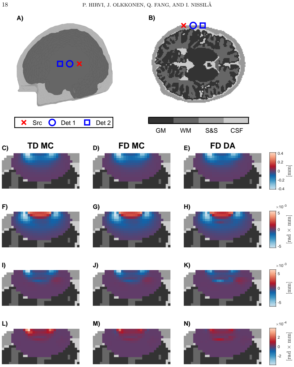
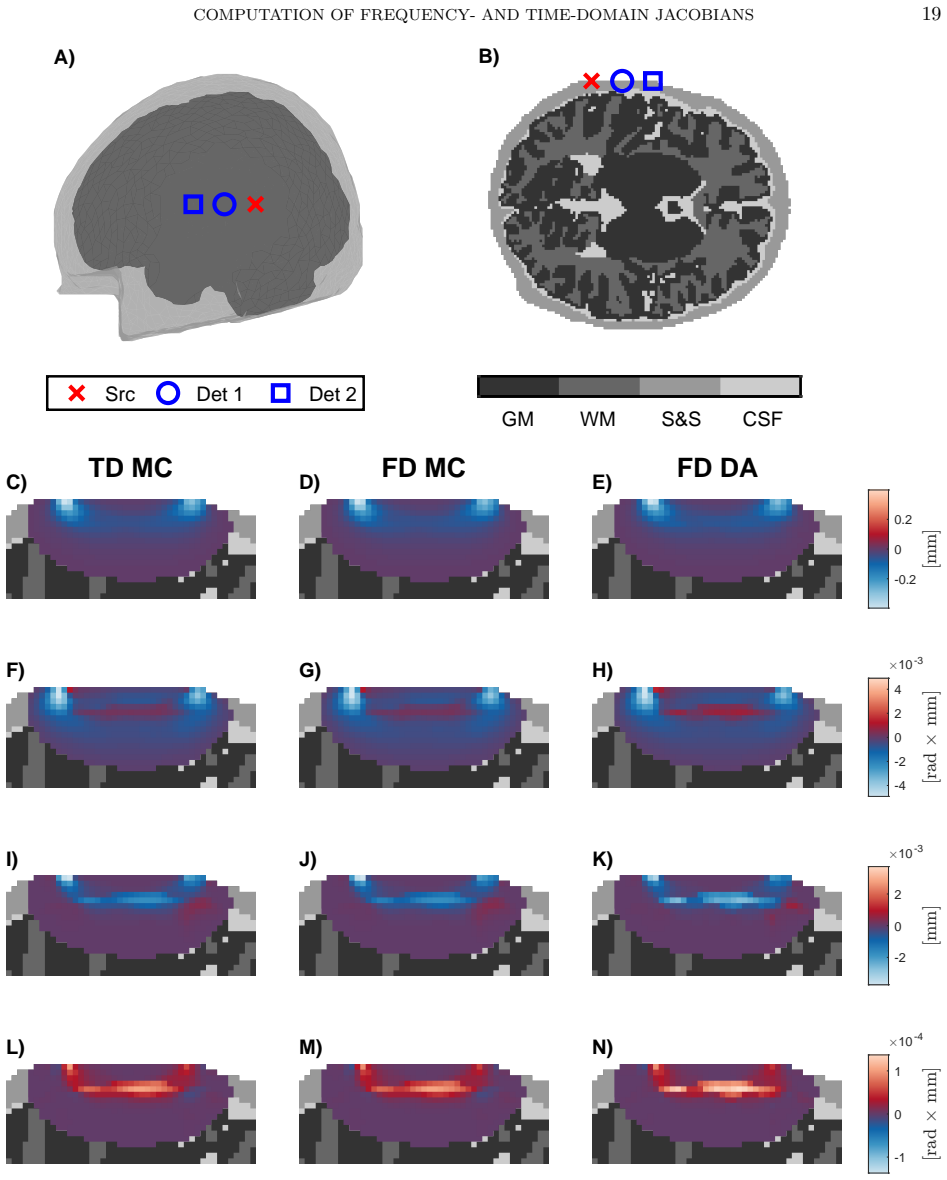
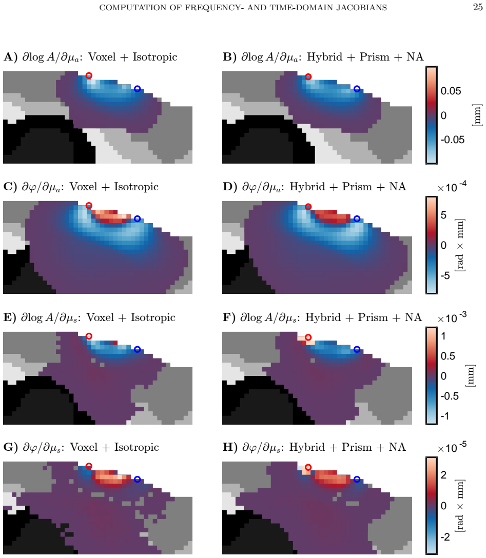
Significance: Jacobians, or spatially resolved sensitivity profiles, are central to image reconstruction in model-based optical tomography of biological tissue. Although Monte Carlo (MC) simulations are the gold standard for modeling light transport in turbid media, methodology for frequency- and time-domain Jacobians remains incomplete. Aim: This work extends MC to directly compute absorption and scattering Jacobians for frequency-domain (amplitude and phase) and time-domain (intensity and mean time-of-flight) measurements and prism-terminated optical fiber detectors. Approach: Jacobians are derived in the perturbation MC framework and implemented in the high-performance, open-source Monte Carlo eXtreme (MCX) simulator. Results are validated against the diffusion approximation (DA) solved using the finite element method in neonatal head models. MC with split voxels on curved surfaces is extended to Jacobian computation. The detector model is implemented in post-processing and compared with isotropic reception at surface. Results: MC- and DA-derived Jacobians show excellent agreement only in high-scattering regimes, highlighting the importance of MC for low-scattering domains. The detector model reduces surface sensitivity and marginally increases sensitivity to deeper tissues at short (< 2 cm) source-detector separations. Conclusion: A complete theoretical framework and MC software for computing frequency- and time-domain Jacobians is provided. Realistic detector modeling is encouraged for short-separation channels.
Editorial analysis
A structured set of objections, weighed in public.
Referee Report
Summary. The manuscript develops a perturbation Monte Carlo framework to compute absorption and scattering Jacobians for frequency-domain (amplitude and phase) and time-domain (intensity and mean time-of-flight) measurements in optical tomography. It implements this in the MCX simulator, extends split-voxel handling for curved surfaces to Jacobians, and models prism-terminated fiber detectors via post-processing. Validation is performed against finite-element solutions of the diffusion approximation in neonatal head models, with results showing good agreement in high-scattering regimes but divergence in low-scattering ones, and the detector model affecting surface vs. deep tissue sensitivity at short source-detector separations.
Significance. If the computed Jacobians are accurate, this work fills a gap in MC-based sensitivity profile computation for time- and frequency-domain optical tomography, particularly useful in low-scattering or short-separation scenarios where the diffusion approximation breaks down. The open-source implementation in MCX is a strength, enabling reproducible use in image reconstruction. The emphasis on realistic detector modeling is valuable for practical applications.
major comments (2)
- [Results] The claim of 'excellent agreement' between MC- and DA-derived Jacobians in high-scattering regimes lacks specific quantitative error metrics (e.g., relative L2 norms, mean absolute deviations, or correlation coefficients) or details on the exact source-detector geometries, scattering coefficients, and neonatal head model parameters tested. Without these, it is difficult to assess the degree of agreement and the precise conditions under which divergence occurs in low-scattering regimes, which is central to the argument for using MC.
- [Approach / Detector model] The post-processing implementation of the prism-terminated fiber detector model is compared only to isotropic surface reception, showing marginal changes in depth sensitivity at separations <2 cm. However, since the paper argues for the necessity of MC in low-scattering domains (where angular acceptance effects are more pronounced due to broader path-length distributions), a direct comparison to full Monte Carlo simulation with explicit modeling of the fiber acceptance cone is needed to validate that the post-processing does not introduce errors in the reported Jacobians for the frequency- and time-domain quantities.
minor comments (2)
- [Abstract] The abstract states that 'MC with split voxels on curved surfaces is extended to Jacobian computation' but provides no details on the implementation or any validation that this extension preserves Jacobian accuracy.
- [Theory] Notation for the frequency-domain and time-domain quantities (e.g., how amplitude/phase and intensity/mean TOF are denoted in the Jacobian definitions) could be clarified with explicit equations early in the manuscript to aid readability.
Simulated Author's Rebuttal
We thank the referee for the constructive comments and positive assessment of the significance of our work. We address each major comment point by point below.
read point-by-point responses
-
Referee: [Results] The claim of 'excellent agreement' between MC- and DA-derived Jacobians in high-scattering regimes lacks specific quantitative error metrics (e.g., relative L2 norms, mean absolute deviations, or correlation coefficients) or details on the exact source-detector geometries, scattering coefficients, and neonatal head model parameters tested. Without these, it is difficult to assess the degree of agreement and the precise conditions under which divergence occurs in low-scattering regimes, which is central to the argument for using MC.
Authors: We agree that quantitative metrics and precise parameter details are needed to substantiate the agreement claim. In the revised manuscript, we will report relative L2 norms and mean absolute deviations for the absorption and scattering Jacobians (both amplitude/phase and intensity/mean-time) across the tested conditions. We will also explicitly state the source-detector separations (1–4 cm), the neonatal head model layer thicknesses and optical properties (as in Table 1), and the range of reduced scattering coefficients (0.5–15 mm^{-1}). These additions will quantify the excellent agreement (errors typically <5 %) in high-scattering regimes and the divergence observed at lower scattering values. revision: yes
-
Referee: [Approach / Detector model] The post-processing implementation of the prism-terminated fiber detector model is compared only to isotropic surface reception, showing marginal changes in depth sensitivity at separations <2 cm. However, since the paper argues for the necessity of MC in low-scattering domains (where angular acceptance effects are more pronounced due to broader path-length distributions), a direct comparison to full Monte Carlo simulation with explicit modeling of the fiber acceptance cone is needed to validate that the post-processing does not introduce errors in the reported Jacobians for the frequency- and time-domain quantities.
Authors: The post-processing reweights each detected photon's contribution by the cosine of its exit angle relative to the prism acceptance cone; this is exact within the ray-optics limit used by MCX and does not alter the underlying path-length or momentum-transfer statistics that enter the frequency- and time-domain Jacobians. Nevertheless, we acknowledge that an explicit full-MC implementation of the prism would provide additional reassurance in low-scattering regimes. In the revision we will add a dedicated paragraph deriving the post-processing formula, discussing its validity when path-length distributions broaden, and reporting a limited numerical check against a cone-filtered photon set extracted from the same MCX runs. Full prism geometry tracing inside the MC kernel is outside the current MCX feature set and would require substantial new development; we therefore treat the post-processing validation as partial. revision: partial
Circularity Check
No significant circularity; derivation extends standard perturbation MC independently
full rationale
The paper derives frequency- and time-domain Jacobians via the established perturbation Monte Carlo framework and implements the extension in MCX, with direct validation against an independent finite-element diffusion-approximation solver on neonatal head models. No quoted equations reduce the Jacobians to fitted parameters renamed as predictions, self-definitional loops, or load-bearing self-citations whose content is unverified outside the present work. The post-processing detector model is an implementation detail compared to isotropic reception, not a circular renaming or ansatz smuggled via prior self-work. The central result (MC-DA agreement only in high-scattering regimes) rests on explicit numerical comparison rather than construction from the paper's own inputs.
Axiom & Free-Parameter Ledger
axioms (1)
- domain assumption The perturbation Monte Carlo framework accurately captures first-order changes in detected signals due to small perturbations in absorption and scattering coefficients.
Reference graph
Works this paper leans on
-
[1]
Optical tomography in medical imaging.Inverse problems, 15(2):R41, 1999
Simon R Arridge. Optical tomography in medical imaging.Inverse problems, 15(2):R41, 1999
1999
-
[2]
Diffuse optical imaging
Ilkka Nissil¨ a, Tommi Noponen, Jenni Heino, Timo Kajava, and Toivo Katila. Diffuse optical imaging. InAdvances in electromagnetic fields in living systems, pages 77–129. Springer, 2005
2005
-
[3]
High-density diffuse optical tomography for imaging human brain function.Review of Scientific Instruments, 90(5), 2019
Muriah D Wheelock, Joseph P Culver, and Adam T Eggebrecht. High-density diffuse optical tomography for imaging human brain function.Review of Scientific Instruments, 90(5), 2019
2019
-
[4]
Small animal fluorescence and bioluminescence tomography: a review of approaches, algorithms and technology update.Physics in Medicine & Biology, 59(1):R1–R64, 2014
Chinmay Darne, Yujie Lu, and Eva M Sevick-Muraca. Small animal fluorescence and bioluminescence tomography: a review of approaches, algorithms and technology update.Physics in Medicine & Biology, 59(1):R1–R64, 2014
2014
-
[5]
Optical tomographic imaging of small animals.Current Opinion in Biotechnology, 16(1):79–88,
Andreas H Hielscher. Optical tomographic imaging of small animals.Current Opinion in Biotechnology, 16(1):79–88,
-
[6]
Analytical biotechnology
-
[7]
Review of optical breast imaging and spec- troscopy.Journal of Biomedical Optics, 21(9):091311–091311, 2016
Dirk Grosenick, Herbert Rinneberg, Rinaldo Cubeddu, and Paola Taroni. Review of optical breast imaging and spec- troscopy.Journal of Biomedical Optics, 21(9):091311–091311, 2016
2016
-
[8]
Overview of diffuse optical tomography and its clinical applications.Journal of Biomedical Optics, 21(9):091312–091312, 2016
Yoko Hoshi and Yukio Yamada. Overview of diffuse optical tomography and its clinical applications.Journal of Biomedical Optics, 21(9):091312–091312, 2016
2016
-
[9]
Recent advances in diffuse optical imaging.Physics in Medicine & Biology, 50(4):R1–R43, 2005
Adam P Gibson, Jeremy C Hebden, and Simon R Arridge. Recent advances in diffuse optical imaging.Physics in Medicine & Biology, 50(4):R1–R43, 2005
2005
-
[10]
Evaluation of optical properties of highly scattering media by moments of distributions of times of flight of photons.Applied Optics, 42(28):5785–5792, 2003
Adam Liebert, Heidrun Wabnitz, Dirk Grosenick, Michael M¨ oller, Rainer Macdonald, and Herbert Rinneberg. Evaluation of optical properties of highly scattering media by moments of distributions of times of flight of photons.Applied Optics, 42(28):5785–5792, 2003
2003
-
[11]
McBride, Brian W
Troy O. McBride, Brian W. Pogue, Ulf L. ¨Osterberg, and Keith D. Paulsen. Separation of Absorption and Scattering Heterogeneities in NIR Tomographic Imaging of Tissue. InBiomedical Optical Spectroscopy and Diagnostics, page TuC2. Optica Publishing Group, 2000
2000
-
[12]
Frequency domain optical tomography using a Monte Carlo perturbation method.Optics Communications, 364:165–176, 2016
Toshihiro Yamamoto and Hiroki Sakamoto. Frequency domain optical tomography using a Monte Carlo perturbation method.Optics Communications, 364:165–176, 2016
2016
-
[13]
Stochastic Gauss-Newton method for estimating absorption and scattering in optical tomography with the Monte Carlo method for light transport
Jonna Kangasniemi, Meghdoot Mozumder, Aki Pulkkinen, and Tanja Tarvainen. Stochastic Gauss-Newton method for estimating absorption and scattering in optical tomography with the Monte Carlo method for light transport. Biomedical Optics Express, 15(8):4925–4942, 2024
2024
-
[14]
Nonuniqueness in diffusion-based optical tomography.Optics Letters, 23(11):882–884, 1998
Simon R Arridge and William RB Lionheart. Nonuniqueness in diffusion-based optical tomography.Optics Letters, 23(11):882–884, 1998
1998
-
[15]
Phd dissertation, Helsinki University of Technology, Department of Engineering Physics and Mathematics, Espoo, Finland, December 2004
Ilkka Nissil¨ a.Instrumentation for medical optical tomography with applications. Phd dissertation, Helsinki University of Technology, Department of Engineering Physics and Mathematics, Espoo, Finland, December 2004
2004
-
[16]
Instrumentation for the accurate measurement of phase and amplitude in optical tomography.Review of Scientific Instruments, 73(9):3306–3312, 2002
Ilkka Nissil¨ a, Kalle Kotilahti, Kim Fallstr¨ om, and Toivo Katila. Instrumentation for the accurate measurement of phase and amplitude in optical tomography.Review of Scientific Instruments, 73(9):3306–3312, 2002
2002
-
[17]
Instrumentation and calibration methods for the multichannel measurement of phase and amplitude in optical tomography.Review of Scientific Instruments, 76(044302), 2005
Ilkka Nissil¨ a, Tommi Noponen, Kalle Kotilahti, Toivo Katila, Lauri Lipi¨ ainen, Tanja Tarvainen, Martin Schweiger, and Simon Arridge. Instrumentation and calibration methods for the multichannel measurement of phase and amplitude in optical tomography.Review of Scientific Instruments, 76(044302), 2005
2005
-
[18]
Nissil¨ a, Jeremy C
Ilkka T. Nissil¨ a, Jeremy C. Hebden, David Jennions, Jenni Heino, Martin Schweiger, Kalle M. Kotilahti, Tommi E.J. Noponen, Adam P. Gibson, Seppo J¨ arvenp¨ a¨ a, Lauri Lipi¨ ainen, and Toivo E. Katila. Comparison between a time- domain and a frequency-domain system for optical tomography.Journal of Biomedical Optics, 11(6):064015, 2006
2006
-
[19]
Kernel flow: a high channel count scalable time-domain functional near-infrared spectroscopy system.Journal of Biomedical Optics, 27(7):074710, 2022
Han Y Ban, Geoffrey M Barrett, Alex Borisevich, Ashutosh Chaturvedi, Jacob L Dahle, Hamid Dehghani, Julien Dubois, Ryan M Field, Viswanath Gopalakrishnan, Andrew Gundran, Michael Henninger, Wilson C Ho, Howard D Hughes, Rong Jin, Julian Kates-Harbec, Thanh Landy, Michael Leggiero, Gabriel Lerner, Zahra M Aghajan, Michael Moon, Isai Olvera, Sangyong Park, ...
2022
-
[20]
Eggebrecht, and Hamid Dehghani
Matthaios Doulgerakis, Adam T. Eggebrecht, and Hamid Dehghani. High-density functional diffuse optical tomography based on frequency-domain measurements improves image quality and spatial resolution.Neurophotonics, 6(3):035007, 2019
2019
-
[21]
Tuulari, Linnea Karlsson, Antti Hannukainen, Hasse Karlsson, and Nuutti Hyv¨ onen
Pauliina Hirvi, Ilkka Nissil¨ a, Ambika Maria, Qianqian Fang, Kalle Kotilahti, Juha Heiskala, Jetro J. Tuulari, Linnea Karlsson, Antti Hannukainen, Hasse Karlsson, and Nuutti Hyv¨ onen. Improved utilization of frequency-domain data for optical tomographic imaging of the human brain. In Sergio Fantini and Paola Taroni, editors,Optical Tomography COMPUTATIO...
2023
-
[22]
Image reconstruction for novel time domain near infrared optical tomography: towards clinical applications
Jingjing Jiang, Aldo Di Costanzo Mata, Scott Lindner, Chao Zhang, Edoardo Charbon, Martin Wolf, and Alexander Kalyanov. Image reconstruction for novel time domain near infrared optical tomography: towards clinical applications. Biomedical Optics Express, 11(8):4723–4734, Aug 2020
2020
-
[23]
Depth-selective data analysis for time-domain fNIRS: moments vs
Heidrun Wabnitz, Davide Contini, Lorenzo Spinelli, Alessandro Torricelli, and Adam Liebert. Depth-selective data analysis for time-domain fNIRS: moments vs. time windows.Biomedical Optics Express, 11(8):4224–4243, 2020
2020
-
[24]
Calibration techniques and datatype extraction for time-resolved optical tomography.Review of Scientific Instruments, 71(9):3415–3427, 2000
Elizabeth M C Hillman, Jeremy C Hebden, Florian E W Schmidt, Simon R Arridge, Martin Schweiger, Hamid Dehghani, and David T Delpy. Calibration techniques and datatype extraction for time-resolved optical tomography.Review of Scientific Instruments, 71(9):3415–3427, 2000
2000
-
[25]
Angelo Sassaroli, Giles Blaney, and Sergio Fantini. Novel data types for frequency-domain diffuse optical spectroscopy and imaging of tissues: characterization of sensitivity and contrast-to-noise ratio for absorption perturbations.Biomedical Optics Express, 14(5):2091–2116, 2023
2091
-
[26]
Estimation of optical absorption in anisotropic background.Inverse Problems, 18(3):559, 2002
Jenni Heino and Erkki Somersalo. Estimation of optical absorption in anisotropic background.Inverse Problems, 18(3):559, 2002
2002
-
[27]
Monte Carlo simulation of photon migration in 3D turbid media accelerated by graphics processing units.Optics Express, 17(22):20178–20190, 2009
Qianqian Fang and David A Boas. Monte Carlo simulation of photon migration in 3D turbid media accelerated by graphics processing units.Optics Express, 17(22):20178–20190, 2009
2009
-
[28]
A Monte Carlo model of light propagation in tissue
Scott A Prahl, Marleen Keijzer, Steven L Jacques, and Ashley J Welch. A Monte Carlo model of light propagation in tissue. In Gerhard Mueller, David Sliney, and Roy Potter, editors,Dosimetry of laser radiation in medicine and biology, volume 10305, pages 101–111. SPIE, 1989
1989
-
[29]
Equivalence of four Monte Carlo methods for photon migration in turbid media
Angelo Sassaroli and Fabrizio Martelli. Equivalence of four Monte Carlo methods for photon migration in turbid media. JOSA A, 29(10):2110–2117, 2012
2012
-
[30]
Affective and non-affective touch evoke differential brain responses in 2-month-old infants.NeuroImage, 169:162–171, 2018
Emma H J¨ onsson, Kalle Kotilahti, Juha Heiskala, Helena Backlund Wasling, H˚ akan Olausson, Ilona Croy, Hanna Mus- taniemi, Petri Hiltunen, Jetro J Tuulari, Noora M Scheinin, et al. Affective and non-affective touch evoke differential brain responses in 2-month-old infants.NeuroImage, 169:162–171, 2018
2018
-
[31]
Imaging affective and non-affective touch processing in two-year-old children.NeuroImage, 251:118983, 2022
Ambika Maria, Pauliina Hirvi, Kalle Kotilahti, Juha Heiskala, Jetro J Tuulari, Linnea Karlsson, Hasse Karlsson, and Ilkka Nissil¨ a. Imaging affective and non-affective touch processing in two-year-old children.NeuroImage, 251:118983, 2022
2022
-
[32]
Maternal prenatal depressive symptoms and child brain responses to affective touch at two years of age
Shashank Shekhar, Pauliina Hirvi, Ambika Maria, Kalle Kotilahti, Jetro J Tuulari, Linnea Karlsson, Hasse Karlsson, and Ilkka Nissil¨ a. Maternal prenatal depressive symptoms and child brain responses to affective touch at two years of age. Journal of Affective Disorders, 356:177–189, 2024
2024
-
[33]
Hemodynamic responses to emotional speech in two-month-old infants imaged using diffuse optical tomography.Scientific reports, 9(1):4745, 2019
Shashank Shekhar, Ambika Maria, Kalle Kotilahti, Minna Huotilainen, Juha Heiskala, Jetro J Tuulari, Pauliina Hirvi, Linnea Karlsson, Hasse Karlsson, and Ilkka Nissil¨ a. Hemodynamic responses to emotional speech in two-month-old infants imaged using diffuse optical tomography.Scientific reports, 9(1):4745, 2019
2019
-
[34]
Relationship between maternal pregnancy-related anxiety and infant brain responses to emotional speech–a pilot study.Journal of Affective Disorders, 262:62–70, 2020
Ambika Maria, Ilkka Nissil¨ a, Shashank Shekhar, Kalle Kotilahti, Jetro J Tuulari, Pauliina Hirvi, Minna Huotilainen, Juha Heiskala, Linnea Karlsson, and Hasse Karlsson. Relationship between maternal pregnancy-related anxiety and infant brain responses to emotional speech–a pilot study.Journal of Affective Disorders, 262:62–70, 2020
2020
-
[35]
An application of perturbation Monte Carlo in optical tomography
Kalle Kotilahti, Juha Heiskala, and Ilkka Nissila. An application of perturbation Monte Carlo in optical tomography. In 2005 IEEE Engineering in Medicine and Biology 27th Annual Conference, pages 274–277. IEEE, 2006
2005
-
[36]
Effect of task-related extracerebral circulation on diffuse optical tomography: experimental data and simulations on the forehead.Biomedical Optics Express, 4(3):412–426, 2013
Tiina N¨ asi, Hanna M¨ aki, Petri Hiltunen, Juha Heiskala, Ilkka Nissil¨ a, Kalle Kotilahti, and Risto J Ilmoniemi. Effect of task-related extracerebral circulation on diffuse optical tomography: experimental data and simulations on the forehead.Biomedical Optics Express, 4(3):412–426, 2013
2013
-
[37]
Simultaneously acquired magnetoencephalography and diffuse optical tomography data reveals correlated somatosen- sory activity.Human Brain Mapping, 46:e70293, 2025
Salla Autti, Pauliina Hirvi, Mariia Keitaanniemi, Hanna Mustaniemi, Kalle Kotilahti, Hanna Renvall, and Ilkka Nissil¨ a. Simultaneously acquired magnetoencephalography and diffuse optical tomography data reveals correlated somatosen- sory activity.Human Brain Mapping, 46:e70293, 2025
2025
-
[38]
Optical tomographic imaging of activation of the infant auditory cortex using perturbation Monte Carlo with anatomical a priori informa- tion
Juha Heiskala, Kalle Kotilahti, Lauri Lipi¨ ainen, Petri Hiltunen, P Ellen Grant, and Ilkka Nissil¨ a. Optical tomographic imaging of activation of the infant auditory cortex using perturbation Monte Carlo with anatomical a priori informa- tion. InEuropean Conference on Biomedical Optics, page 6629 29. Optica Publishing Group, 2007
2007
-
[39]
Probabilistic atlas can improve recon- struction from optical imaging of the neonatal brain.Optics Express, 17(17):14977–14992, 2009
Juha Heiskala, Mika Pollari, Marjo Mets¨ aranta, P Ellen Grant, and Ilkka Nissil¨ a. Probabilistic atlas can improve recon- struction from optical imaging of the neonatal brain.Optics Express, 17(17):14977–14992, 2009
2009
-
[40]
Time-gated perturbation Monte Carlo for whole body functional imaging in small animals
Jin Chen and Xavier Intes. Time-gated perturbation Monte Carlo for whole body functional imaging in small animals. Optics Express, 17(22):19566–19579, 2009
2009
-
[41]
Website for MCX Software, 2009
Qianqian Fang. Website for MCX Software, 2009. URL: https://mcx.space Accessed: 2026-04-14
2009
-
[42]
MCX Software in GitHub, 2009
Qianqian Fang. MCX Software in GitHub, 2009. URL: https://github.com/fangq/mcx, Accessed: 2026-04-14
2009
-
[43]
Direct approach to compute Jacobians for diffuse optical tomography using perturbation Monte Carlo-based photon ‘replay’.Biomedical Optics Express, 9(10):4588–4603, 2018
Ruoyang Yao, Xavier Intes, and Qianqian Fang. Direct approach to compute Jacobians for diffuse optical tomography using perturbation Monte Carlo-based photon ‘replay’.Biomedical Optics Express, 9(10):4588–4603, 2018
2018
-
[44]
PhD thesis, Helsinki University of Technology, Department of Biomedical Engineering and Computational Science, 2009
Juha Heiskala.Accurate modelling of tissue properties in diffuse optical imaging of the human brain. PhD thesis, Helsinki University of Technology, Department of Biomedical Engineering and Computational Science, 2009. https: //urn.fi/URN:ISBN:978-952-248-059-0
2009
-
[45]
Effects of atlas- based anatomy on modelled light transport in the neonatal head.Physics in Medicine & Biology, 68(13):135019, 2023
Pauliina Hirvi, Topi Kuutela, Qianqian Fang, Antti Hannukainen, Nuutti Hyv¨ onen, and Ilkka Nissil¨ a. Effects of atlas- based anatomy on modelled light transport in the neonatal head.Physics in Medicine & Biology, 68(13):135019, 2023
2023
-
[46]
Application limits of the scaling relations for monte carlo simulations in diffuse optics
Caterina Amendola, Giulia Maffeis, Andrea Farina, Lorenzo Spinelli, Alessandro Torricelli, Antonio Pifferi, Angelo Sas- saroli, Duccio Fanelli, Federico Tommasi, and Fabrizio Martelli. Application limits of the scaling relations for monte carlo simulations in diffuse optics. part 1: theory.Opt. Express, 32(1):125–150, Jan 2024
2024
-
[47]
The Toast++ software suite for forward and inverse modeling in optical tomog- raphy.Journal of Biomedical Optics, 19(4):040801, 2014
Martin Schweiger and Simon R Arridge. The Toast++ software suite for forward and inverse modeling in optical tomog- raphy.Journal of Biomedical Optics, 19(4):040801, 2014
2014
-
[48]
Near infrared optical tomography using NIRFAST: Algorithm for numerical 32P
Hamid Dehghani, Matthew E Eames, Phaneendra K Yalavarthy, Scott C Davis, Subhadra Srinivasan, Colin M Carpenter, Brian W Pogue, and Keith D Paulsen. Near infrared optical tomography using NIRFAST: Algorithm for numerical 32P. HIRVI, J. OLKKONEN, Q. FANG, AND I. NISSIL ¨A model and image reconstruction.Communications in Numerical Methods in Engineering, 25...
2009
-
[49]
NIR- FASTerFF: an accessible, cross-platform Python package for fast photon modeling.Journal of Biomedical Optics, 30(11):115001, 2025
Jiaming Cao, Samuel Montero-Hernandez, Rickson C Mesquita, Adam T Eggebrecht, and Hamid Dehghani. NIR- FASTerFF: an accessible, cross-platform Python package for fast photon modeling.Journal of Biomedical Optics, 30(11):115001, 2025
2025
-
[50]
PhD thesis, Thayer School of Engineering, Dartmouth College, Hanover, New Hampshire, December 2004
Qianqian Fang.Computational Methods for Microwave Medical Imaging. PhD thesis, Thayer School of Engineering, Dartmouth College, Hanover, New Hampshire, December 2004
2004
-
[51]
Qianqian Fang, Stefan A Carp, Juliette Selb, Greg Boverman, Quan Zhang, Daniel B Kopans, Richard H Moore, Eric L Miller, Dana H Brooks, and David A Boas. Combined optical imaging and mammography of the healthy breast: Optical contrast derived from breast structure and compression.IEEE Transactions on Medical Imaging, 28(1):30– 42, 2009
2009
-
[52]
Redbird Software in GitHub, 2005
Qianqian Fang. Redbird Software in GitHub, 2005. URL: https://github.com/fangq/redbird, Accessed: 2026-04-14
2005
-
[53]
H´ ector A Garc´ ıa, Demi´ an A Vera, Nicol´ as A Carbone, Mar´ ıa V Waks-Serra, and Juan A Pomarico. Analytical sensitivity factors from distributions of time of flight of photons for near-infrared spectroscopy studies in multilayered turbid media.Journal of Biomedical Optics, 30(1):015002, 2025
2025
-
[54]
The finite element method for the propagation of light in scattering media: boundary and source conditions.Medical physics, 22(11):1779–1792, 1995
Martin Schweiger, Simon R Arridge, Mutsuhisa Hiraoka, and David T Delpy. The finite element method for the propagation of light in scattering media: boundary and source conditions.Medical physics, 22(11):1779–1792, 1995
1995
-
[55]
Optical tomography in the presence of void regions.J
Hamid Dehghani, Simon R Arridge, Martin Schweiger, and David T Delpy. Optical tomography in the presence of void regions.J. Opt. Soc. Am. A, 17(9):1659–1670, Sep 2000
2000
-
[56]
Revisiting equivalent optical properties for cerebrospinal fluid to improve diffusion-based modeling accuracy in the brain.Neurophotonics, 12(1):015009, 2025
Aiden Vincent Lewis and Qianqian Fang. Revisiting equivalent optical properties for cerebrospinal fluid to improve diffusion-based modeling accuracy in the brain.Neurophotonics, 12(1):015009, 2025
2025
-
[57]
Hybrid mesh and voxel based monte carlo algorithm for accurate and efficient photon transport modeling in complex bio-tissues.Biomedical Optics Express, 11(11):6262–6270, 2020
Shijie Yan and Qianqian Fang. Hybrid mesh and voxel based monte carlo algorithm for accurate and efficient photon transport modeling in complex bio-tissues.Biomedical Optics Express, 11(11):6262–6270, 2020
2020
-
[58]
Edge-promoting reconstruction of absorption and diffusivity in optical tomography.Inverse Problems, 32(1):015008, 2015
Antti Hannukainen, Lauri Harhanen, Nuutti Hyv¨ onen, and Helle Majander. Edge-promoting reconstruction of absorption and diffusivity in optical tomography.Inverse Problems, 32(1):015008, 2015
2015
-
[59]
Diffuse radiation in the galaxy.Astrophysical Journal, vol
Louis G Henyey and Jesse L Greenstein. Diffuse radiation in the galaxy.Astrophysical Journal, vol. 93, p. 70-83 (1941)., 93:70–83, 1941
1941
-
[60]
ValoMC: a Monte Carlo software and MATLAB toolbox for simulating light transport in biological tissue.OSA Continuum, 2(3):957–972, 2019
Aleksi A Leino, Aki Pulkkinen, and Tanja Tarvainen. ValoMC: a Monte Carlo software and MATLAB toolbox for simulating light transport in biological tissue.OSA Continuum, 2(3):957–972, 2019
2019
-
[61]
Perturbation Monte Carlo methods to solve inverse photon migration problems in heterogeneous tissues
Carole K Hayakawa, Jerome Spanier, Fr´ ed´ eric Bevilacqua, Andrew K Dunn, Joon S You, Bruce J Tromberg, and Vasan Venugopalan. Perturbation Monte Carlo methods to solve inverse photon migration problems in heterogeneous tissues. Optics Letters, 26(17):1335–1337, 2001
2001
-
[62]
Monte Carlo procedure for investigating light propagation and imaging of highly scattering media.Applied Optics, 37(31):7392–7400, 1998
Angelo Sassaroli, Costantino Blumetti, Fabrizio Martelli, Lucia Alianelli, Daniele Contini, Andrea Ismaelli, and Giovanni Zaccanti. Monte Carlo procedure for investigating light propagation and imaging of highly scattering media.Applied Optics, 37(31):7392–7400, 1998
1998
-
[63]
Leino, Tuomas Lunttila, Meghdoot Mozumder, Aki Pulkkinen, and Tanja Tarvainen
Aleksi A. Leino, Tuomas Lunttila, Meghdoot Mozumder, Aki Pulkkinen, and Tanja Tarvainen. Perturbation Monte Carlo Method for Quantitative Photoacoustic Tomography.IEEE Transactions on Medical Imaging, 39(10):2985–2995, 2020
2020
-
[64]
Juha Heiskala, Petri Hiltunen, and Ilkka Nissil¨ a. Significance of background optical properties, time-resolved information and optode arrangement in diffuse optical imaging of term neonates.Physics in Medicine & Biology, 54(3):535, 2009
2009
-
[65]
Evaluation of temporal moments and Fourier transformed data in time-domain diffuse optical tomography.Journal of the Optical Society of America A, 37(12):1845–1856, 2020
Meghdoot Mozumder and Tanja Tarvainen. Evaluation of temporal moments and Fourier transformed data in time-domain diffuse optical tomography.Journal of the Optical Society of America A, 37(12):1845–1856, 2020
2020
-
[66]
Determining changes in NIR absorption using a layered model of thehuman head.Physics in Medicine & Biology, 46(3):879, 2001
Jens Steinbrink, Heidrun Wabnitz, Hellmuth Obrig, Arno Villringer, and Herbert Rinneberg. Determining changes in NIR absorption using a layered model of thehuman head.Physics in Medicine & Biology, 46(3):879, 2001
2001
-
[67]
The theoretical basis for the determination of optical pathlengths in tissue: temporal and frequency analysis.Physics in Medicine & Biology, 37(7):1531, jul 1992
Simon R Arridge, Mark Cope, and David T Delpy. The theoretical basis for the determination of optical pathlengths in tissue: temporal and frequency analysis.Physics in Medicine & Biology, 37(7):1531, jul 1992
1992
-
[68]
PhD Dissertation, University College London, Department of Medical Physics and Bioengineering, London, United Kingdom, April 1991
Mark Cope.The development of a near infrared spectroscopy system and its application for non invasive monitoring of cerebral blood and tissue oxygenation in the newborn infants. PhD Dissertation, University College London, Department of Medical Physics and Bioengineering, London, United Kingdom, April 1991
1991
-
[69]
SIAM, 2011
Pierre Grisvard.Elliptic Problems in Nonsmooth Domains. SIAM, 2011
2011
-
[70]
Haskell, Lars O
Richard C. Haskell, Lars O. Svaasand, Tsong-Tseh Tsay, Ti-Chen Feng, Matthew S. McAdams, and Bruce J. Tromberg. Boundary conditions for the diffusion equation in radiative transfer.J. Opt. Soc. Am. A, 11(10):2727–2741, Oct 1994
1994
-
[71]
Photon-measurement density functions
Simon R Arridge and M Schweiger. Photon-measurement density functions. Part 2: Finite-element-method calculations. Applied Optics, 34(34):8026–8037, 1995
1995
-
[72]
Collins-Jones, Tomoki Arichi, Tanya Poppe, Addison Billing, Jiaxin Xiao, Lorenzo Fabrizi, Sabrina Brigadoi, Jeremy C
Liam H. Collins-Jones, Tomoki Arichi, Tanya Poppe, Addison Billing, Jiaxin Xiao, Lorenzo Fabrizi, Sabrina Brigadoi, Jeremy C. Hebden, Clare E. Elwell, and Robert J. Cooper. Construction and validation of a database of head models for functional imaging of the neonatal brain.Human Brain Mapping, 42(3):567–586, 2021
2021
-
[73]
Aada Hakula, Pauliina Hirvi, and Nuutti Hyv¨ onen. A bilinear inverse problem with forward operator inaccuracy applied to neonatal atlas-based diffuse optical tomography, March 2026. arXiv:2603.18980
-
[74]
Monte Carlo prediction of near-infrared light propagation in realistic adult and neonatal head models.Applied Optics, 42(16):2881–2887, Jun 2003
Yuich Fukui, Yusaku Ajichi, and Eiji Okada. Monte Carlo prediction of near-infrared light propagation in realistic adult and neonatal head models.Applied Optics, 42(16):2881–2887, Jun 2003
2003
-
[75]
Demo script for computing Jacobians in MCXLAB, 2025
Pauliina Hirvi and Qianqian Fang. Demo script for computing Jacobians in MCXLAB, 2025. Accessed: 2026-04-14
2025
-
[76]
Tom Gustafsson and G. D. McBain. scikit-fem: A Python package for finite element assembly.Journal of Open Source Software, 5(52):2369, 2020
2020
-
[77]
Optical brain imaging in vivo: techniques and applications from animal to man.Journal of Biomedical Optics, 12(5):051402–051402, 2007
Elizabeth MC Hillman. Optical brain imaging in vivo: techniques and applications from animal to man.Journal of Biomedical Optics, 12(5):051402–051402, 2007
2007
-
[78]
Diffuse optics for tissue monitoring and tomography
Turgut Durduran, Regine Choe, Wesley B Baker, and Arjun G Yodh. Diffuse optics for tissue monitoring and tomography. Reports on Progress in Physics, 73(7):076701, 2010
2010
-
[79]
Significance of tissue anisotropy in optical tomography COMPUTATION OF FREQUENCY- AND TIME-DOMAIN JACOBIANS33 of the infant brain.Applied Optics, 46(10):1633–1640, 2007
Juha Heiskala, Tuomas Neuvonen, P Ellen Grant, and Ilkka Nissil¨ a. Significance of tissue anisotropy in optical tomography COMPUTATION OF FREQUENCY- AND TIME-DOMAIN JACOBIANS33 of the infant brain.Applied Optics, 46(10):1633–1640, 2007
2007
-
[80]
Light scattering changes follow evoked potentials from hippocampal Schaeffer collateral stimulation.Journal of Neurophysiology, 78(3):1707–1713, 1997
David M Rector, Gina R Poe, Morten P Kristensen, and Ronald M Harper. Light scattering changes follow evoked potentials from hippocampal Schaeffer collateral stimulation.Journal of Neurophysiology, 78(3):1707–1713, 1997
1997
discussion (0)
Sign in with ORCID, Apple, or X to comment. Anyone can read and Pith papers without signing in.