Recognition: unknown
X-ray dark-field imaging from intensity flow: A Fokker-Planck approach to grating interferometry
Pith reviewed 2026-05-10 14:54 UTC · model grok-4.3
The pith
A Fokker-Planck equation method retrieves transmission and dark-field X-ray images with fewer artifacts than conventional fitting.
A machine-rendered reading of the paper's core claim, the machinery that carries it, and where it could break.
Core claim
By treating the intensity variations across the gratings as governed by the Fokker-Planck equation, the authors derive new formulas to extract transmission and dark-field information. Experimental validation shows the images are consistent with conventional retrieval while being smoother and less affected by grating perturbations or reduced flux, with particular benefits for noisy, fast-acquisition data.
What carries the argument
The X-ray Fokker-Planck equation modeling the propagation of intensity through the interferometer setup to derive signal separation formulas.
Load-bearing premise
Intensity measurements in the grating setup follow the Fokker-Planck equation closely enough that the derived formulas cleanly isolate transmission and dark-field without interference from unmodeled setup-specific effects.
What would settle it
Experimental data from a grating with known scratches where the Fokker-Planck images show no reduction in artifacts compared to the conventional method would indicate the claim does not hold.
Figures


read the original abstract
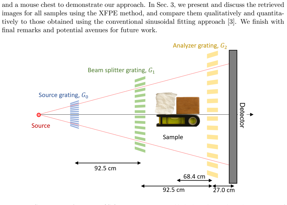
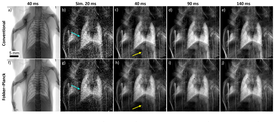
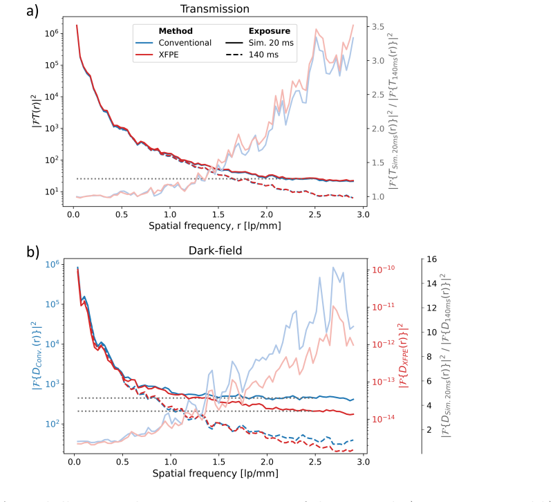
Grating interferometry is a promising diagnostic technique that enables simultaneous acquisition of three complementary, synergistic X-ray images: transmission, differential phase, and dark-field. Its key advantage over other setups is its ability to use large pixels and, hence, large-area detectors, as well as its compatibility with low-coherence, compact X-ray sources, both of which are key factors for human-scale imaging. It has already demonstrated strong potential for chest imaging applications, including the diagnosis of pulmonary emphysema, fibrosis, and cancer. To retrieve transmission, differential phase, and dark-field images from data, an algorithm is required to separate the distinct mechanisms contributing to measured contrast. Since its realization, this image-retrieval step has remained fundamentally unchanged. In this work, we develop a novel transmission- and dark-field retrieval algorithm for grating-interferometry derived from the X-ray Fokker-Planck equation. To demonstrate and validate our Fokker-Planck algorithm, we apply it to experimental measurements of a test sample and to data from a mouse chest acquired with varying exposure times and added Poisson noise. The retrieved images were qualitatively and quantitatively compared with those retrieved using a conventional sinusoidal-fitting approach. Across both samples, the Fokker--Planck method produced images consistent with conventional retrieval, with a comparable signal-to-noise ratio. Notably, our Fokker-Planck method suppresses artefacts arising in the conventional approach under grating perturbations (e.g., structural defects like scratches) and reduced flux or visibility, yielding smoother and more reproducible images. Additionally, we demonstrate that our Fokker-Planck method has an advantage over the conventional dark-field retrieval method for fast sample imaging with short exposure times and high noise.
Editorial analysis
A structured set of objections, weighed in public.
Referee Report
Summary. The paper develops a novel retrieval algorithm for transmission and dark-field images in X-ray grating interferometry by solving the Fokker-Planck equation for intensity flow. It reports that the method yields images consistent with the conventional sinusoidal-fitting approach on both a test sample and mouse chest data, with comparable SNR, while suppressing artifacts from grating defects, reduced flux/visibility, and high noise (short exposures), offering advantages for fast imaging.
Significance. If the central claim holds, the approach could improve robustness in clinical grating interferometry applications such as pulmonary imaging, where grating imperfections and low-dose constraints are common. The experimental validation under controlled perturbations and noise is a concrete strength, as is the focus on practical advantages over the long-standard retrieval method.
major comments (2)
- [Abstract and Results] Abstract and results (comparison with conventional method): The claims of 'qualitative and quantitative agreement' and 'comparable signal-to-noise ratio' are not supported by any reported numerical error metrics, difference maps, statistical tests, or explicit SNR values; without these, it is impossible to evaluate whether the observed smoothness reflects improved fidelity or implicit regularization.
- [Theory] Theory section (Fokker-Planck derivation): The manuscript does not demonstrate that the derived retrieval expressions reduce, by the paper's own equations, to the transmission and dark-field coefficients that would be obtained by fitting the same measured intensities; this leaves open whether unmodeled grating-specific propagation or higher-order scattering terms bias the separation.
minor comments (1)
- [Methods] Methods: Specify the precise form of the Fokker-Planck equation employed, including any boundary conditions or approximations for the grating interferometer geometry.
Simulated Author's Rebuttal
We thank the referee for their thorough review and constructive feedback on our manuscript. We address each of the major comments below and have made revisions to strengthen the paper as suggested.
read point-by-point responses
-
Referee: [Abstract and Results] Abstract and results (comparison with conventional method): The claims of 'qualitative and quantitative agreement' and 'comparable signal-to-noise ratio' are not supported by any reported numerical error metrics, difference maps, statistical tests, or explicit SNR values; without these, it is impossible to evaluate whether the observed smoothness reflects improved fidelity or implicit regularization.
Authors: We agree with the referee that additional quantitative metrics would enhance the rigor of the comparison. In the revised manuscript, we now include explicit SNR values computed for regions of interest in both the test sample and mouse chest data, difference maps showing the pixel-wise differences between the Fokker-Planck and conventional retrieval methods, and root-mean-square deviation metrics. These additions confirm the qualitative and quantitative agreement while demonstrating that the reduced artifacts and smoothness arise from the method's robustness to noise and perturbations rather than over-regularization. revision: yes
-
Referee: [Theory] Theory section (Fokker-Planck derivation): The manuscript does not demonstrate that the derived retrieval expressions reduce, by the paper's own equations, to the transmission and dark-field coefficients that would be obtained by fitting the same measured intensities; this leaves open whether unmodeled grating-specific propagation or higher-order scattering terms bias the separation.
Authors: We thank the referee for highlighting this important point regarding the theoretical consistency. Upon review, we recognize that an explicit limiting case was not presented. In the revised version, we have added a new paragraph in the Theory section that derives the reduction of our Fokker-Planck-based expressions to the conventional sinusoidal fitting coefficients under the assumptions of the standard grating interferometry model (small phase gradients and negligible higher-order scattering). This shows that our method is consistent with the conventional approach in the appropriate limit, with differences arising only from the handling of perturbations and noise. revision: yes
Circularity Check
Fokker-Planck derivation yields independent retrieval formulas
full rationale
The paper derives its transmission and dark-field retrieval algorithm directly by solving the X-ray Fokker-Planck equation for intensity flow through the grating interferometer. The resulting closed-form expressions are obtained from the differential equation under stated assumptions rather than by fitting parameters to the measured intensities or by re-expressing fitted quantities as predictions. No self-definitional loops, fitted-input-as-prediction steps, or load-bearing self-citations appear in the derivation chain. Validation consists of qualitative and quantitative comparison against the conventional sinusoidal-fitting method on experimental data, but the formulas themselves are not shown to reduce to those data by construction. This is a standard first-principles derivation with external experimental checks, producing a self-contained result.
Axiom & Free-Parameter Ledger
axioms (1)
- domain assumption X-ray intensity propagation and scattering in grating interferometry obey the Fokker-Planck equation.
Reference graph
Works this paper leans on
-
[1]
Atsushi Momose et al.Jpn. J. Appl. Phys., 42(7B):L866, 2003
2003
-
[2]
Express, 13(16):6296–6304, 2005
Timm Weitkamp et al.Opt. Express, 13(16):6296–6304, 2005
2005
-
[3]
Mater., 7:134–137, 2008
Franz Pfeiffer et al.Nat. Mater., 7:134–137, 2008
2008
-
[4]
Phys., 2(4):258–261, 2006
Franz Pfeiffer et al.Nat. Phys., 2(4):258–261, 2006
2006
-
[5]
Synchrotron Radiat., 16(1):43–47, 2009
Martin Bech et al.J. Synchrotron Radiat., 16(1):43–47, 2009
2009
-
[6]
Rep., 7(1):14477, 2017
Christoph Jud et al.Sci. Rep., 7(1):14477, 2017
2017
-
[7]
Atsushi Momose et al.Philos. Trans. R. Soc. A, 372(2010):20130023, 2014
2010
-
[8]
Health, 3(11):e733–e744, 2021
Konstantin Willer et al.Lancet Digit. Health, 3(11):e733–e744, 2021
2021
-
[9]
Radiol., 58(11):775–781, 2023
Theresa Urban et al.Invest. Radiol., 58(11):775–781, 2023
2023
-
[10]
Manuel Viermetz et al.Proc. Natl. Acad. Sci. U.S.A., 119(8):e2118799119, 2022
2022
-
[11]
Chem., 181:118052, 2024
Zhili Wang.TrAC, Trends Anal. Chem., 181:118052, 2024
2024
-
[12]
Phys., 39(1):424–428, 2012
Nicholas Bevins et al.Med. Phys., 39(1):424–428, 2012
2012
-
[13]
Phys., 38(7):4133–4140, 2011
Thomas Weber et al.Med. Phys., 38(7):4133–4140, 2011
2011
-
[14]
Express, 25(6):6349–6364, 2017
Carolina Arboleda et al.Opt. Express, 25(6):6349–6364, 2017
2017
-
[15]
Rep., 15(1):14223, 2025
Alexandre Pereira et al.Sci. Rep., 15(1):14223, 2025
2025
-
[16]
Imaging, 4(4):58, 2018
Volker Ludwig et al.J. Imaging, 4(4):58, 2018
2018
-
[17]
Phys., 52(4):2145–2154, 2025
Karin Hellerhoff et al.Med. Phys., 52(4):2145–2154, 2025
2025
-
[18]
Sebastian Kaeppler et al.J. Med. Imaging, 4(3):034005, 2017
2017
-
[19]
Phd thesis, University of Helsinki, Helsinki, Finland, 2025
Henrik M¨ akinen. Phd thesis, University of Helsinki, Helsinki, Finland, 2025
2025
-
[20]
Wolfgang Noichl et al.IEEE Trans. Med. Imaging, 43(1):28–38, 2023
2023
-
[21]
Express, 33(1):1345–1358, 2025
Simon Spindler et al.Opt. Express, 33(1):1345–1358, 2025
2025
-
[22]
Rep., 14(1):32169, 2024
Sami Wirtensohn et al.Sci. Rep., 14(1):32169, 2024
2024
-
[23]
Rep., 9(1):17537, 2019
David M Paganin and Kaye S Morgan.Sci. Rep., 9(1):17537, 2019. 16
2019
-
[24]
Rep., 9(1):17465, 2019
Kaye S Morgan and David M Paganin.Sci. Rep., 9(1):17465, 2019
2019
-
[25]
M R Teague.J. Opt. Soc. Am., 73(11):1434–1441, 1983
1983
-
[26]
Masanori Mitome.Microscopy, 70(1):69–74, 2021
2021
-
[27]
Lasers Eng., 135:106187, 2020
Chao Zuo et al.Opt. Lasers Eng., 135:106187, 2020
2020
-
[28]
Microsc., 206(1):33–40, 2002
David M Paganin et al.J. Microsc., 206(1):33–40, 2002
2002
-
[29]
Thomas A Leatham et al.IEEE Trans. Med. Imaging, 42(6), 2023
2023
-
[30]
Express, 32(3):4588–4602, 2024
Thomas A Leatham et al.Opt. Express, 32(3):4588–4602, 2024
2024
-
[31]
Jannis N Ahlers et al.Optica, 11(8):1182–1191, 2024
2024
-
[32]
Lett., 50(7):2171–2174, 2025
Jannis N Ahlers et al.Opt. Lett., 50(7):2171–2174, 2025
2025
-
[33]
Opt., 22(12):125604, 2020
Konstantin M Pavlov et al.J. Opt., 22(12):125604, 2020
2020
-
[34]
Konstantin M Pavlov et al.Phys. Rev. A, 104(5):053505, 2021
2021
-
[35]
Samantha J Alloo et al.J. Med. Imaging, 9:031502–031502, 2022
2022
-
[36]
Rep., 13(1):5424, 2023
Samantha J Alloo et al.Sci. Rep., 13(1):5424, 2023
2023
-
[37]
Mario A Beltran et al.Optica, 10(4):422–429, 2023
2023
-
[38]
Express, 33(2):3577–3600, 2025
Samantha J Alloo et al.Opt. Express, 33(2):3577–3600, 2025
2025
-
[39]
Jayvan Liu et al.Phys. Rev. A, 112(6):063530, 2025
2025
-
[40]
Scr., 100(7):075566, 2025
Samantha J Alloo and Kaye S Morgan.Phys. Scr., 100(7):075566, 2025
2025
-
[41]
Express, 30(7):10899–10918, 2022
Ying Ying How and Kaye S Morgan.Opt. Express, 30(7):10899–10918, 2022
2022
-
[42]
Marie-Christine Zdora et al.Phys. Rev. Lett., 118(20):203903, 2017
2017
-
[43]
Rep., 4(1):7243, 2014
Markus Strobl.Sci. Rep., 4(1):7243, 2014
2014
-
[44]
Synchrotron Radiat., 27:494–502, 2020
Zhili Wang et al.J. Synchrotron Radiat., 27:494–502, 2020
2020
-
[45]
Express, 31(1):635–650, 2022
Fabio De Marco et al.Opt. Express, 31(1):635–650, 2022
2022
-
[46]
Werneri A Lindberg et al.Phys. Med. Biol., 70(19):195015, 2025
2025
-
[47]
Alloo et al.J
Samantha J. Alloo et al.J. Synchrotron Radiat., 33(2), 2026
2026
-
[48]
Rep., 14(1):17807, 2024
Michelle K Croughan et al.Sci. Rep., 14(1):17807, 2024
2024
-
[49]
Kaye S Morgan et al.Appl. Phys. Lett., 100(12), 2012
2012
-
[50]
Phys., 37(11):6047–6054, 2010
Eric E Bennett et al.Med. Phys., 37(11):6047–6054, 2010
2010
-
[51]
Lett., 36(1):55–57, 2010
Kaye S Morgan et al.Opt. Lett., 36(1):55–57, 2010
2010
-
[52]
Konstantin M Pavlov et al.Phys. Rev. Appl., 13(5):054023, 2020
2020
-
[53]
Oxford University Press, 2006
David M Paganin.Coherent X-ray Optics. Oxford University Press, 2006
2006
-
[54]
Express, 28(11):16363–16384, 2020
Koh Hashimoto et al.Opt. Express, 28(11):16363–16384, 2020. 17
2020
-
[55]
Synchrotron Rad., 27(5):1395–1414, 2020
Benedikt G¨ unther et al.J. Synchrotron Rad., 27(5):1395–1414, 2020
2020
-
[56]
Regine Gradl et al.IEEE Trans. Med. Imaging, 38(2):649–656, 2018
2018
-
[57]
A Fokker–Planck approach to grating interferometry
Samantha J. Alloo. Resources for Alloo et al. “...A Fokker–Planck approach to grating interferometry”.https://zenodo.org/records/18395476, 2026. Accessed: 1-May-2026
-
[58]
Express, 18(16):16890–16901, 2010
Wataru Yashiro et al.Opt. Express, 18(16):16890–16901, 2010
2010
-
[59]
Express, 23(7):9233–9251, 2015
Wataru Yashiro and Atsushi Momose.Opt. Express, 23(7):9233–9251, 2015
2015
-
[60]
Cardiothorac
Florian T Gassert et al.Radiol. Cardiothorac. Imaging, 7(6):e240560, 2025
2025
-
[61]
Radiol., 4:1487895, 2025
Florian T Gassert et al.Front. Radiol., 4:1487895, 2025
2025
-
[62]
Michael Chabior et al.J. Appl. Phys., 110(5):053105, 2011
2011
-
[63]
Phys., 44(6):2453–2465, 2017
Xu Ji et al.Med. Phys., 44(6):2453–2465, 2017
2017
-
[64]
Status Solidi A, 204(8):2746–2752, 2007
Peter Modregger et al.Phys. Status Solidi A, 204(8):2746–2752, 2007
2007
-
[65]
Rep., 10(1):7890, 2020
Timur Gureyev et al.Sci. Rep., 10(1):7890, 2020
2020
-
[66]
Martin Bech et al.Phys. Med. Biol., 55(18):5529–5539, 2010
2010
-
[67]
Opt., 50(22):4310–4319, 2011
Susanna K Lynch et al.Appl. Opt., 50(22):4310–4319, 2011
2011
-
[68]
Lett., 112(6):68002, 2016
F Prade et al.Europhys. Lett., 112(6):68002, 2016
2016
-
[69]
David M Paganin et al.Phys. Rev. A, 108(1):013517, 2023
2023
-
[70]
Hunter C Meyer et al.arXiv preprint:2509.16503, 2025
work page internal anchor Pith review Pith/arXiv arXiv 2025
-
[71]
Jakob Haeusele et al.IEEE Trans. Med. Imaging, 42(10):2876–2885, 2023
2023
-
[72]
Tech., 15(8):937–945, 1980
Eckhart F¨ orster et al.Krist. Tech., 15(8):937–945, 1980
1980
-
[73]
Phys., 28(8):1610–1619, 2001
A Olivo et al.Med. Phys., 28(8):1610–1619, 2001
2001
-
[74]
Opt., 22(11):115607, 2020
David M Paganin et al.J. Opt., 22(11):115607, 2020. 18
2020
discussion (0)
Sign in with ORCID, Apple, or X to comment. Anyone can read and Pith papers without signing in.