Recognition: unknown
TRACED: In vivo imaging of extracellular intrinsic diffusivity, tortuosity, cell size distribution and cell density in human glioma patients
Pith reviewed 2026-05-08 01:57 UTC · model grok-4.3
The pith
TRACED is a biophysical model enabling simultaneous in vivo quantification of intracellular volume fraction, cell size distribution, extracellular intrinsic diffusivity, and tortuosity in glioma patients using diffusion MRI.
A machine-rendered reading of the paper's core claim, the machinery that carries it, and where it could break.
Core claim
TRACED incorporates diffusion time dependence in cell distributions to quantify pathologically-relevant properties in solid tumors. Neural networks were trained on Monte Carlo diffusion simulations using sphere distribution-based geometries to enable rapid computation of time-dependent diffusion MRI signals in cell populations of variable cell size. Data from eight glioma patients was fitted using a physics-informed transfer learning pipeline called Sim2PINN, enabling the simultaneous in vivo quantification of intracellular volume fraction, cell size distribution, extracellular intrinsic diffusivity, and tortuosity.
What carries the argument
The TRACED biophysical model that incorporates diffusion time dependence in cell distributions, implemented via neural networks trained on Monte Carlo simulations of sphere distribution-based geometries and fitted with the Sim2PINN physics-informed transfer learning pipeline.
Load-bearing premise
Sphere distribution-based geometries in the Monte Carlo simulations accurately represent the complex, non-spherical microstructure of glioma tissue.
What would settle it
Direct comparison showing that TRACED-derived cell size distributions and densities mismatch image-localized histology measurements in additional glioma patients.
Figures

read the original abstract
The lack of analytical models describing diffusion time dependence at intermediate time scales in complex tissue microstructure limits the accurate quantification of extracellular diffusivity and tissue microstructure. We introduce TRACED, a biophysical model that incorporates diffusion time dependence in cell distributions to quantify pathologically-relevant properties in solid tumors. Neural networks were trained on Monte Carlo diffusion simulations using sphere distribution-based geometries to enable the rapid computation of time-dependent diffusion MRI signals in cell populations of variable cell size. Model sensitivity and fit performance were assessed via simulation. Diffusion data from eight mixed-grade glioma patients was fitted using the TRACED model. Data fitting was performed using a novel physics-informed transfer learning pipeline, Sim2PINN. In two patients, cell size measurements were compared directly with image-localized histology. Simulation results indicate improved parameter estimation compared to the simple two-compartment model. TRACED enabled the simultaneous in vivo quantification of intracellular volume fraction, cell size distribution, extracellular intrinsic diffusivity, and tortuosity in glioma patients. Neural network implementations of diffusion time-dependence and tortuosity showed behavior consistent with coarse-graining and effective medium theory, respectively. Future work will explore the clinical utility of TRACED parameters in additional patients.
Editorial analysis
A structured set of objections, weighed in public.
Referee Report
Summary. The manuscript introduces TRACED, a biophysical model that incorporates diffusion time dependence into cell size distributions for analyzing diffusion MRI signals in complex tissue. Neural networks are trained on Monte Carlo simulations using polydisperse sphere geometries to enable rapid signal computation and parameter fitting. The model is applied to diffusion data from eight mixed-grade glioma patients via a physics-informed transfer learning pipeline (Sim2PINN), with direct cell-size comparison to image-localized histology in two patients. The central claim is that TRACED enables simultaneous in vivo quantification of intracellular volume fraction, cell size distribution, extracellular intrinsic diffusivity, and tortuosity, with improved performance over a simple two-compartment model and consistency with coarse-graining and effective medium theory.
Significance. If the forward model is shown to be faithful to real glioma microstructure, TRACED would represent a meaningful advance in non-invasive tumor imaging by providing maps of pathologically relevant parameters beyond standard ADC or two-compartment fits. The explicit incorporation of time dependence, the use of neural networks for efficient inversion, and the reported consistency of the learned tortuosity and time-dependence behaviors with established physical limits are strengths. The small patient cohort and limited histology validation, however, constrain the immediate translational significance.
major comments (2)
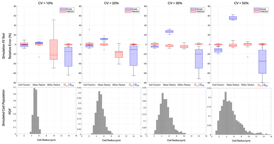
- [Methods (Monte Carlo geometry generation)] Methods section on Monte Carlo simulations: the training geometries are restricted to polydisperse spheres. Glioma cells are pleomorphic with irregular shapes and clustered extracellular spaces; if the intermediate-time diffusion features differ between spherical and realistic geometries, the fitted extracellular diffusivity, tortuosity, and cell-size distribution parameters cannot be interpreted as the claimed microstructural quantities. This assumption is load-bearing for the in vivo maps. A quantitative comparison of simulated signals (or recovered parameters) between sphere distributions and more realistic geometries (e.g., ellipsoids or histology-segmented cells) is required.
- [Results (simulation performance and histology comparison)] Results (simulation assessment and patient data fitting): the statements that TRACED shows 'improved parameter estimation' and enables 'simultaneous in vivo quantification' are not supported by reported quantitative metrics. No bias, variance, or RMSE values are given for parameter recovery across the simulation test set, nor are error bars or correlation statistics provided for the two-patient histology comparison of cell-size means. Without these, the superiority claim and the reliability of the patient maps cannot be evaluated.
minor comments (2)
- [Methods (Sim2PINN pipeline)] The description of the Sim2PINN transfer-learning procedure would benefit from an explicit statement of the physics-informed loss terms and how they enforce consistency with the underlying diffusion equation.
- [Model description] Notation for the cell-size distribution parameters (mean, variance, etc.) should be defined with equations in the model section to avoid ambiguity when reporting fitted maps.
Simulated Author's Rebuttal
We thank the referee for their constructive and detailed review of our manuscript. The comments highlight important aspects of model assumptions and quantitative reporting that we address below. We believe incorporating these points will strengthen the presentation of TRACED.
read point-by-point responses
-
Referee: Methods section on Monte Carlo simulations: the training geometries are restricted to polydisperse spheres. Glioma cells are pleomorphic with irregular shapes and clustered extracellular spaces; if the intermediate-time diffusion features differ between spherical and realistic geometries, the fitted extracellular diffusivity, tortuosity, and cell-size distribution parameters cannot be interpreted as the claimed microstructural quantities. This assumption is load-bearing for the in vivo maps. A quantitative comparison of simulated signals (or recovered parameters) between sphere distributions and more realistic geometries (e.g., ellipsoids or histology-segmented cells) is required.
Authors: We agree that the restriction to polydisperse spherical geometries represents a significant modeling assumption. Spheres were chosen to enable efficient Monte Carlo simulation of size distributions and to train the neural network on a well-defined parameter space while capturing key time-dependent effects. Real glioma cells are indeed pleomorphic, and differences in shape could influence intermediate-time diffusion features. A full quantitative comparison against ellipsoidal or histology-segmented geometries would require new, computationally intensive simulations and is not feasible within the current study timeline. In the revised manuscript we will expand the Discussion to explicitly state this limitation, reference relevant effective-medium and coarse-graining literature for non-spherical cells, and clarify that the reported parameters are effective quantities under the spherical approximation. We will also list realistic-geometry validation as an important direction for future work. revision: partial
-
Referee: Results (simulation assessment and patient data fitting): the statements that TRACED shows 'improved parameter estimation' and enables 'simultaneous in vivo quantification' are not supported by reported quantitative metrics. No bias, variance, or RMSE values are given for parameter recovery across the simulation test set, nor are error bars or correlation statistics provided for the two-patient histology comparison of cell-size means. Without these, the superiority claim and the reliability of the patient maps cannot be evaluated.
Authors: We thank the referee for this observation. The original manuscript presented simulation results primarily through visual comparisons and qualitative statements of improvement. In the revised version we will add a dedicated table reporting bias, variance, and RMSE for each recovered parameter (intracellular volume fraction, cell-size distribution moments, extracellular intrinsic diffusivity, and tortuosity) on the held-out simulation test set, with direct numerical comparison to the two-compartment model. For the two-patient histology validation we will include error bars on the mean cell-size estimates and report quantitative agreement metrics (Pearson correlation coefficient, slope, and p-value) between TRACED-derived and histologically measured cell sizes. These additions will provide the quantitative support needed to substantiate the performance claims. revision: yes
Circularity Check
No significant circularity; derivation is self-contained forward modeling
full rationale
The paper defines TRACED as a biophysical model whose signals are generated by Monte Carlo simulations on explicit sphere-distribution geometries, trains NNs on those simulations, and inverts patient data via Sim2PINN transfer learning to obtain parameter maps. All claimed quantities (intracellular volume fraction, cell-size distribution, extracellular diffusivity, tortuosity) are defined inside the model and recovered by standard least-squares or NN inversion; no equation or step equates a derived prediction back to its own fitted inputs by construction. No self-citation is invoked as a uniqueness theorem or load-bearing premise, and the geometry assumption is stated explicitly rather than smuggled. The derivation chain therefore remains independent of the target patient results.
Axiom & Free-Parameter Ledger
free parameters (2)
- cell size distribution parameters
- tortuosity and extracellular diffusivity scalars
axioms (1)
- domain assumption Sphere distribution-based geometries represent glioma tissue microstructure
Reference graph
Works this paper leans on
-
[1]
intrinsic
Athinoula A. Martinos Center for Biomedical Imaging, Department of Radiology, Massachusetts General Hospital, Boston, United States of America 2. Harvard-MIT Program in Health Sciences and Technology, Massachusetts Institute of Technology, Cambridge, United States of America 3. Harvard Medical School, Boston, United States of America 4. Department of Neur...
2000
-
[2]
Di8usion Imaging for Tumor Grading of Supratentorial Brain Tumors in the First Year of Life
Kralik SF , Taha A, Kamer AP , Cardinal JS, Seltman TA, Ho CY . Di8usion Imaging for Tumor Grading of Supratentorial Brain Tumors in the First Year of Life. American Journal of Neuroradiology. 2014;35(4):815-823. doi:10.3174/ajnr.A3757 2. Shin N, Kang TW, Min JH, et al. Utility of Di8usion-Weighted MRI for Detection of Locally Recurrent Pancreatic Cancer ...
-
[3]
A Model of E8ective Di8usion and Tortuosity in the Extracellular Space of the Brain
Hrabe J, Hrabĕtová S, Segeth K. A Model of E8ective Di8usion and Tortuosity in the Extracellular Space of the Brain. Biophysical Journal. 2004;87(3):1606-1617. doi:10.1529/biophysj.103.039495 9. Xiong H, Wilson BA, Ge X, et al. Glioblastoma Margin as a Di8usion Barrier Revealed by Photoactivation of Plasmonic Nanovesicles. Nano Lett. 2024;24(5):1570-1578....
-
[4]
Mapping immune cell infiltration using restricted di8usion MRI
Yeh F , Liu L, Hitchens TK, Wu YL. Mapping immune cell infiltration using restricted di8usion MRI. Magnetic Resonance in Med. 2017;77(2):603-612. doi:10.1002/mrm.26143 17. Panagiotaki E, Walker-Samuel S, Siow B, et al. Noninvasive Quantification of Solid Tumor Microstructure Using VERDICT MRI. Cancer Research. 2014;74(7):1902-1912. doi:10.1158/0008-5472.CAN...
-
[5]
Revealing mesoscopic structural universality with di8usion
Novikov DS, Jensen JH, Helpern JA, Fieremans E. Revealing mesoscopic structural universality with di8usion. Proc Natl Acad Sci USA. 2014;111(14):5088-5093. doi:10.1073/pnas.1316944111 33. Novikov DS, Fieremans E, Jespersen SN, Kiselev VG. Quantifying brain microstructure with di8usion MRI: Theory and parameter estimation. NMR in Biomedicine. 2019;32(4):e3...
-
[6]
Bartelink IH, Jones EF , Shahidi-Latham SK, et al. Tumor Drug Penetration Measurements Could Be the Neglected Piece of the Personalized Cancer Treatment Puzzle. Clin Pharmacol Ther. 2019;106(1):148-163. doi:10.1002/cpt.1211 42. Marin BM, Porath KA, Jain S, et al. Heterogeneous delivery across the blood-brain barrier limits the e8icacy of an EGFR-targeting...
-
[7]
Quantitative Analysis of the Correlation between Cell Size and Cellular Uptake of Particles
Khetan J, Shahinuzzaman M, Barua S, Barua D. Quantitative Analysis of the Correlation between Cell Size and Cellular Uptake of Particles. Biophysical Journal. 2019;116(2):347-359. doi:10.1016/j.bpj.2018.11.3134 51. Miotto M, Scalise S, Leonetti M, Ruocco G, Peruzzi G, Gosti G. A size-dependent division strategy accounts for leukemia cell size heterogeneit...
-
[8]
Iwanaga T, Usher W, Herman J. Toward SALib 2.0: Advancing the accessibility and interpretability of global sensitivity analyses. SESMO. 2022;4:18155. doi:10.18174/sesmo.18155 59. Setsompop K, Kimmlingen R, Eberlein E, et al. Pushing the limits of in vivo di8usion MRI for the Human Connectome Project. NeuroImage. 2013;80:220-233. doi:10.1016/j.neuroimage.2...
-
[9]
Paszke A, Gross S, Massa F , et al. PyTorch: An Imperative Style, High-Performance Deep Learning Library. 2019. doi:10.48550/ARXIV .1912.01703 67. Bankhead P , Loughrey MB, Fernández JA, et al. QuPath: Open source software for digital pathology image analysis. Sci Rep. 2017;7(1):16878. doi:10.1038/s41598-017-17204-5 68. Wicksell SD. THE CORPUSCLE PROBLEM....
work page internal anchor Pith review doi:10.48550/arxiv 2019
-
[10]
Latour LL, Svoboda K, Mitra PP , Sotak CH. Time-dependent di8usion of water in a biologicalmodel system. Proc Natl Acad Sci USA. 1994;91(4):1229-1233. doi:10.1073/pnas.91.4.1229 76. Mohiuddin E, Wakimoto H. Extracellular matrix in glioblastoma: opportunities for emerging therapeutic approaches. Am J Cancer Res. 2021;11(8):3742-3754. 77. Foo TKF , Tan ET, ...
-
[11]
and Soures, Nicholas and Kudithipudi, Dhireesha , year=
Ven GM van de, Soures N, Kudithipudi D. Continual Learning and Catastrophic Forgetting. In: ; 2025:153-168. doi:10.1016/B978-0-443-15754-7.00073-0 2. Bankhead P , Loughrey MB, Fernández JA, et al. QuPath: Open source software for digital pathology image analysis. Sci Rep. 2017;7(1):16878. doi:10.1038/s41598-017-17204-5 3. Guo AC, Cummings TJ, Dash RC, Pro...
discussion (0)
Sign in with ORCID, Apple, or X to comment. Anyone can read and Pith papers without signing in.