Recognition: unknown
Device-Induced Thrombus Formation in Cerebral Aneurysms: Linking Patient-Specific Clot Modeling and Functional Occlusion to Virtual Angiographic Assessment
Pith reviewed 2026-05-07 12:41 UTC · model grok-4.3
The pith
A coupled simulation framework shows early thrombus formation drives much of the perfusion suppression and altered contrast washout seen in virtual angiograms after aneurysm device placement.
A machine-rendered reading of the paper's core claim, the machinery that carries it, and where it could break.
Core claim
Coupling acute fibrin thrombus formation with virtual angiography under pulsatile hemodynamics reveals that early clot growth contributes substantially to functional occlusion, producing visible reductions in perfusion and shifts in contrast washout patterns even when devices leave some residual contrast access, with these signatures appearing directly in the simulated DSA-like images.
What carries the argument
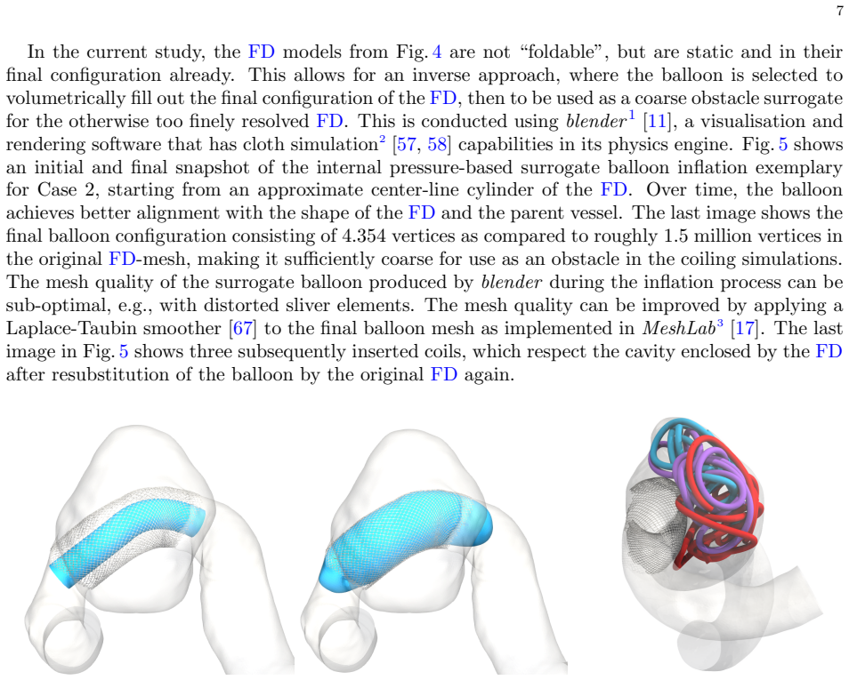
The coupled acute fibrin thrombus formation model and virtual contrast transport simulation that converts device-induced clotting into clinically interpretable angiographic signals.
If this is right
- Early thrombus formation substantially augments the perfusion suppression achieved by inflow reduction alone.
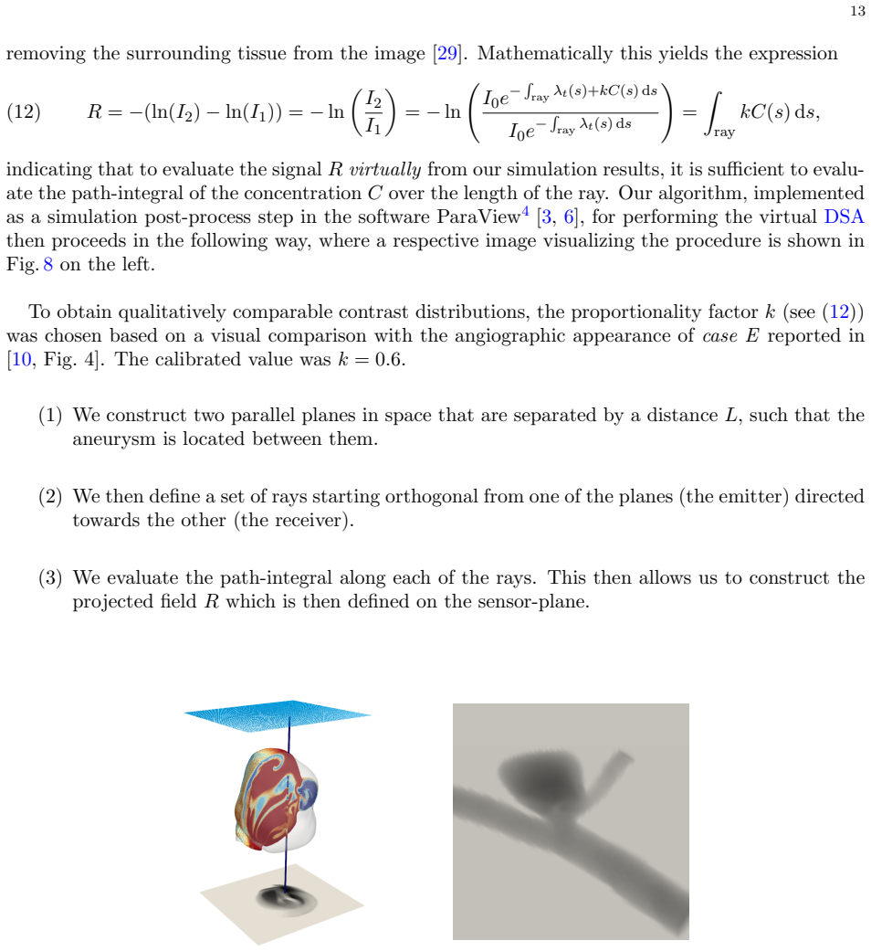
- Residual contrast access and trapping can persist after device placement and are visible in virtual images.
- Altered washout patterns produced by thrombus growth are directly reflected in the simulated angiographic sequences.
- Vortical flow structures promote device-induced thrombosis in at least some aneurysm morphologies.
- The framework supplies a route for evaluating occlusion outcomes through metrics already familiar in clinical DSA assessment.
Where Pith is reading between the lines
- Patient-specific versions of the model could be used to rank which device type is most likely to produce rapid functional occlusion for a given aneurysm shape.
- Linking the simulation output to real-time DSA data streams might help explain why certain treatments achieve incomplete isolation.
- Extending the thrombus model beyond the acute phase could allow prediction of long-term occlusion durability.
- The same coupling technique could be applied to other vascular sites where devices are used to induce localized clotting.
Load-bearing premise
The acute fibrin thrombus formation model and virtual contrast transport simulation accurately represent real in-vivo processes and DSA imaging without direct experimental or clinical validation.
What would settle it
Side-by-side comparison of the generated virtual angiograms against actual post-treatment DSA sequences from patients or animal models that have the same aneurysm geometries and device placements.
Figures


read the original abstract
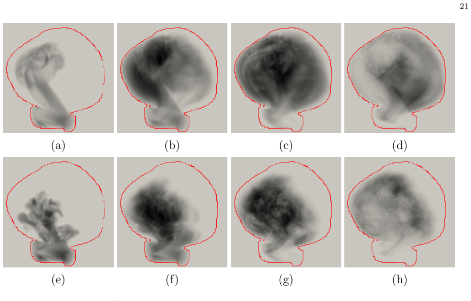
Endovascular treatment of cerebral aneurysms aims to achieve functional occlusion and isolation of the aneurysm sac from bloodflow. In clinical practice, treatment success is assessed primarily through digital subtraction angiography (DSA), which visualizes contrast-agent inflow and washout but does not directly resolve thrombus formation driving early occlusion. We present a computational framework that couples acute fibrin thrombus formation with virtual angiography, enabling early thrombus growth to be interpreted through clinically familiar DSA-like imaging. Three common treatment strategies: endovascular coiling, flow diversion, and stent-assisted coiling, are modeled under pulsatile hemodynamics and linked to simulated contrast transport. Across three representative aneurysm morphologies, the simulations demonstrate that while devices reduce inflow, residual contrast access and trapping may persist, with early thrombus formation contributing substantially to perfusion suppression and altered washout patterns. These effects are clearly reflected in the virtual angiographic imaging. The importance of vortical structures in device-induced thrombosis is highligthed in one of the cases. By seeking to align modelling and simulation tools with clinically-relevant metrics, with a particular focus on occlusion outcome, this work presents a good starting point for bridging the gap between these two paradigms.
Editorial analysis
A structured set of objections, weighed in public.
Referee Report
Summary. The manuscript presents a computational framework coupling patient-specific acute fibrin thrombus formation modeling with virtual angiography to assess functional occlusion in cerebral aneurysms treated by endovascular coiling, flow diversion, or stent-assisted coiling. Under pulsatile hemodynamics, simulations for three representative aneurysm morphologies demonstrate that device-induced inflow reduction is augmented by early thrombus growth, which substantially suppresses perfusion and alters contrast washout patterns as visualized in the simulated DSA-like imaging; vortical flow structures are noted as important in one morphology.
Significance. If the coupled model holds, the work offers a clinically aligned way to interpret DSA findings in terms of underlying thrombus dynamics rather than device geometry alone, potentially aiding prediction of early occlusion outcomes. The explicit linkage of thrombus kinetics to virtual angiographic metrics is a constructive step toward bridging computational hemodynamics with treatment assessment paradigms.
major comments (3)
- [Abstract/Methods] Abstract and Methods: The central claim that 'early thrombus formation contributing substantially to perfusion suppression' is not supported by any reported parameter values, rate constants for fibrin formation or platelet adhesion, sensitivity analysis, or direct validation against experimental/clinical thrombus growth rates or DSA washout data; without these the attribution of effects to thrombus versus device inflow reduction cannot be evaluated.
- [Results] Results: No quantitative metrics (e.g., time to occlusion, residual contrast volume, or washout half-times) comparing device-only versus device-plus-thrombus cases are provided across the three morphologies, leaving the 'substantially' qualifier and the virtual angiographic reflection of thrombus effects unquantified.
- [Methods] Methods: The virtual contrast transport simulation and its coupling to the thrombus model lack benchmarking against in-vivo or in-vitro DSA imaging under device-altered flows; this directly affects the reliability of the claimed alignment with clinical assessment.
minor comments (2)
- [Abstract] Abstract: Typo in 'highligthed' (should be 'highlighted').
- [Abstract] Abstract: The self-referential phrase 'this work presents a good starting point' should be revised to an objective statement about the framework's scope and limitations.
Simulated Author's Rebuttal
We thank the referee for the constructive and detailed comments. We have addressed each major point below, providing clarifications and committing to revisions that strengthen the manuscript without overstating the current results.
read point-by-point responses
-
Referee: [Abstract/Methods] Abstract and Methods: The central claim that 'early thrombus formation contributing substantially to perfusion suppression' is not supported by any reported parameter values, rate constants for fibrin formation or platelet adhesion, sensitivity analysis, or direct validation against experimental/clinical thrombus growth rates or DSA washout data; without these the attribution of effects to thrombus versus device inflow reduction cannot be evaluated.
Authors: We agree that explicit parameter reporting is essential. The thrombus model employs rate constants for fibrin formation and platelet adhesion drawn from established literature on acute thrombus kinetics under shear. In the revised manuscript we will add a table in Methods listing all parameter values, their literature sources, and the governing equations. A full sensitivity analysis and direct experimental/clinical validation of growth rates against DSA data are beyond the scope of this computational framework; we will explicitly note this limitation in the Discussion and qualify the 'substantially' claim as arising from comparative device-only versus device-plus-thrombus simulations rather than absolute validation. revision: partial
-
Referee: [Results] Results: No quantitative metrics (e.g., time to occlusion, residual contrast volume, or washout half-times) comparing device-only versus device-plus-thrombus cases are provided across the three morphologies, leaving the 'substantially' qualifier and the virtual angiographic reflection of thrombus effects unquantified.
Authors: We accept this observation. The existing simulation datasets contain the necessary time-resolved contrast fields. In the revised Results we will add quantitative metrics—washout half-times, residual contrast volume at 5 s and 10 s post-injection, and effective perfusion suppression ratios—for device-only versus device-plus-thrombus cases across all three morphologies. These will be presented in a new table and referenced in the text to make the contribution of early thrombus growth explicit and quantifiable. revision: yes
-
Referee: [Methods] Methods: The virtual contrast transport simulation and its coupling to the thrombus model lack benchmarking against in-vivo or in-vitro DSA imaging under device-altered flows; this directly affects the reliability of the claimed alignment with clinical assessment.
Authors: The contrast transport is solved via an advection-diffusion equation on the thrombus-modified velocity field using standard numerical methods previously validated for aneurysm CFD. We will expand the Methods section to include explicit references to prior benchmarking studies of similar contrast-transport models and to describe the one-way coupling procedure in greater detail. New in-vivo or in-vitro DSA benchmarking under device conditions is not feasible within the present study; we will acknowledge this as a limitation and identify it as future work. revision: partial
- Direct validation of thrombus growth rates and DSA washout patterns against experimental or clinical data under device-altered flows
- Comprehensive sensitivity analysis of all thrombus-model parameters
Circularity Check
No significant circularity; derivation self-contained in simulation framework
full rationale
The accessible manuscript text consists of the abstract and high-level description of a coupled computational framework for thrombus formation and virtual angiography. No equations, parameter-fitting procedures, or derivation steps are quoted that reduce predictions to inputs by construction. Claims about thrombus contribution to occlusion are presented as outcomes of the simulations across morphologies, without self-definitional loops, fitted inputs renamed as predictions, or load-bearing self-citations that collapse the central result. The work explicitly positions itself as a starting point without invoking uniqueness theorems or ansatzes from prior author work as forcing mechanisms. This is the normal case of an independent modeling study whose results can be benchmarked externally.
Axiom & Free-Parameter Ledger
free parameters (2)
- Thrombus formation rate constants
- Contrast transport coefficients
axioms (2)
- domain assumption Thrombus model captures real acute fibrin formation dynamics in aneurysms
- domain assumption Virtual angiography reproduces key features of clinical DSA
Reference graph
Works this paper leans on
-
[1]
In-silico trial of intracranial flow diverters replicates and expands insights from conventional clinical trials.Nature Communications, 12(1):3861, 2021
2021
-
[2]
Rahim Abo Kasem, Zachary Hubbard, Conor Cunningham, Hani Almorawed, Julio Isidor, Imad Samman Tahhan, Mohammad-Mahdi Sowlat, Sofia Babool, Layal Abodest, and Alejandro M Spiotta. Comparison of flow diverter alone versus flow diverter with coiling for large and giant intracranial aneurysms: systematic review and meta- analysis of observational studies.Jour...
2026
-
[3]
Paraview: An end-user tool for large data visualization
James Ahrens, Berk Geveci, and Charles Law. Paraview: An end-user tool for large data visualization. In Visualization Handbook. Elsevier, 2005
2005
-
[4]
Predictors of aneurysm occlusion following treatment with the WEB device: systematic review and case series.Neurosurgical Review, 45(2):925–936, 2022
Fadi Al Saiegh, Lohit Velagapudi, Omaditya Khanna, Ahmad Sweid, Nikolaos Mouchtouris, Michael P Baldas- sari, Thana Theofanis, Rizwan Tahir, Victoria Schunemann, Carrie Andrews, Lucas Philipp, Nohra Chalouhi, Stavropoula I Tjoumakaris, David Hasan, M Reid Gooch, Nabeel A Herial, Robert H Rosenwasser, and Pascal Jabbour. Predictors of aneurysm occlusion fo...
2022
-
[5]
Unauthorized use, distribution or duplication is prohibited
Ansys, Inc., Southpointe, 2600 Ansys Drive, Canonsburg, PA 15317, USA.Ansys Fluent User’s Guide, release 2025 r2 edition, 7 2025.©2025 Ansys, Inc. Unauthorized use, distribution or duplication is prohibited
2025
-
[6]
Kitware, 2015
Utkarsh Ayachit.The ParaView Guide: A Parallel Visualization Application. Kitware, 2015
2015
-
[7]
H. Baek, M. V. Jayaraman, P. D. Richardson, and G. E. Karniadakis. Flow instability and wall shear stress variation in intracranial aneurysms.Journal of The Royal Society Interface, 7(47):967–988, 12 2009
2009
-
[8]
Matthew T Bender, Geoffrey P Colby, Li-Mei Lin, Bowen Jiang, Erick M Westbroek, Risheng Xu, Jessica K Campos, Judy Huang, Rafael J Tamargo, and Alexander L Coon. Predictors of cerebral aneurysm persistence and occlusion after flow diversion: a single-institution series of 445 cases with angiographic follow-up.Journal of Neurosurgery, 130(1):259–267, 2018
2018
-
[9]
Virtual stenting of intracranial aneurysms – explicit versus implicit approaches
Philipp Berg and G´ abor Janiga. Virtual stenting of intracranial aneurysms – explicit versus implicit approaches. In Conference on Modelling Fluid Flow (CMFF’18): The 17th International Conference on Fluid Flow Technologies, 2018
2018
-
[10]
Multiple aneurysms anatomy challenge 2018 (match): phase i: segmentation.Cardiovascular engineering and technology, 9(4):565–581, 2018
Philipp Berg, Samuel Voß, Sylvia Saalfeld, G´ abor Janiga, Aslak W Bergersen, Kristian Valen-Sendstad, Jan Bruening, Leonid Goubergrits, Andreas Spuler, Nicole M Cancelliere, et al. Multiple aneurysms anatomy challenge 2018 (match): phase i: segmentation.Cardiovascular engineering and technology, 9(4):565–581, 2018
2018
-
[11]
Blender Foundation, Stichting Blender Foundation, Amsterdam, 2025
Blender Online Community.Blender - a 3D modelling and rendering package. Blender Foundation, Stichting Blender Foundation, Amsterdam, 2025
2025
-
[12]
Brindise, Sean Rothenberger, Benjamin Dickerhoff, Susanne Schnell, Michael Markl, David Saloner, Vitaliy L
Melissa C. Brindise, Sean Rothenberger, Benjamin Dickerhoff, Susanne Schnell, Michael Markl, David Saloner, Vitaliy L. Rayz, and Pavlos P. Vlachos. Multi-modality cerebral aneurysm haemodynamic analysis: in vivo 4d flow mri, in vitro volumetric particle velocimetry and in silico computational fluid dynamics.Journal of The Royal Society Interface, 16(158):...
2019
-
[13]
Cebral, Fernando Mut, Rainald L¨ ohner, Laurel Marsh, Alireza Chitsaz, Cem Bilgin, Esref Bayraktar, David Kallmes, and Ramanathan Kadirvel
Juan R. Cebral, Fernando Mut, Rainald L¨ ohner, Laurel Marsh, Alireza Chitsaz, Cem Bilgin, Esref Bayraktar, David Kallmes, and Ramanathan Kadirvel. Modeling fibrin accumulation on flow-diverting devices for intracranial aneurysms.International Journal for Numerical Methods in Biomedical Engineering, 40(12):e3883, 2024
2024
-
[14]
A new aneurysm occlusion classification after the impact of flow modification.American Journal of Neuroradiology, 37(1):19–24, 2016
HS Cekirge and I Saatci. A new aneurysm occlusion classification after the impact of flow modification.American Journal of Neuroradiology, 37(1):19–24, 2016
2016
-
[15]
Comparison of flow diversion and coiling in large unruptured intracranial saccular aneurysms.Stroke, 44(8):2150–2154, 2013
Nohra Chalouhi, Stavropoula Tjoumakaris, Robert M Starke, L Fernando Gonzalez, Ciro Randazzo, David Hasan, Jeffrey F McMahon, Saurabh Singhal, Lea A Moukarzel, Aaron S Dumont, et al. Comparison of flow diversion and coiling in large unruptured intracranial saccular aneurysms.Stroke, 44(8):2150–2154, 2013
2013
-
[16]
Future directions of flow diverter therapy.Neurosurgery, 86(Supplement 1):S106–S116, 2020
Albert Ho Yuen Chiu and Timothy John Phillips. Future directions of flow diverter therapy.Neurosurgery, 86(Supplement 1):S106–S116, 2020
2020
-
[17]
Meshlab: an open-source mesh processing tool
Paolo Cignoni, Marco Callieri, Massimiliano Corsini, Matteo Dellepiane, Fabio Ganovelli, Guido Ranzuglia, et al. Meshlab: an open-source mesh processing tool. InEurographics Italian chapter conference, volume 2008, pages 129–136. Salerno, 2008
2008
-
[18]
Czaja, G
B. Czaja, G. Z´ avodszky, V. Azizi Tarksalooyeh, and A. G. Hoekstra. Cell-resolved blood flow simulations of sac- cular aneurysms: effects of pulsatility and aspect ratio.Journal of The Royal Society Interface, 15(146):20180485, 2018
2018
-
[19]
Engineering design of optimal strategies for blood clot dissolution.Annual review of biomedical engineering, 1(1):427–461, 1999
Scott L Diamond. Engineering design of optimal strategies for blood clot dissolution.Annual review of biomedical engineering, 1(1):427–461, 1999
1999
-
[20]
Inner clot diffusion and permeation during fibrinolysis.Biophysical journal, 65(6):2622–2643, 1993
Scott L Diamond and Sriram Anand. Inner clot diffusion and permeation during fibrinolysis.Biophysical journal, 65(6):2622–2643, 1993
1993
-
[21]
Computational and experimental investigation of particulate matter deposition in cerebral side aneurysms.Journal of The Royal Society Interface, 17(169):20200510, 2020
Mark Epshtein and Netanel Korin. Computational and experimental investigation of particulate matter deposition in cerebral side aneurysms.Journal of The Royal Society Interface, 17(169):20200510, 2020. 26
2020
-
[22]
S. P. Ferns, J. J. Schneiders, M. Siebes, R. van den Berg, E. T. van Bavel, and C. B. Majoie. Intracranial blood-flow velocity and pressure measurements using an intra-arterial dual-sensor guidewire.American Journal of Neuroradiology, 31:324–326, 2010
2010
-
[23]
The current landscape of intracranial aneurysms in Africa: management outcomes, challenges, and strategies, a narrative review.Neurosurgical Review, 46(1):1–16, 2023
Tomas Ferreira, Wireko Andrew Awuah, Joecelyn Kirani Tan, Favour Tope Adebusoye, Syed Hasham Ali, Ha- reesha Rishab Bharadwaj, Adrenito Nicolas, Carolina Fernandes, Muhammad Jawad Zahid, and Toufik Abdul- Rahman. The current landscape of intracranial aneurysms in Africa: management outcomes, challenges, and strategies, a narrative review.Neurosurgical Rev...
2023
-
[24]
Ford, G.R
M.D. Ford, G.R. Stuhne, H.N. Nikolov, D.F. Habets, S.P. Lownie, D.W. Holdsworth, and D.A. Steinman. Vir- tual angiography for visualization and validation of computational models of aneurysm hemodynamics.IEEE Transactions on Medical Imaging, 24(12):1586–1592, 2005
2005
-
[25]
Numerical simulation of endovascular treatment options for cerebral aneurysms.GAMM-Mitteilungen, page e202370007, 2024
Martin Frank, Fabian Holzberger, Medeea Horvat, Jan Kirschke, Matthias Mayr, Markus Muhr, Natalia Neb- ulishvili, Alexander Popp, Julian Schwarting, and Barbara Wohlmuth. Numerical simulation of endovascular treatment options for cerebral aneurysms.GAMM-Mitteilungen, page e202370007, 2024
2024
-
[26]
Saccular intracranial aneurysm: pathology and mechanisms.Acta Neuropathologica, 123(6):773–786, 2012
Juhana Fr¨ osen, Riikka Tulamo, Anders Paetau, Elisa Laaksamo, Miikka Korja, Aki Laakso, Mika Niemel¨ a, and Juha Hernesniemi. Saccular intracranial aneurysm: pathology and mechanisms.Acta Neuropathologica, 123(6):773–786, 2012
2012
-
[27]
Goubergrits, J
L. Goubergrits, J. Schaller, U. Kertzscher, N. van den Bruck, K. Poethkow, Ch. Petz, H.-Ch. Hege, and A. Spuler. Statistical wall shear stress maps of ruptured and unruptured middle cerebral artery aneurysms.Journal of The Royal Society Interface, 9(69):677–688, 09 2011
2011
-
[28]
Electrothrombosis of saccular aneurysms via endovascular approach.Journal of neurosurgery, 75:1–7, 1991
Guido Guglielmi, Fernando Vinuela, Ivan Sepetka, and Velio Macellari. Electrothrombosis of saccular aneurysms via endovascular approach.Journal of neurosurgery, 75:1–7, 1991
1991
-
[29]
Method of pro- cessing images for digital subtraction angiography, 2007
Sylvain Justin Georges Andr´ e Haupert, Peter Maria Johannes Rongen, and Herman Stegehuis. Method of pro- cessing images for digital subtraction angiography, 2007. Filed 2002-09-04; priority 2001-09-04
2007
-
[30]
W. H. Ho, I. J. Tshimanga, M. N. Ngoepe, M. C. Jermy, and P. H. Geoghegan. Evaluation of a Desktop 3D Printed Rigid Refractive-Indexed-Matched Flow Phantom for PIV Measurements on Cerebral Aneurysms.Cardiovascular Engineering and Technology, pages 24–28, 2019
2019
-
[31]
Pathological findings of saccular cere- bral aneurysms—impact of subintimal fibrin deposition on aneurysm rupture.Neurosurgical Review, 38(3):531– 540, 2015
Masaaki Hokari, Naoki Nakayama, Hiroshi Nishihara, and Kiyohiro Houkin. Pathological findings of saccular cere- bral aneurysms—impact of subintimal fibrin deposition on aneurysm rupture.Neurosurgical Review, 38(3):531– 540, 2015
2015
-
[32]
Fabian Holzberger, Markus Muhr, and Barbara Wohlmuth. A comprehensive numerical approach to coil place- ment in cerebral aneurysms: mathematical modeling and in silico occlusion classification.Biomechanics and Modeling in Mechanobiology, 23(6):2063–2089, Dec 2024
2063
-
[33]
Malan, Wei Hua Ho, and Male- bogo N
Struan Hume, Jean Marc Ilunga Tshimanga, Patrick Geoghegan, Arnaud G. Malan, Wei Hua Ho, and Male- bogo N. Ngoepe. Effect of Pulsatility on the Transport of Thrombin in an Idealized Cerebral Aneurysm Geometry. Symmetry, 14(1):1–18, 2022
2022
-
[34]
Struan Robertson Hume. Computational model of thrombosis in cerebral aneurysms for predicting clotting out- comes in flow diverter treated patient-derived geometries validated with novel piv-based ln vitro clotting flow experiment. 2024
2024
-
[35]
Numerical Solution of Advection-Diffusion-Reaction Equations
Willem Hundsdorfer. Numerical Solution of Advection-Diffusion-Reaction Equations
-
[36]
Rajagopal
Alena Jarol´ ımov´ a, Jaroslav Hron, Karel T˚ uma, Radom´ ır Chabiniok, Josef M´ alek, and Kumbakonam R. Rajagopal. Evidence arguing against the validity of the no-slip boundary condition: blood flow in vivo.arXiv preprint, 2025
2025
-
[37]
Clinical Practice Guideline for the Management of Intracranial Aneurysms.Neurointervention, 9(2):63–71, 9 2014
Hae Woong Jeong, Jung Hwa Seo, Sung Tae Kim, Cheol Kyu Jung, and Sang-il Suh. Clinical Practice Guideline for the Management of Intracranial Aneurysms.Neurointervention, 9(2):63–71, 9 2014
2014
-
[38]
A Mechano-Chemical Computational Model of Deep Vein Thrombosis.Frontiers in Physics, 10, 6 2022
Qudus Jimoh-Taiwo, Rashid Haffejee, and Malebogo Ngoepe. A Mechano-Chemical Computational Model of Deep Vein Thrombosis.Frontiers in Physics, 10, 6 2022
2022
-
[39]
An experimental study on thrombogenicity of various metallic microcoils with or without thrombogenic coatings.Investigative Radiology, 33(7):407–410, July 1998
Tae Sung Kim, Jae Hyung Park, Yoonshin Lee, Jin Wook Chung, and Man Chung Han. An experimental study on thrombogenicity of various metallic microcoils with or without thrombogenic coatings.Investigative Radiology, 33(7):407–410, July 1998
1998
-
[40]
Cen- trum voor Wiskunde en Informatica Amsterdam, 1993
Barry Koren.A robust upwind discretization method for advection, diffusion and source terms, volume 45. Cen- trum voor Wiskunde en Informatica Amsterdam, 1993
1993
-
[41]
R. M. W. Kremers, B. de Laat, R. J. Wagenvoord, and H. C. Hemker. Computational modelling of clot devel- opment in patient-specific cerebral aneurysm cases: rebuttal.Journal of Thrombosis and Haemostasis, 15:399, 2017
2017
-
[42]
Graduate Texts in Physics
Timm Kr¨ uger, Halim Kusumaatmaja, Alexandr Kuzmin, Orest Shardt, Goncalo Silva, and Erlend Magnus Viggen.The Lattice Boltzmann Method: Principles and Practice. Graduate Texts in Physics. Springer Interna- tional Publishing, Cham, 2017
2017
-
[43]
Thrombus organization and healing in the swine 27 experimental aneurysm model
Daniel Lee, Ichiro Yuki, Yuichi Murayama, Alexander Chiang, Ichiro Nishimura, Harry V Vinters, Chiachien J Wang, Yih-Lin Nien, Akira Ishii, Fernando Vi˜ nuela, et al. Thrombus organization and healing in the swine 27 experimental aneurysm model. part i. a histological and molecular analysis.Journal of neurosurgery, 107(1):94– 108, 2007
2007
-
[44]
Zhang, Vincent Nguyen, Julian Han, Jeremiah N
Keng Siang Lee, John J.Y. Zhang, Vincent Nguyen, Julian Han, Jeremiah N. Johnson, Ramez Kirollos, and Mario Teo. The evolution of intracranial aneurysm treatment techniques and future directions.Neurosurgical Review, 45(1):1–25, 2022
2022
-
[45]
Comparison of flow diversion alone or combined with coiling for treatment of intracranial very large and giant aneurysms.Journal of Clinical Neuroscience, 140:111548, 2025
Yuanzhi Li, Feng Fan, Zhen Chen, Chao Liu, Tao Quan, Yongjie Yuan, Xiaozheng Ling, Shuo Liu, Hang Zhang, Yu Fu, and Sheng Guan. Comparison of flow diversion alone or combined with coiling for treatment of intracranial very large and giant aneurysms.Journal of Clinical Neuroscience, 140:111548, 2025
2025
-
[46]
Accelerated simulation method- ologies for computational vascular flow modelling.Journal of the Royal Society Interface, 21(211):20230565, 2024
Michael MacRaild, Ali Sarrami-Foroushani, Toni Lassila, and Alejandro F Frangi. Accelerated simulation method- ologies for computational vascular flow modelling.Journal of the Royal Society Interface, 21(211):20230565, 2024
2024
-
[47]
Recurrence of endovascularly and mi- crosurgically treated intracranial aneurysms—review of the putative role of aneurysm wall biology.Neurosurgical review, 42(1):49–58, 2019
Serge Marbacher, Mika Niemel¨ a, Juha Hernesniemi, and Juhana Fr¨ os´ en. Recurrence of endovascularly and mi- crosurgically treated intracranial aneurysms—review of the putative role of aneurysm wall biology.Neurosurgical review, 42(1):49–58, 2019
2019
-
[48]
An update to the raymond–roy occlusion classification of intracranial aneurysms treated with coil embolization.Journal of NeuroInterventional Surgery, 7(7):496–502, 2015
Justin R Mascitelli, Henry Moyle, Eric K Oermann, Maritsa F Polykarpou, Aanand A Patel, Amish H Doshi, Yakov Gologorsky, Joshua B Bederson, and Aman B Patel. An update to the raymond–roy occlusion classification of intracranial aneurysms treated with coil embolization.Journal of NeuroInterventional Surgery, 7(7):496–502, 2015
2015
-
[49]
H Meng, VM Tutino, J Xiang, and AJAJoN Siddiqui. High wss or low wss? complex interactions of hemodynamics with intracranial aneurysm initiation, growth, and rupture: toward a unifying hypothesis.American Journal of Neuroradiology, 35(7):1254–1262, 2014
2014
-
[50]
Mkhize, Victor M
Nomasonto N. Mkhize, Victor M. Mngomezulu, and Thandi E. Buthelezi. Accuracy of CT angiography for detecting ruptured intracranial aneurysms.South African Journal of Radiology, 27(1):1–6, 2023
2023
-
[51]
The clotting system–a major player in wound healing.Haemophilia, 18:11–16, 2012
Dougald M Monroe and Maureane Hoffman. The clotting system–a major player in wound healing.Haemophilia, 18:11–16, 2012
2012
-
[52]
M. N. Ngoepe and Yiannis Ventikos. Computational modelling of clot development in patient-specific cerebral aneurysm cases.Journal of Thrombosis and Haemostasis, 14(2):262–272, 2016
2016
-
[53]
Ngoepe, Alejandro F
Malebogo N. Ngoepe, Alejandro F. Frangi, James V. Byrne, and Yiannis Ventikos. Thrombosis in cerebral aneurysms and the computational modeling thereof: A review.Frontiers in Physiology, 9(APR):1–22, 2018
2018
-
[54]
Thrombin–Fibrinogen In Vitro Flow Model of Thrombus Growth in Cerebral Aneurysms.TH Open, 05(02), 2021
Malebogo N Ngoepe, Etheresia Pretorius, Ilunga J Tshimanga, Zahra Shaikh, Yiannis Ventikos, and Wei Hua Ho. Thrombin–Fibrinogen In Vitro Flow Model of Thrombus Growth in Cerebral Aneurysms.TH Open, 05(02), 2021
2021
-
[55]
Influence of vortical structures on fibrin clot formation in cerebral aneurysms: A two-dimensional computational study.Journal of Biomechanics, 165:111994, 2024
Tinashe Ngwenya, Divan Grundlingh, and Malebogo N Ngoepe. Influence of vortical structures on fibrin clot formation in cerebral aneurysms: A two-dimensional computational study.Journal of Biomechanics, 165:111994, 2024
2024
-
[56]
Chubin Ou, Wei Huang, and Matthew Ming-Fai Yuen. A computational model based on fibrin accumulation for the prediction of stasis thrombosis following flow-diverting treatment in cerebral aneurysms.Medical & Biological Engineering & Computing, 55(1):89–99, 1 2017
2017
-
[57]
Collision and self-collision handling in cloth model dedicated to design garments
Xavier Provot. Collision and self-collision handling in cloth model dedicated to design garments. InComputer Animation and Simulation’97: Proceedings of the Eurographics Workshop in Budapest, Hungary, September 2–3, 1997, pages 177–189. Springer, 1997
1997
-
[58]
Deformation constraints in a mass-spring model to describe rigid cloth behaviour
Xavier Provot et al. Deformation constraints in a mass-spring model to describe rigid cloth behaviour. InGraphics interface, pages 147–147. Canadian Information Processing Society, 1995
1995
-
[59]
Upright catheter-based cerebral angiography.Journal of vascular and interventional neurology, 9(6):14, 2017
Adnan I Qureshi, Muhammad A Saleem, Omer Naveed, Mohtasim A Qureshi, and Shawn S Wallery. Upright catheter-based cerebral angiography.Journal of vascular and interventional neurology, 9(6):14, 2017
2017
-
[60]
Ali Sarrami-Foroushani, Toni Lassila, Seyed Mostafa Hejazi, Sanjoy Nagaraja, Andrew Bacon, and Alejandro F. Frangi. A computational model for prediction of clot platelet content in flow-diverted intracranial aneurysms. Journal of Biomechanics, 91:7–13, 2019
2019
-
[61]
Flow diversion vs
Matteo Scalise, Leonardo Di Cosmo, Carlo Cossa, Nicol` o Andreella, Camilla Micieli, Stefano Bendoni, Roberto Stefini, and Delia Cannizzaro. Flow diversion vs. coiling for large and giant intracranial aneurysms: A systematic review and meta-analysis.Journal of Clinical Medicine, 15(4), 2026
2026
-
[62]
Julian Schwarting, Fabian Holzberger, Markus Muhr, Martin Renz, Tobias Boeckh-Behrens, Barbara Wohlmuth, and Jan Kirschke. Numerical simulation of individual coil placement-a proof-of-concept study for the prediction of recurrence after aneurysm coiling.arXiv preprint arXiv:2403.06889, 2024
-
[63]
Inter- species differences in coagulation profile.Thrombosis and haemostasis, 100(09):397–404, 2008
Jolanta M Siller-Matula, Roberto Plasenzotti, Alexander Spiel, Peter Quehenberger, and Bernd Jilma. Inter- species differences in coagulation profile.Thrombosis and haemostasis, 100(09):397–404, 2008
2008
-
[64]
Shear stress and aneurysms: a review
Brittany Staarmann, Matthew Smith, and Charles J Prestigiacomo. Shear stress and aneurysms: a review. Neurosurgical focus, 47(1):E2, 2019. 28
2019
-
[65]
European stroke organization guidelines for the management of intracranial aneurysms and subarachnoid haemorrhage
Thorsten Steiner, Seppo Juvela, Andreas Unterberg, Carla Jung, Michael Forsting, and Gabriel Rinkel. European stroke organization guidelines for the management of intracranial aneurysms and subarachnoid haemorrhage. Cerebrovascular Diseases, 35(2):93–112, 2013
2013
-
[66]
The pig as a model for human wound healing.Wound repair and regeneration, 9(2):66–76, 2001
Tory P Sullivan, William H Eaglstein, Stephen C Davis, and Patricia Mertz. The pig as a model for human wound healing.Wound repair and regeneration, 9(2):66–76, 2001
2001
-
[67]
A signal processing approach to fair surface design
Gabriel Taubin. A signal processing approach to fair surface design. InProceedings of the 22nd annual conference on Computer graphics and interactive techniques, pages 351–358, 1995
1995
-
[68]
Guidelines for the Management of Patients With Unruptured Intracranial Aneurysms.Stroke, 46(8), 2015
B Gregory Thompson, Robert D Brown, Sepideh Amin-Hanjani, Joseph P Broderick, Kevin M Cockroft, E Sander Connolly, Gary R Duckwiler, Catherine C Harris, Virginia J Howard, S Claiborne (Clay) Johnston, Philip M Meyers, Andrew Molyneux, Christopher S Ogilvy, Andrew J Ringer, and James Torner. Guidelines for the Management of Patients With Unruptured Intracr...
2015
-
[69]
Andrew T Treweeke, Benjamin H Maskrey, Kirsty Hickson, John H Miller, Stephen J Leslie, and Ian L Meg- son. Iodixanol has a favourable fibrinolytic profile compared to iohexol in cardiac patients undergoing elective angiography: A double-blind, randomized, parallel group study.Plos one, 11(1):e0147196, 2016
2016
-
[70]
Francis Turjman, Olivier Levrier, Xavier Combaz, Alain Bonaf´ e, Alessandra Biondi, Hubert Desal, Serge Brac- ard, Charbel Mounayer, Roberto Riva, Francois Chapuis, Laure Huot, Xavier Armoiry, and Benjamin Gory. EVIDENCE Trial: design of a phase 2, randomized, controlled, multicenter study comparing flow diversion and traditional endovascular strategy in ...
2014
-
[71]
Identification of vortex structures in a cohort of 204 intracranial aneurysms.Journal of The Royal Society Interface, 14(130):20170021, 2017
Nicole Varble, Gabriel Trylesinski, Jianping Xiang, Kenneth Snyder, and Hui Meng. Identification of vortex structures in a cohort of 204 intracranial aneurysms.Journal of The Royal Society Interface, 14(130):20170021, 2017
2017
-
[72]
@ neurist complex information processing toolchain for the integrated management of cerebral aneurysms
MC Villa-Uriol, G Berti, DR Hose, A Marzo, A Chiarini, J Penrose, J Pozo, JG Schmidt, P Singh, R Lycett, et al. @ neurist complex information processing toolchain for the integrated management of cerebral aneurysms. Interface Focus, 1(3):308–319, 2011
2011
-
[73]
Real-time modeling of vascular flow for angiography simulation
Xunlei Wu, J´ er´ emie Allard, and St´ ephane Cotin. Real-time modeling of vascular flow for angiography simulation. InInternational Conference on Medical Image Computing and Computer-Assisted Intervention, pages 557–565. Springer, 2007
2007
-
[74]
Embolic agents: coils
Nicholas Xiao and Robert J Lewandowski. Embolic agents: coils. InSeminars in interventional radiology, vol- ume 39, pages 113–118. Thieme Medical Publishers, Inc., 2022
2022
-
[75]
Brindise, Sean M
Jiacheng Zhang, Melissa C. Brindise, Sean M. Rothenberger, Michael Markl, Vitaliy L. Rayz, and Pavlos P. Vlachos. A multi-modality approach for enhancing 4d flow magnetic resonance imaging via sparse representation. Journal of The Royal Society Interface, 19(186):20210751, 01 2022. E-mail address:holf@cit.tum.de E-mail address:struan.hume@uct.ac.za E-mail...
2022
discussion (0)
Sign in with ORCID, Apple, or X to comment. Anyone can read and Pith papers without signing in.