Recognition: 2 theorem links
· Lean TheoremMicroDiffuse3D: A Foundation Model for 3D Microscopy Imaging Restoration
Pith reviewed 2026-05-12 01:15 UTC · model grok-4.3
The pith
A pretrained foundation model restores high-quality 3D structure from sparse and degraded microscopy measurements.
A machine-rendered reading of the paper's core claim, the machinery that carries it, and where it could break.
Core claim
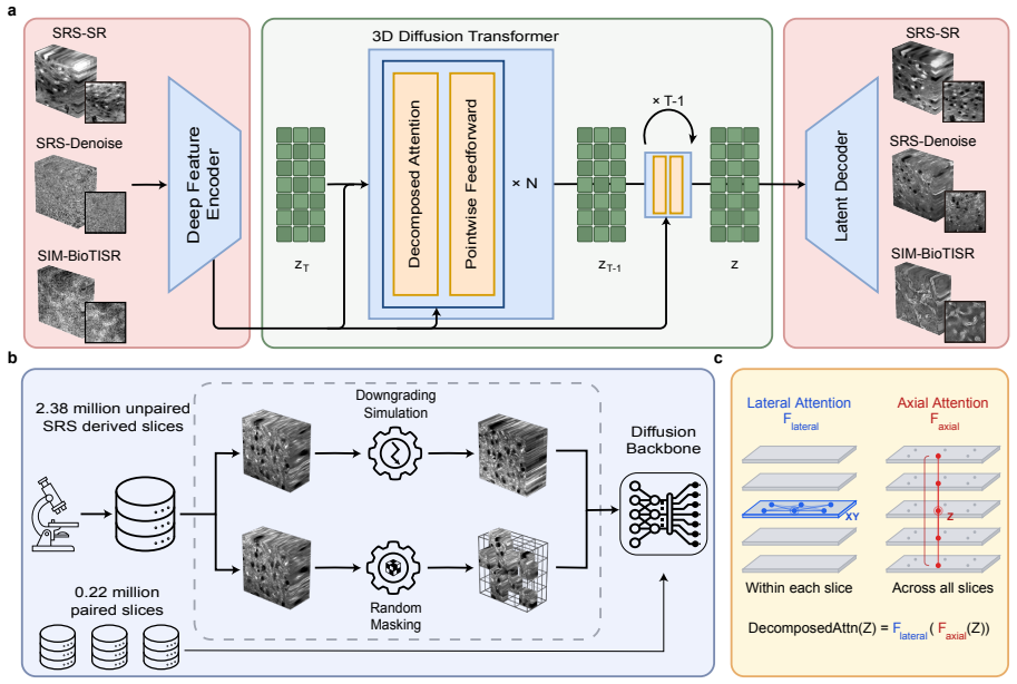
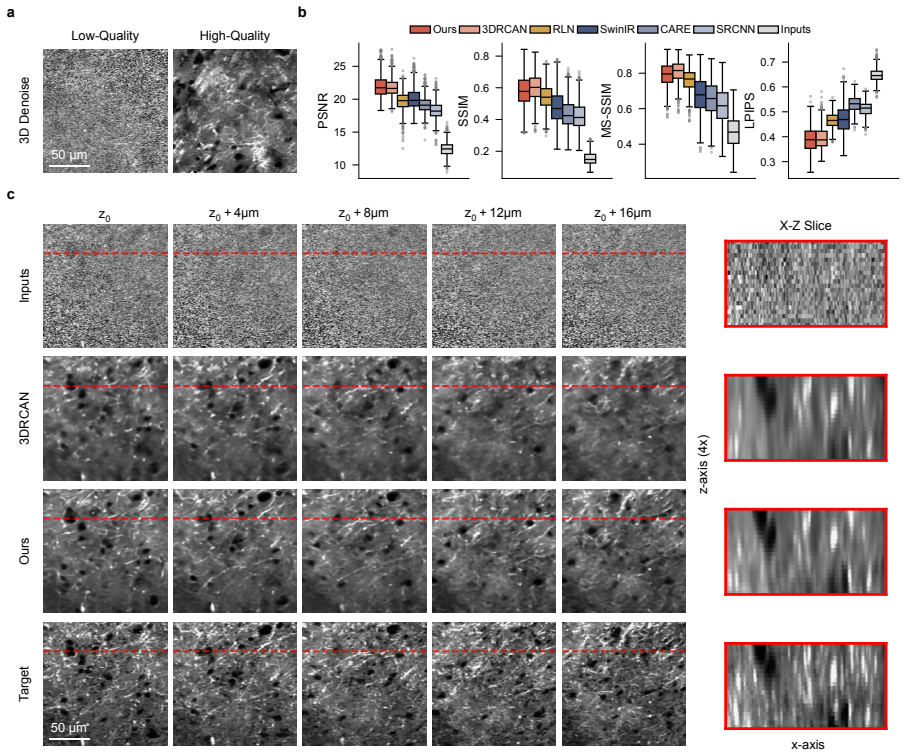
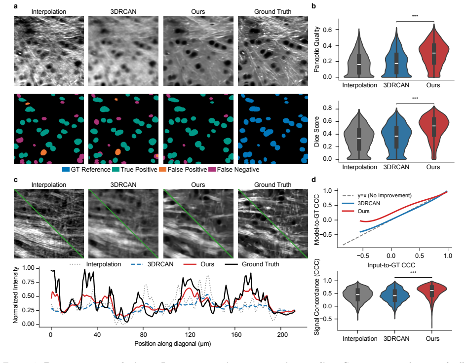
MicroDiffuse3D is a pretrained foundation model for 3D microscopy image restoration that recovers high-quality volumetric structure from degraded low-resolution measurements acquired at substantially higher throughput. Evaluated on three settings—16-fold sparse super-resolution, joint resolution-plus-noise degradation, and low-SNR denoising—the model outperforms strong baselines. In the sparse super-resolution regime it yields clearer continuity across depth with fewer artifacts.
What carries the argument
MicroDiffuse3D, a pretrained foundation model that learns to map degraded 3D inputs onto restored high-quality volumetric outputs.
If this is right
- Pretrained 3D restoration becomes a broadly applicable strategy for overcoming throughput and SNR limits in volumetric chemical imaging.
- High-resolution analysis becomes feasible at scales and speeds that were previously difficult to achieve.
- Downstream tasks such as segmentation gain from the improved depth continuity and reduced artifacts.
Where Pith is reading between the lines
- The same pretraining strategy could be applied to other 3D modalities that face similar acquisition-speed versus resolution trade-offs.
- Lower-resolution hardware combined with this restoration step could reduce equipment costs while maintaining usable output quality.
- Faster low-resolution scans followed by model restoration might enable real-time or live-sample 3D workflows.
Load-bearing premise
The pretrained foundation model generalizes effectively to the three specific 3D degradation regimes in chemical microscopy without overfitting to the training distribution or requiring heavy task-specific adaptation.
What would settle it
Testing the model on a fourth unseen degradation type or on data acquired from an independent microscope system and checking whether performance still exceeds baselines would falsify the claim of broad applicability.
Figures

read the original abstract
Chemical imaging enables label-free visualization of cells, tissues and living systems while providing direct biochemical information that is difficult to obtain with conventional fluorescence microscopy. Despite its promise in applications ranging from intraoperative diagnosis to drug-response analysis, its broader use remains limited by slow data acquisition, particularly for three-dimensional imaging. Here we present MicroDiffuse3D, a pretrained foundation model for 3D microscopy image restoration that recovers high-quality volumetric structure from degraded low-resolution measurements acquired at substantially higher throughput. We evaluated MicroDiffuse3D across three challenging restoration settings, including 3D super-resolution under 16-fold volumetric sparsity, joint degradation in resolution and noise, and 3D denoising in the low signal-to-noise ratio (SNR) regime, where the model delivered clear gains over strong baselines. Under the sparse 3D super-resolution setting, MicroDiffuse3D produced clearer continuity across depth with fewer artifacts and improved segmentation quality by 10.58% and line-profile concordance by 15.59%. Together, our results establish pretrained 3D restoration as a broadly applicable strategy for overcoming the throughput and SNR limitations in volumetric chemical imaging, enabling high-resolution analysis at scales and speeds that were previously difficult to achieve.
Editorial analysis
A structured set of objections, weighed in public.
Referee Report
Summary. The manuscript introduces MicroDiffuse3D, a pretrained foundation model for 3D microscopy image restoration in chemical imaging. It claims to recover high-quality volumetric structure from degraded low-resolution measurements and reports gains over baselines across three settings: 16-fold volumetric sparse super-resolution (with clearer depth continuity, fewer artifacts, +10.58% segmentation quality, and +15.59% line-profile concordance), joint resolution+noise degradation, and low-SNR denoising. The work positions pretrained 3D restoration as a broadly applicable strategy to overcome throughput and SNR limits.
Significance. If the reported gains are shown to stem from pretraining-enabled generalization rather than task-specific factors, the work could meaningfully advance high-throughput 3D chemical microscopy for intraoperative diagnosis and drug-response studies by enabling high-resolution analysis at previously inaccessible scales and speeds.
major comments (2)
- [Abstract] The abstract reports quantitative gains (10.58% segmentation quality, 15.59% line-profile concordance) but supplies no information on training datasets, model architecture, baseline implementations, statistical testing, or cross-validation procedures. This information is load-bearing for the central claim that pretraining confers robustness to the three specific 3D degradation regimes.
- [Experiments] No ablation removing the pretraining step, pretraining corpus diversity metrics, or explicit distribution-shift tests are described. Without these, the reported improvements in depth continuity and artifact reduction cannot be confidently attributed to foundation-model generalization rather than architecture or in-distribution fine-tuning.
minor comments (1)
- [Abstract] The abstract would be clearer if it briefly noted the number of test volumes or the specific nature of the strong baselines used for comparison.
Simulated Author's Rebuttal
We thank the referee for the constructive feedback on our manuscript. We address the major comments point by point below, agreeing where revisions are warranted to better support our claims about MicroDiffuse3D.
read point-by-point responses
-
Referee: [Abstract] The abstract reports quantitative gains (10.58% segmentation quality, 15.59% line-profile concordance) but supplies no information on training datasets, model architecture, baseline implementations, statistical testing, or cross-validation procedures. This information is load-bearing for the central claim that pretraining confers robustness to the three specific 3D degradation regimes.
Authors: We agree that the abstract would be strengthened by including concise references to these elements. In the revised manuscript we will expand the abstract to note the pretraining on diverse 3D chemical microscopy volumes, the 3D diffusion-based architecture, the baseline methods used, and the cross-validation with statistical testing employed. Full details remain in the Methods and Experiments sections. revision: yes
-
Referee: [Experiments] No ablation removing the pretraining step, pretraining corpus diversity metrics, or explicit distribution-shift tests are described. Without these, the reported improvements in depth continuity and artifact reduction cannot be confidently attributed to foundation-model generalization rather than architecture or in-distribution fine-tuning.
Authors: We acknowledge that an explicit ablation removing the pretraining step is absent from the current manuscript. We will add a discussion of this limitation in the revised Experiments section and, where computationally feasible, include a limited ablation on a data subset to help isolate the pretraining contribution. Corpus diversity metrics appear in the supplementary materials, and the three distinct degradation regimes (sparse super-resolution, joint degradation, and low-SNR denoising) function as distribution-shift tests; we will clarify this attribution and add explicit shift analysis in the revision. revision: partial
Circularity Check
No circularity: empirical performance claims with no derivations or self-referential reductions
full rationale
The paper reports empirical results from training and evaluating a pretrained 3D restoration model on three degradation regimes, with gains quantified via segmentation quality (+10.58%) and line-profile concordance (+15.59%) on held-out test settings. No equations, first-principles derivations, fitted parameters renamed as predictions, or load-bearing self-citations appear in the provided text. The central claims rest on experimental comparisons rather than any step that reduces by construction to its own inputs, satisfying the criteria for a self-contained non-circular finding.
Axiom & Free-Parameter Ledger
free parameters (1)
- Neural network weights and training hyperparameters
axioms (1)
- domain assumption Deep neural networks pretrained on large image corpora can serve as effective priors for restoring degraded 3D microscopy volumes.
Lean theorems connected to this paper
-
IndisputableMonolith/Foundation/AlexanderDuality.leanalexander_duality_circle_linking unclear?
unclearRelation between the paper passage and the cited Recognition theorem.
MicroDiffuse3D operates as a transformer-based Latent Diffusion Model... decomposed lateral (XY-plane) and axial (Z-axis) attention mechanisms
-
IndisputableMonolith/Cost/FunctionalEquation.leanwashburn_uniqueness_aczel unclear?
unclearRelation between the paper passage and the cited Recognition theorem.
Built on large-scale pretraining over a curated corpus of 2.55 million microscopy images... dynamic physical degradation simulations and 3D block-masking
What do these tags mean?
- matches
- The paper's claim is directly supported by a theorem in the formal canon.
- supports
- The theorem supports part of the paper's argument, but the paper may add assumptions or extra steps.
- extends
- The paper goes beyond the formal theorem; the theorem is a base layer rather than the whole result.
- uses
- The paper appears to rely on the theorem as machinery.
- contradicts
- The paper's claim conflicts with a theorem or certificate in the canon.
- unclear
- Pith found a possible connection, but the passage is too broad, indirect, or ambiguous to say the theorem truly supports the claim.
Reference graph
Works this paper leans on
-
[2]
Science376(6594), 5197 (2022) https://doi.org/10.1126/science
Ji-Xin Cheng and X. Sunney Xie. “Vibrational spectroscopic imaging of living systems: An emerging platform for biology and medicine”. In:Science350.6264 (2015), aaa8870.doi:10.1126/science. aaa8870.url:http://science.sciencemag.org/content/sci/350/6264/aaa8870.full.pdf
-
[3]
Bond-selective imaging by optically sensing the mid-infrared photothermal effect
Yeran Bai, Jiaze Yin, and Ji-Xin Cheng. “Bond-selective imaging by optically sensing the mid-infrared photothermal effect”. In:Science Advances7.20 (2021), eabg1559.doi:doi:10.1126/sciadv.abg1559. url:https://www.science.org/doi/abs/10.1126/sciadv.abg1559
-
[4]
A 20-year journey on the invention of vibrational photothermal microscopy
J. X. Cheng. “A 20-year journey on the invention of vibrational photothermal microscopy”. In:Nat Methods22.5 (2025). 1548-7105 Cheng, Ji-Xin Orcid: 0000-0002-5607-6683 R35 GM 136223/U.S. De- partment of Health & Human Services — NIH — National Institute of General Medical Sciences (NIGMS)/ Journal Article United States 2025/05/14 Nat Methods. 2025 May;22(...
-
[5]
Daniel A. Orringer et al. “Rapid intraoperative histology of unprocessed surgical specimens via fibre- laser-based stimulated Raman scattering microscopy”. In:Nature Biomedical Engineering1.2 (2017), p. 0027.issn: 2157-846X.doi:10.1038/s41551-016-0027
-
[6]
The TRIPOD-LLM reporting guideline for studies using large language models
Todd C. Hollon et al. “Near real-time intraoperative brain tumor diagnosis using stimulated Raman histology and deep neural networks”. In:Nature Medicine26.1 (2020), pp. 52–58.issn: 1546-170X.doi: 10.1038/s41591- 019- 0715- 9.url:https://doi.org/10.1038/s41591- 019- 0715- 9%20https: //escholarship.org/content/qt2bx2h425/qt2bx2h425.pdf?t=qeu5ld
-
[8]
Optical imaging of metabolic dynamics in animals
Lingyan Shi et al. “Optical imaging of metabolic dynamics in animals”. In:Nature Communications 9.1 (2018), p. 2995.issn: 2041-1723.doi:10.1038/s41467-018-05401-3.url:https://doi.org/ 10.1038/s41467-018-05401-3
work page doi:10.1038/s41467-018-05401-3.url:https://doi.org/ 2018
-
[9]
Spectral tracing of deuterium for imaging glucose metabolism
Luyuan Zhang et al. “Spectral tracing of deuterium for imaging glucose metabolism”. In:Nature Biomedical Engineering3.5 (2019), pp. 402–413.issn: 2157-846X.doi:10.1038/s41551-019-0393-4. url:https://doi.org/10.1038/s41551-019-0393-4
-
[10]
B. S. Wong et al. “Facilitated Transport of EGFR Inhibitors Plays an Important Role in Their Cellular Uptake”. In:Analytical Chemistry96.4 (2024), pp. 1547–1555.issn: 0003-2700.doi:10.1021/acs. analchem.3c04242. 18
work page doi:10.1021/acs 2024
-
[11]
Fiona Xi Xu et al. “Assessing drug uptake and response differences in 2D and 3D cellular environments using stimulated Raman scattering microscopy”. In:bioRxiv(2024), p. 2024.04.22.590622.doi:10. 1101/2024.04.22.590622.url:https://www.biorxiv.org/content/biorxiv/early/2024/04/26/ 2024.04.22.590622.full.pdf
work page 2024
-
[12]
Quantitative Stimulated Raman Scattering Microscopy: Promises and Pitfalls
Bryce Manifold and Dan Fu. “Quantitative Stimulated Raman Scattering Microscopy: Promises and Pitfalls”. In:Annual Reviews of Analytical Chemistry15.1 (2022), pp. 269–289.doi:10 . 1146 / annurev - anchem - 061020 - 015110.url:https : / / www . annualreviews . org / doi / abs / 10 . 1146 / annurev-anchem-061020-015110
work page 2022
-
[13]
Review of bio-optical imaging systems with a high space-bandwidth product
Jongchan Park et al. “Review of bio-optical imaging systems with a high space-bandwidth product”. In:Advanced Photonics3.4 (2021), p. 044001.doi:10 . 1117 / 1 . AP . 3 . 4 . 044001.url:https : //doi.org/10.1117/1.AP.3.4.044001
-
[14]
AI Feynman: A physics-inspired method for symbolic regression.Science Advances, 6(16):eaay2631, 2020
Wei Min and Xin Gao. “Absolute signal of stimulated Raman scattering microscopy: A quantum electrodynamics treatment”. In:Science Advances10.50 (2024), eadm8424.doi:10.1126/sciadv. adm8424. eprint:https : / / www . science . org / doi / pdf / 10 . 1126 / sciadv . adm8424.url:https : //www.science.org/doi/abs/10.1126/sciadv.adm8424
-
[15]
Content-aware image restoration: pushing the limits of fluorescence microscopy
Martin Weigert, Uwe Schmidt, Tobias Boothe, et al. “Content-aware image restoration: pushing the limits of fluorescence microscopy”. In:Nature Methods15 (2018), pp. 1090–1097.doi:10 . 1038 / s41592-018-0216-7
work page 2018
-
[16]
Deep learning enables cross-modality super-resolution in fluorescence microscopy
Hongda Wang et al. “Deep learning enables cross-modality super-resolution in fluorescence microscopy”. In:Nature Methods16 (2019), pp. 103–110.doi:10.1038/s41592-018-0239-0
-
[17]
Evaluation and development of deep neural networks for image super-resolution in optical microscopy
Chang Qiao et al. “Evaluation and development of deep neural networks for image super-resolution in optical microscopy”. In:Nature Methods18.2 (2021), pp. 194–202.doi:10.1038/s41592-020-01048- 5
-
[18]
Chang Qiao, Yunmin Zeng, Qian Meng, et al. “Zero-shot learning enables instant denoising and super- resolution in optical fluorescence microscopy”. In:Nature Communications15 (2024), p. 4180.doi: 10.1038/s41467-024-48575-9
-
[19]
Incorporating the image formation process into deep learning improves network performance
Yijun Li, Yijun Su, Min Guo, et al. “Incorporating the image formation process into deep learning improves network performance”. In:Nature Methods19 (2022), pp. 1427–1437.doi:10.1038/s41592- 022-01652-7
-
[20]
Jiji Chen et al. “Three-dimensional residual channel attention networks denoise and sharpen fluores- cence microscopy image volumes”. In:Nature Methods18.6 (2021), pp. 678–687
work page 2021
-
[21]
Kyungryun Lee and Won-Ki Jeong. “Reference-free Axial Super-resolution of 3D Microscopy Images using Implicit Neural Representation with a 2D Diffusion Prior”. In:Medical Image Computing and Computer Assisted Intervention – MICCAI 2024. arXiv:2408.08616. 2024
-
[22]
Hyoungjun Park et al. “Deep learning enables reference-free isotropic super-resolution for volumetric fluorescence microscopy”. In:Nature Communications13 (2022), p. 3297.doi:10.1038/s41467-022- 30949-6
-
[23]
Kai Ning, Bin Lu, Xin Wang, et al. “Deep self-learning enables fast, high-fidelity isotropic resolution restoration for volumetric fluorescence microscopy”. In:Light: Science & Applications12 (2023), p. 204. doi:10.1038/s41377-023-01230-2
-
[24]
Sushama Joshi, Amanda Forjaz, K. S. Han, et al. “InterpolAI: deep learning-based optical flow inter- polation and restoration of biomedical images for improved 3D tissue mapping”. In:Nature Methods 22 (2025), pp. 1556–1567.doi:10.1038/s41592-025-02712-4
-
[25]
Image Quality Assessment: From Error Visibility to Structural Similarity
Zhou Wang et al. “Image Quality Assessment: From Error Visibility to Structural Similarity”. In: IEEE Transactions on Image Processing13.4 (2004), pp. 600–612
work page 2004
-
[26]
Multi-Scale Structural Similarity for Image Quality Assessment
Zhou Wang, Eero P. Simoncelli, and Alan C. Bovik. “Multi-Scale Structural Similarity for Image Quality Assessment”. In:The Thirty-Seventh Asilomar Conference on Signals, Systems & Computers. Vol. 2. 2003, pp. 1398–1402. 19
work page 2003
-
[27]
The Unreasonable Effectiveness of Deep Features as a Perceptual Metric
Richard Zhang et al. “The Unreasonable Effectiveness of Deep Features as a Perceptual Metric”. In: Proceedings of the IEEE/CVF Conference on Computer Vision and Pattern Recognition. 2018, pp. 586– 595
work page 2018
-
[28]
Scalable Diffusion Models with Transformers
William Peebles and Saining Xie. “Scalable Diffusion Models with Transformers”. In:Proceedings of the IEEE/CVF International Conference on Computer Vision. 2023, pp. 4195–4205
work page 2023
-
[29]
William Peebles and Saining Xie. “Scalable Diffusion Models with Transformers”. In:Proceedings of the IEEE/CVF International Conference on Computer Vision (ICCV). 2023, pp. 4195–4205.doi: 10.1109/ICCV51070.2023.00387
-
[30]
Predicting transcriptional outcomes of novel multigene perturba- tions with GEARS
Chang Qiao et al. “A neural network for long-term super-resolution imaging of live cells with reliable confidence quantification”. In:Nature Biotechnology44.1 (2026), pp. 110–119.doi:10.1038/s41587- 025-02553-8
-
[31]
Cellpose: A Generalist Algorithm for Cellular Segmentation
Carsen Stringer et al. “Cellpose: A Generalist Algorithm for Cellular Segmentation”. In:Nature Meth- ods18.1 (2021), pp. 100–106
work page 2021
-
[32]
Cellpose-SAM: superhuman generalization for cellular segmentation
Marius Pachitariu, Michael Rariden, and Carsen Stringer. “Cellpose-SAM: superhuman generalization for cellular segmentation”. In:BioRxiv(2025), pp. 2025–04
work page 2025
-
[33]
Alexander Kirillov et al. “Panoptic Segmentation”. In:Proceedings of the IEEE/CVF Conference on Computer Vision and Pattern Recognition (CVPR). 2019, pp. 9404–9413
work page 2019
-
[34]
V-Net: Fully Convolutional Neural Net- works for Volumetric Medical Image Segmentation
Fausto Milletari, Nassir Navab, and Seyed-Ahmad Ahmadi. “V-Net: Fully Convolutional Neural Net- works for Volumetric Medical Image Segmentation”. In:2016 Fourth International Conference on 3D Vision (3DV). IEEE. 2016, pp. 79–87
work page 2016
-
[35]
A Concordance Correlation Coefficient to Evaluate Reproducibility
Lawrence I-Kuei Lin. “A Concordance Correlation Coefficient to Evaluate Reproducibility”. In:Bio- metrics45.1 (1989), pp. 255–268
work page 1989
-
[36]
Haonan Lin et al. “Spectroscopic stimulated Raman scattering imaging of highly dynamic specimens through matrix completion”. In:Light: Science & Applications7.5 (2018), pp. 17179–17179.issn: 2047-7538.doi:10.1038/lsa.2017.179.url:https://doi.org/10.1038/lsa.2017.179
work page doi:10.1038/lsa.2017.179.url:https://doi.org/10.1038/lsa.2017.179 2018
-
[37]
Super-resolution SRS microscopy with A-PoD
Hongje Jang et al. “Super-resolution SRS microscopy with A-PoD”. In:Nature Methods20.3 (2023), pp. 448–458.issn: 1548-7105.doi:10.1038/s41592-023-01779-1.url:https://doi.org/10.1038/ s41592-023-01779-1
work page doi:10.1038/s41592-023-01779-1.url:https://doi.org/10.1038/ 2023
-
[38]
Denoising of stimulated Raman scattering microscopy images via deep learning
Bryce Manifold et al. “Denoising of stimulated Raman scattering microscopy images via deep learning”. In:Biomedical Optics Express10.8 (2019), pp. 3860–3874.doi:10.1364/BOE.10.003860.url:http: //www.osapublishing.org/boe/abstract.cfm?URI=boe-10-8-3860
-
[39]
Md Inzamam Ul Haque et al. “Deep learning-driven super-resolution in Raman hyperspectral imaging: Efficient high-resolution reconstruction from low-resolution data”. In:Applied Physics Letters125.20 (2024), p. 204104.issn: 0003-6951.doi:10.1063/5.0228645.url:https://doi.org/10.1063/5. 0228645
work page doi:10.1063/5.0228645.url:https://doi.org/10.1063/5 2024
-
[40]
Super-resolution vibrational imaging based on photoswitchable Raman probe
Jingwen Shou et al. “Super-resolution vibrational imaging based on photoswitchable Raman probe”. In:Science Advances9.24 (), eade9118.doi:10.1126/sciadv.ade9118.url:https://doi.org/10. 1126/sciadv.ade9118
-
[41]
Label-free super-resolution stimulated Raman scattering imaging of biomedical specimens
Julien Guilbert et al. “Label-free super-resolution stimulated Raman scattering imaging of biomedical specimens”. In:Advanced Imaging1.11 (Jan. 2024), p. 011004.url:https://www.researching.cn/ articles/OJfff2f087cb065461
work page 2024
-
[42]
4Pi stimulated Raman scattering for label-free super-resolution chemical imaging
Jonathan I. Kim et al. “4Pi stimulated Raman scattering for label-free super-resolution chemical imaging”. In:Science Advances12.1 (), eaec0523.doi:10 . 1126 / sciadv . aec0523.url:https : //doi.org/10.1126/sciadv.aec0523
-
[43]
Mean Flows for One-step Generative Modeling
Zhengyang Geng et al. “Mean Flows for One-step Generative Modeling”. In:The Thirty-ninth Annual Conference on Neural Information Processing Systems. 2025.url:https://openreview.net/forum? id=uWj4s7rMnR
work page 2025
-
[44]
Generative Modeling via Drifting
Mingyang Deng et al. “Generative Modeling via Drifting”. In:arXiv preprint arXiv:2602.04770(2026). 20
work page internal anchor Pith review arXiv 2026
-
[45]
Learning a Deep Convolutional Network for Image Super-Resolution
Chao Dong et al. “Learning a Deep Convolutional Network for Image Super-Resolution”. In:European Conference on Computer Vision (ECCV). 2014, pp. 184–199
work page 2014
-
[46]
SwinIR: Image Restoration Using Swin Transformer
Jingyun Liang et al. “SwinIR: Image Restoration Using Swin Transformer”. In:IEEE/CVF Interna- tional Conference on Computer Vision Workshops (ICCVW). 2021, pp. 1833–1844. 21 Supplementary Figure 1:Point spread function illustration. A, Differences in the point spread function between high-resolution (HR) and low-resolution (LR) objectives.The LR objective...
work page 2021
discussion (0)
Sign in with ORCID, Apple, or X to comment. Anyone can read and Pith papers without signing in.