Recognition: 2 theorem links
· Lean TheoremImage-Based Whole-Heart Cardiac Flow Simulations in Health and Congenital Heart Disease
Pith reviewed 2026-05-12 02:59 UTC · model grok-4.3
The pith
An image-based whole-heart CFD framework with ML segmentation and RIS valve modeling reproduces physiologic pressures, valve timing, and flow structures in healthy and pediatric CHD patients, showing qualitative agreement with imaging and quantitative agreement with catheterization.
A machine-rendered reading of the paper's core claim, the machinery that carries it, and where it could break.
Core claim
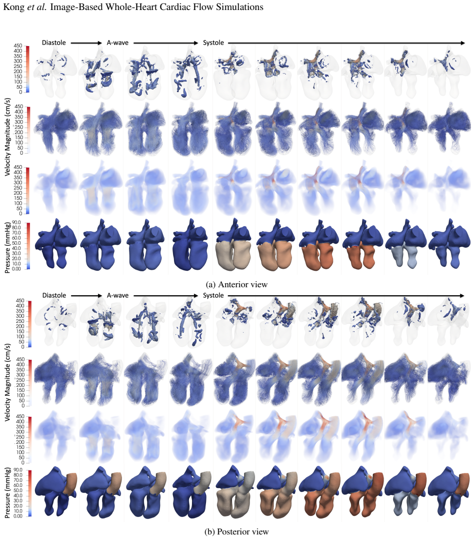
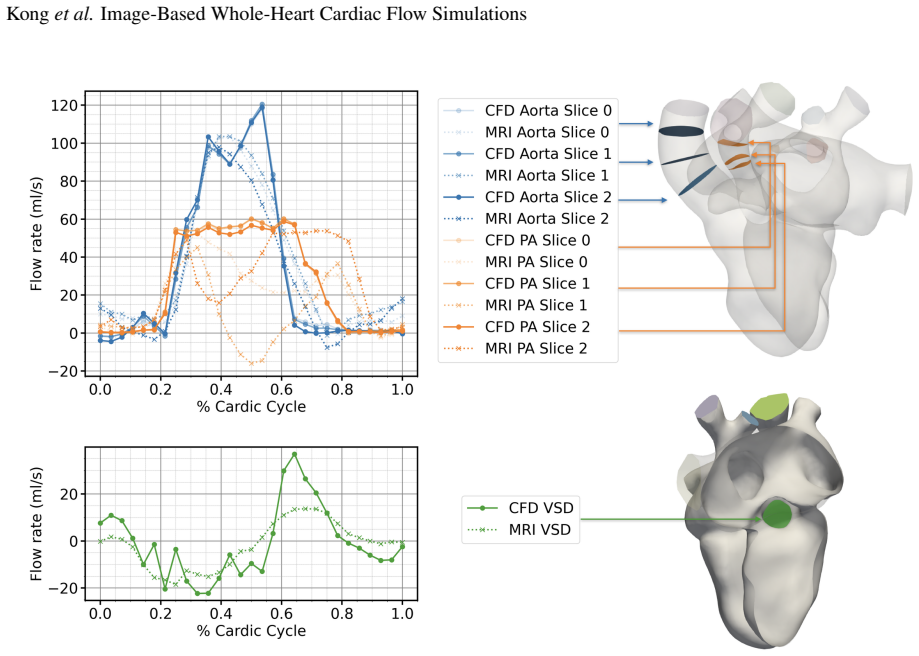
In the CHD case, simulated chamber and vessel pressures showed agreement with cardiac catheterization measurements. Simulated flow fields were qualitatively consistent with 4D-Flow MRI, while providing higher-resolution visualization of flow structures that were partially obscured by imaging artifacts.
Load-bearing premise
The resistive immersed surfaces model accurately captures physiologically realistic opening and closing dynamics for all four valves using only image-derived chamber motion and without patient-specific valve geometry or direct flow measurements through the valves.
Figures


read the original abstract
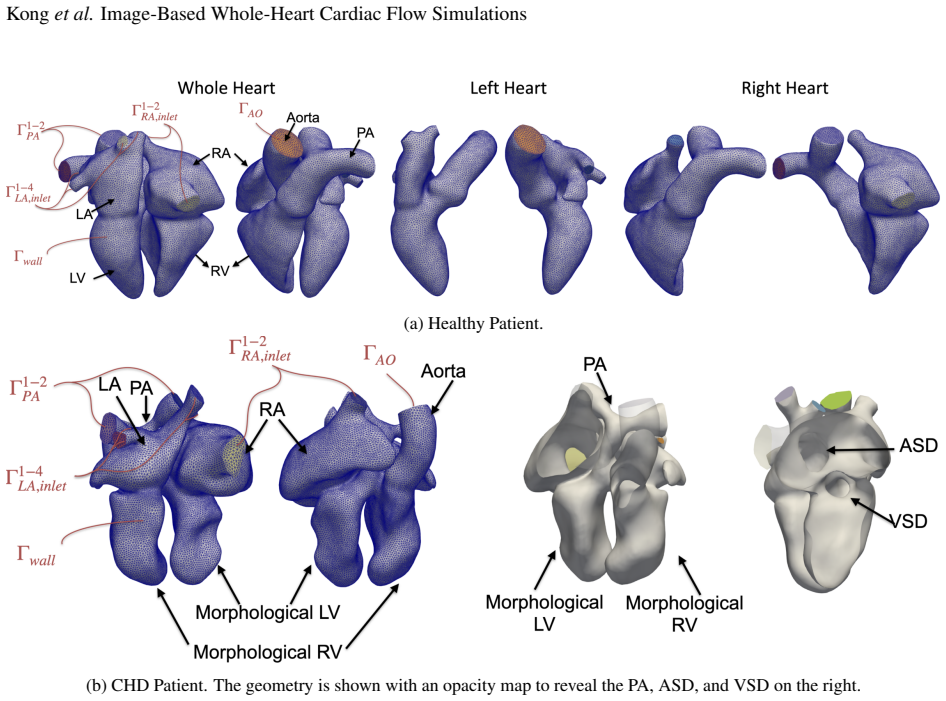
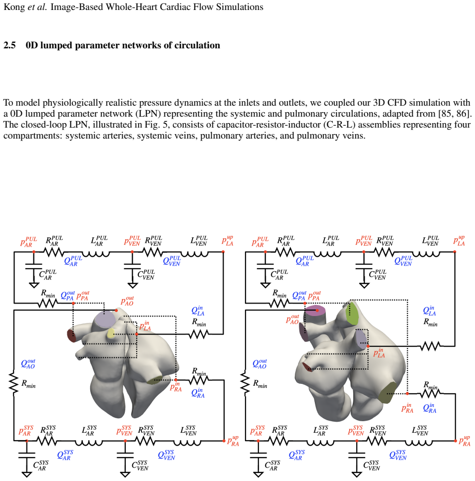
Intracardiac flow patterns are shaped by the coupled motion of the cardiac chambers and heart valves and provide important information about cardiac function. However, clinical flow imaging remains limited by exam times, noise, resolution, and incomplete details of the three-dimensional flow. Computational fluid dynamics (CFD) can potentially provide detailed flow quantification and predictive insight into treatment outcomes, but clinical translation requires frameworks that reproduce patient-specific measurements while balancing physiological realism, computational cost, and modeling effort. Herein, we present an image-based, patient-specific computational framework for simulating whole-heart intracardiac hemodynamics that balances physiological fidelity with computational efficiency. The framework first employs machine learning-based segmentation and mesh propagation to reconstruct moving cardiac anatomies from time-resolved images. CFD simulations are then performed to resolve blood flow in deforming domains, while resistive immersed surfaces (RIS) are used to model all four cardiac valves with physiologically realistic opening and closing dynamics. The framework was applied to model hemodynamics in a healthy adult and a pediatric patient with complex congenital heart disease (CHD). In the healthy case, the simulations reproduced physiologic pressure-volume behavior, valve timing, and ventricular vortex formation. In the CHD case, simulated chamber and vessel pressures showed agreement with cardiac catheterization measurements. Simulated flow fields were qualitatively consistent with 4D-Flow MRI, while providing higher-resolution visualization of flow structures that were partially obscured by imaging artifacts. Comparison between the healthy and CHD cases further revealed altered diastolic flow organization and elevated normalized viscous dissipation in the CHD heart.
Editorial analysis
A structured set of objections, weighed in public.
Referee Report
Summary. The manuscript presents an image-based patient-specific CFD framework for whole-heart intracardiac hemodynamics. It reconstructs moving cardiac geometries via machine learning segmentation and mesh propagation from time-resolved images, solves Navier-Stokes equations in deforming domains, and employs resistive immersed surfaces (RIS) to model physiologically realistic opening/closing of all four valves without explicit patient-specific valve geometry. Demonstrated on one healthy adult (reproducing physiologic PV loops, valve timing, and ventricular vortices) and one pediatric CHD patient (pressure agreement with catheterization; qualitative flow consistency with 4D-Flow MRI plus higher-resolution visualization of structures obscured by artifacts), the work also contrasts altered diastolic flow organization and elevated normalized viscous dissipation in the CHD case.
Significance. If the central claims hold, the framework offers an efficient route to detailed, patient-specific whole-heart flow fields from standard clinical images, filling a gap between limited-resolution 4D-Flow MRI and more expensive fully resolved valve models. The RIS approach for valves is a pragmatic strength that reduces modeling effort while still capturing timing effects. Direct comparison to both invasive catheterization and non-invasive MRI in a complex CHD anatomy is valuable for assessing translational potential. The single-case demonstration and lack of quantitative error metrics currently limit the strength of the evidence for predictive fidelity.
major comments (1)
- [CHD case results] CHD case results: The reported agreement between simulated chamber/vessel pressures and catheterization measurements is central to the claim that the framework reproduces patient-specific hemodynamics. However, chamber volumes are image-prescribed, so pressures depend sensitively on net inflow/outflow timing and resistance; the manuscript provides no sensitivity analysis on the free RIS resistance and opening parameters, no patient-specific valve geometry, and no direct transvalvular flow measurements to confirm that the RIS dynamics are physiologically realistic rather than adjusted to match the pressure data.
minor comments (2)
- [Abstract] Abstract: The statement that simulated pressures 'showed agreement' with catheterization would be strengthened by reporting at least one quantitative metric (e.g., RMSE or peak-pressure difference) rather than a qualitative descriptor.
- [Methods] Figure captions and methods: Clarify whether the RIS parameters were held fixed across both subjects or adjusted per case; if fixed, state the values and any literature basis.
Simulated Author's Rebuttal
We thank the referee for the constructive and detailed review. The feedback highlights an important aspect of the CHD case validation. We respond point-by-point below and outline revisions that will strengthen the manuscript without altering its core claims or methods.
read point-by-point responses
-
Referee: [CHD case results] CHD case results: The reported agreement between simulated chamber/vessel pressures and catheterization measurements is central to the claim that the framework reproduces patient-specific hemodynamics. However, chamber volumes are image-prescribed, so pressures depend sensitively on net inflow/outflow timing and resistance; the manuscript provides no sensitivity analysis on the free RIS resistance and opening parameters, no patient-specific valve geometry, and no direct transvalvular flow measurements to confirm that the RIS dynamics are physiologically realistic rather than adjusted to match the pressure data.
Authors: We agree that the pressure agreement is a key result and that a sensitivity analysis would better demonstrate robustness. The RIS resistance and opening parameters were selected from literature values for physiologic valve timing and transvalvular pressure drops (Methods section), then verified to produce valve opening/closing consistent with the time-resolved images and to yield pressure-volume loops matching expected physiology in the healthy case. In the CHD case, the same parameter set produced chamber and vessel pressures in agreement with the available catheterization data. We acknowledge that the original manuscript did not include a formal sensitivity study. In revision we will add a dedicated subsection reporting the effect of varying RIS resistance and timing parameters over physiologic ranges on the resulting pressures and flow structures; this will show that the reported agreement is not overly sensitive to small changes. The RIS formulation is intentionally geometry-free precisely because patient-specific valve anatomy is rarely available from standard clinical imaging; this is presented as a pragmatic modeling choice rather than a limitation to be overcome with additional data. Direct transvalvular flow measurements were not part of the clinical dataset provided for this patient, so they cannot be added; however, the simulated flow fields remain qualitatively consistent with the available 4D-Flow MRI, including large-scale structures and regions of elevated dissipation. These additions will be incorporated in the revised manuscript. revision: partial
Circularity Check
No significant circularity in derivation chain
full rationale
The paper describes a forward CFD simulation pipeline: machine-learning segmentation produces time-resolved geometries whose motion is prescribed as boundary conditions; the incompressible Navier-Stokes equations are solved in deforming domains; resistive immersed surfaces supply valve resistance without direct fitting to the reported catheterization pressures or 4D-Flow data. Pressure-volume agreement and flow-field consistency are presented as post-hoc validation against independent measurements, not as quantities that reduce to the inputs by construction. No self-definitional equations, fitted-input predictions, or load-bearing self-citation chains appear in the described methodology.
Axiom & Free-Parameter Ledger
free parameters (1)
- RIS valve resistance and opening parameters
axioms (2)
- domain assumption Machine learning segmentation and mesh propagation accurately reconstruct time-resolved deforming cardiac anatomies from images
- domain assumption Resistive immersed surfaces can represent all four cardiac valves with realistic hemodynamics using only chamber motion
Lean theorems connected to this paper
-
IndisputableMonolith/Cost/FunctionalEquation.leanwashburn_uniqueness_aczel unclearresistive immersed surfaces (RIS) are used to model all four cardiac valves with physiologically realistic opening and closing dynamics... fRIS,k = Rk/ϵk δΓk(φk(x)) (u−uALE)
-
IndisputableMonolith/Foundation/RealityFromDistinction.leanreality_from_one_distinction unclearmachine learning-based segmentation and mesh propagation... neural ordinary differential equation (NODE) framework
Reference graph
Works this paper leans on
-
[1]
Philip J. Kilner, Guang-Zhong Yang, A. John Wilkes, Raad H. Mohiaddin, David N. Firmin, and Magdi H. Yacoub. Asymmetric redirection of flow through the heart.Nature, 404(6779):759–761, April 2000. Publisher: Nature Publishing Group
work page 2000
-
[2]
J. O. Mangual, F. Domenichini, and G. Pedrizzetti. Describing the Highly Three Dimensional Right Ventricle Flow.Annals of Biomedical Engineering, 40(8):1790–1801, August 2012
work page 2012
-
[3]
Daniel Rodriguez Muñoz, Michael Markl, José Luis Moya Mur, Alex Barker, Covadonga Fernández-Golfín, Patrizio Lancellotti, and José Luis Zamorano Gómez. Intracardiac flow visualization: current status and future directions.European Heart Journal - Cardiovascular Imaging, 14(11):1029–1038, November 2013
work page 2013
-
[4]
Nature Optimizes the Swirling Flow in the Human Left Ventricle
Gianni Pedrizzetti and Federico Domenichini. Nature Optimizes the Swirling Flow in the Human Left Ventricle. Physical Review Letters, 95(10):108101, September 2005. Publisher: American Physical Society
work page 2005
-
[5]
Jung Hee Seo and Rajat Mittal. Effect of diastolic flow patterns on the function of the left ventricle.Physics of Fluids, 25(11):110801, August 2013
work page 2013
- [6]
-
[7]
S. W. DA VIES, A. L. FUSSELL, S. L. JORDAN, P. A. POOLE-WILSON, and D. P. LIPKIN. Abnormal diastolic filling patterns in chronic heart failure—relationship to exercise capacity.European Heart Journal, 13(6):749–757, June 1992
work page 1992
-
[8]
M Ohno, C P Cheng, and W C Little. Mechanism of altered patterns of left ventricular filling during the development of congestive heart failure.Circulation, 89(5):2241–2250, May 1994. Publisher: American Heart Association
work page 1994
-
[9]
Vincenzo Cicchitti, Francesco Radico, Francesco Bianco, Sabina Gallina, Gianni Tonti, and Raffaele De Caterina. Heart failure due to right ventricular apical pacing: the importance of flow patterns.EP Europace, 18(11):1679– 1688, November 2016
work page 2016
-
[10]
Passing Strange.Circulation: Heart Failure, 3(2):326–331, March 2010
Carl Johan Carlhäll and Ann Bolger. Passing Strange.Circulation: Heart Failure, 3(2):326–331, March 2010. Publisher: American Heart Association. 29 Konget al.Image-Based Whole-Heart Cardiac Flow Simulations
work page 2010
-
[11]
McGarvey, Norihiro Kondo, Manabu Takebe, Kevin J
Jeremy R. McGarvey, Norihiro Kondo, Manabu Takebe, Kevin J. Koomalsingh, Walter R. T. Witschey, Alex J. Barker, Michael Markl, Satoshi Takebayashi, Toru Shimaoka, Joseph H. Gorman, Robert C. Gorman, and James J. Pilla. Directed Epicardial Assistance in Ischemic Cardiomyopathy: Flow and Function Using Cardiac Magnetic Resonance Imaging.The Annals of Thorac...
work page 2013
-
[12]
Bee Ting Chan, Azam Ahmad Bakir, Amr Al Abed, Socrates Dokos, Chin Neng Leong, Ean Hin Ooi, Renly Lim, and Einly Lim. Impact of myocardial infarction on intraventricular vortex and flow energetics assessed using computational simulations.International Journal for Numerical Methods in Biomedical Engineering, 35(6):e3204, 2019. _eprint: https://onlinelibrar...
-
[13]
Gianni Pedrizzetti, Federico Domenichini, and Giovanni Tonti. On the Left Ventricular V ortex Reversal after Mitral Valve Replacement.Annals of Biomedical Engineering, 38(3):769–773, March 2010
work page 2010
-
[14]
Morteza Gharib, Edmond Rambod, Arash Kheradvar, David J. Sahn, and John O. Dabiri. Optimal vortex formation as an index of cardiac health.Proceedings of the National Academy of Sciences, 103(16):6305–6308, April 2006. Publisher: Proceedings of the National Academy of Sciences
work page 2006
-
[15]
Gianni Pedrizzetti, Giovanni La Canna, Ottavio Alfieri, and Giovanni Tonti. The vortex—an early predictor of cardiovascular outcome?Nature Reviews Cardiology, 11(9):545–553, September 2014. Publisher: Nature Publishing Group
work page 2014
-
[16]
Megan K. Russ, Nicole M. Lafata, Scott H. Robertson, and Ehsan Samei. Pulsed wave Doppler ultrasound: Accuracy, variability, and impact of acquisition parameters on flow measurements.Medical Physics, 50(11):6704– 6713, 2023. _eprint: https://aapm.onlinelibrary.wiley.com/doi/pdf/10.1002/mp.16774
-
[17]
Christian Prinz, Reka Faludi, Andrew Walker, Mihaela Amzulescu, Hang Gao, Tokuhisa Uejima, Alan G. Fraser, and Jens-Uwe V oigt. Can echocardiographic particle image velocimetry correctly detect motion patterns as they occur in blood inside heart chambers? A validation study using moving phantoms.Cardiovascular Ultrasound, 10(1):24, June 2012
work page 2012
-
[18]
Kondo Claude Assi, Etienne Gay, Christophe Chnafa, Simon Mendez, Franck Nicoud, Juan F P J Abascal, Pierre Lantelme, François Tournoux, and Damien Garcia. Intraventricular vector flow mapping—a Doppler-based regularized problem with automatic model selection.Physics in Medicine & Biology, 62(17):7131, August 2017. Publisher: IOP Publishing
work page 2017
-
[19]
Nyrnes, Solveig Fadnes, Morten Smedsrud Wigen, Luc Mertens, and Lasse Lovstakken
Siri A. Nyrnes, Solveig Fadnes, Morten Smedsrud Wigen, Luc Mertens, and Lasse Lovstakken. Blood Speckle- Tracking Based on High–Frame Rate Ultrasound Imaging in Pediatric Cardiology.Journal of the American Society of Echocardiography, 33(4):493–503.e5, April 2020
work page 2020
-
[20]
Bissell, Francesca Raimondi, Lamia Ait Ali, Bradley D
Malenka M. Bissell, Francesca Raimondi, Lamia Ait Ali, Bradley D. Allen, Alex J. Barker, Ann Bolger, Nicholas Burris, Carl-Johan Carhäll, Jeremy D. Collins, Tino Ebbers, Christopher J. Francois, Alex Frydrychowicz, Pankaj Garg, Julia Geiger, Hojin Ha, Anja Hennemuth, Michael D. Hope, Albert Hsiao, Kevin Johnson, Sebastian Kozerke, Liliana E. Ma, Michael M...
work page 2023
-
[21]
Arshid Azarine, Philippe Garçon, Audrey Stansal, Nadia Canepa, Giorgios Angelopoulos, Stephane Silvera, Daniel Sidi, Véronique Marteau, and Marc Zins. Four-dimensional Flow MRI: Principles and Cardiovascular Applications.RadioGraphics, 39(3):632–648, May 2019. Publisher: Radiological Society of North America (RSNA)
work page 2019
-
[22]
Ahmet Demirkiran, Pim van Ooij, Jos J M Westenberg, Mark B M Hofman, Hans C van Assen, Linda J Schoonmade, Usman Asim, Carmen P S Blanken, Aart J Nederveen, Albert C van Rossum, and Marco J W Götte. Clinical intra-cardiac 4D flow CMR: acquisition, analysis, and clinical applications.European Heart Journal - Cardiovascular Imaging, 23(2):154–165, February 2022
work page 2022
-
[23]
Steinman, and Umberto Morbiducci
Valentina Mazzi, Diego Gallo, Karol Calò, David A. Steinman, and Umberto Morbiducci. A revised and expanded unified theory linking wall shear stress and vorticity topologies to enable the interpretation of cardiovascular flow disturbances.Physics of Fluids, 37(3):031907, March 2025
work page 2025
-
[24]
Liping Dong, Hairu Li, Xiangli Xu, Min Ren, Weidong Yu, Wenkun Bai, Di Sun, and Jiawei Tian. Analysis of diastolic left ventricular wall shear stress in normal people of different age groups.Frontiers in Cardiovascular Medicine, 9, September 2022
work page 2022
-
[25]
Simon Schmidt, Sebastian Flassbeck, Sonja Schmelter, Ellen Schmeyer, Mark E. Ladd, and Sebastian Schmitter. The impact of 4D flow displacement artifacts on wall shear stress estimation.Magnetic Resonance in Medicine, 85(6):3154–3168, 2021. _eprint: https://onlinelibrary.wiley.com/doi/pdf/10.1002/mrm.28641. 30 Konget al.Image-Based Whole-Heart Cardiac Flow...
-
[26]
Giulia Comunale, Francesca M. Susin, and Jonathan P. Mynard. Ventricular wall stress and wall shear stress homeostasis predicts cardiac remodeling during pregnancy: A modeling study.International Journal for Numerical Methods in Biomedical Engineering, 38(1):e3536, January 2022
work page 2022
- [27]
-
[28]
Mohammed SM ElBaz, Emmeline Calkoen, Jos J Westenberg, Arno Roest, and Rob J van der Geest. Impact of disturbed diastolic vortex formation on viscous energy loss in the left ventricle: Quantitative 4D Flow MRI analysis of healthy controls and repaired atrioventricular septal defect patients.Journal of Cardiovascular Magnetic Resonance, 17(Suppl 1):P24, Fe...
work page 2015
-
[29]
Fanwei Kong and Shawn C. Shadden. Learning Whole Heart Mesh Generation From Patient Images for Computational Simulations.IEEE Transactions on Medical Imaging, 42(2):533–545, February 2023. Conference Name: IEEE Transactions on Medical Imaging
work page 2023
-
[30]
Fanwei Kong and Shawn C. Shadden. Automating Model Generation for Image-Based Cardiac Flow Simulation. Journal of Biomechanical Engineering, 142(111011), September 2020
work page 2020
-
[31]
Vijay Vedula, Richard George, Laurent Younes, and Rajat Mittal. Hemodynamics in the Left Atrium and Its Effect on Ventricular Flow Patterns.Journal of Biomechanical Engineering, 137(11):111003, November 2015
work page 2015
- [32]
-
[33]
Wenbin Mao, Andrés Caballero, Raymond McKay, Charles Primiano, and Wei Sun. Fully-coupled fluid- structure interaction simulation of the aortic and mitral valves in a realistic 3D left ventricle model.PloS One, 12(9):e0184729, 2017
work page 2017
-
[34]
Alfio Quarteroni, Toni Lassila, Simone Rossi, and Ricardo Ruiz-Baier. Integrated Heart—Coupling multiscale and multiphysics models for the simulation of the cardiac function.Computer Methods in Applied Mechanics and Engineering, 314:345–407, February 2017
work page 2017
-
[35]
Simulating cardiac fluid dynamics in the human heart.PNAS Nexus, 3(10):pgae392, October 2024
Marshall Davey, Charles Puelz, Simone Rossi, Margaret Anne Smith, David R Wells, Gregory M Sturgeon, W Paul Segars, John P Vavalle, Charles S Peskin, and Boyce E Griffith. Simulating cardiac fluid dynamics in the human heart.PNAS Nexus, 3(10):pgae392, October 2024
work page 2024
-
[36]
Liuyang Feng, Hao Gao, and Xiaoyu Luo. Whole-heart modelling with valves in a fluid–structure interaction framework.Computer Methods in Applied Mechanics and Engineering, 420:116724, February 2024
work page 2024
-
[37]
Taro Kariya, Takumi Washio, Jun-ichi Okada, Machiko Nakagawa, Masahiro Watanabe, Yoshimasa Kadooka, Shunji Sano, Ryozo Nagai, Seiryo Sugiura, and Toshiaki Hisada. Personalized Perioperative Multi-scale, Multi- physics Heart Simulation of Double Outlet Right Ventricle.Annals of Biomedical Engineering, 48(6):1740–1750, June 2020
work page 2020
-
[38]
Francesco Viola, Giulio Del Corso, and Roberto Verzicco. High-fidelity model of the human heart: An immersed boundary implementation.Physical Review Fluids, 8(10):100502, October 2023. Publisher: American Physical Society
work page 2023
-
[39]
Alberto Zingaro, Michele Bucelli, Roberto Piersanti, Francesco Regazzoni, Luca Dede’, and Alfio Quarteroni. An electromechanics-driven fluid dynamics model for the simulation of the whole human heart.Journal of Computational Physics, 504:112885, May 2024
work page 2024
-
[40]
Michele Bucelli, Alberto Zingaro, Pasquale Claudio Africa, Ivan Fumagalli, Luca Dede’, and Alfio Quarteroni. A mathematical model that integrates cardiac electrophysiology, mechanics, and fluid dynamics: Application to the human left heart.International Journal for Numerical Methods in Biomedical Engineering, 39(3):e3678, March 2023
work page 2023
-
[41]
Alexander D. Kaiser, Nicole K. Schiavone, Christopher J. Elkins, Doff B. McElhinney, John K. Eaton, and Alison L. Marsden. Comparison of Immersed Boundary Simulations of Heart Valve Hemodynamics Against In Vitro 4D Flow MRI Data.Annals of Biomedical Engineering, 51(10):2267–2288, October 2023
work page 2023
-
[42]
Alexander D. Kaiser, Jing Wang, Aaron L. Brown, Enbo Zhu, Tzung Hsiai, and Alison L. Marsden. A fluid- structure interaction model of the zebrafish aortic valve.Journal of Biomechanics, page 112794, 2025
work page 2025
-
[43]
Alberto Zingaro, Michele Bucelli, Ivan Fumagalli, Luca Dede’, and Alfio Quarteroni. Modeling iso- volumetric phases in cardiac flows by an Augmented Resistive Immersed Implicit Surface method.In- ternational Journal for Numerical Methods in Biomedical Engineering, 39(12):e3767, 2023. _eprint: https://onlinelibrary.wiley.com/doi/pdf/10.1002/cnm.3767. 31 Ko...
-
[44]
Fernández, and Jean-Frédéric Gerbeau
Alexandre This, Ludovic Boilevin-Kayl, Miguel A. Fernández, and Jean-Frédéric Gerbeau. Augmented resistive immersed surfaces valve model for the simulation of cardiac hemodynamics with isovolumetric phases.International Journal for Numerical Methods in Biomedical Engineering, 36(3):e3223, 2020. _eprint: https://onlinelibrary.wiley.com/doi/pdf/10.1002/cnm.3223
-
[45]
Marco Fedele, Elena Faggiano, Luca Dedè, and Alfio Quarteroni. A patient-specific aortic valve model based on moving resistive immersed implicit surfaces.Biomechanics and Modeling in Mechanobiology, 16(5):1779–1803, October 2017
work page 2017
-
[46]
Charles S. Peskin. The immersed boundary method.Acta Numerica, 11:479–517, January 2002
work page 2002
-
[47]
Arbitrary Lagrangian–Eulerian Methods - Donea - 2004 - Major Reference Works - Wiley Online Library
work page 2004
- [48]
-
[49]
Jun-ichi Okada, Takumi Washio, Seiryo Sugiura, and Toshiaki Hisada. Clinical and pharmacological application of multiscale multiphysics heart simulator, UT-Heart.The Korean Journal of Physiology & Pharmacology : Official Journal of the Korean Physiological Society and the Korean Society of Pharmacology, 23(5):295–303, September 2019
work page 2019
-
[50]
Elias Karabelas, Stefano Longobardi, Jana Fuchsberger, Orod Razeghi, Cristobal Rodero, Marina Strocchi, Ronak Rajani, Gundolf Haase, Gernot Plank, and Steven Niederer. Global Sensitivity Analysis of Four Chamber Heart Hemodynamics Using Surrogate Models.IEEE Transactions on Biomedical Engineering, 69(10):3216–3223, October 2022
work page 2022
-
[51]
Ricky T. Q. Chen, Yulia Rubanova, Jesse Bettencourt, and David K Duvenaud. Neural Ordinary Differential Equations. InAdvances in Neural Information Processing Systems, volume 31. Curran Associates, Inc., 2018
work page 2018
-
[52]
Chi Zhu, Vijay Vedula, Dave Parker, Nathan Wilson, Shawn Shadden, and Alison Marsden. svFSI: A Multiphysics Package for Integrated Cardiac Modeling.Journal of Open Source Software, 7(78):4118, October 2022
work page 2022
-
[53]
Pietro Marchese, Massimiliano Cantinotti, Jef Van den Eynde, Nadia Assanta, Eliana Franchi, Vitali Pak, Giuseppe Santoro, Martin Koestenberger, and Shelby Kutty. Left ventricular vortex analysis by high-frame rate blood speckle tracking echocardiography in healthy children and in congenital heart disease.IJC Heart & Vasculature, 37:100897, December 2021
work page 2021
-
[54]
Vasanawala, Kate Hanneman, Marcus T
Shreyas S. Vasanawala, Kate Hanneman, Marcus T. Alley, and Albert Hsiao. Congenital heart disease as- sessment with 4D flow MRI.Journal of Magnetic Resonance Imaging, 42(4):870–886, 2015. _eprint: https://onlinelibrary.wiley.com/doi/pdf/10.1002/jmri.24856
-
[55]
Jixiang Liang, Bingheng Lu, Xin Zhao, Jiong Wang, Dianjiang Zhao, Gen Zhang, Bin Zhu, Qiang Ma, Guangyu Pan, and Dianyuan Li. Feasibility analyses of virtual models and 3D printing for surgical simulation of the double-outlet right ventricle.Medical & Biological Engineering & Computing, 60(10):3029–3040, October 2022
work page 2022
-
[56]
Yuki Nakamura, Charles Romans, and Ravi Ashwath. Patient-Specific Patch for an Intra-Atrial Rerouting Procedure Developed Through Surgical Simulation.World Journal for Pediatric & Congenital Heart Surgery, 12(2):234–243, March 2021
work page 2021
-
[57]
Ana Mendez, Gorka Gomez-Ciriza, Marie-Josée Raboisson, Jose Rivas, Antonio Ordoñez, Nancy Poirier, and Israel Valverde. Apical Muscular Ventricular Septal Defects: Surgical Strategy Using Three-Dimensional Printed Model.Seminars in Thoracic and Cardiovascular Surgery, 30(4):450–453, December 2018
work page 2018
-
[58]
Chen, Chin Siang Ong, Nagina Malguria, Luca A
Sarah A. Chen, Chin Siang Ong, Nagina Malguria, Luca A. Vricella, Juan R. Garcia, and Narutoshi Hibino. Digital Design and 3D Printing of Aortic Arch Reconstruction in HLHS for Surgical Simulation and Training. World Journal for Pediatric and Congenital Heart Surgery, 9(4):454–458, July 2018
work page 2018
-
[59]
Mingze Sun, Vincent Reed LaSala, Caroline Giuglaris, David Blitzer, Sophia Jackman, Senay Ustunel, Kavya Rajesh, and David Kalfa. Cardiovascular patches applied in congenital cardiac surgery: Current materials and prospects.Bioengineering & Translational Medicine, 10(1):e10706, September 2024
work page 2024
-
[60]
Alexander D. Kaiser, Moussa A. Haidar, Perry S. Choi, Amit Sharir, Alison L. Marsden, and Michael R. Ma. Simulation-based design of bicuspidization of the aortic valve.The Journal of Thoracic and Cardiovascular Surgery, 168(3):923–932.e4, September 2024
work page 2024
-
[61]
Giancarlo Pennati, Chiara Corsini, Tain-Yen Hsia, and Francesco Migliavacca. Computational fluid dynamics models and congenital heart diseases.Frontiers in Pediatrics, 1:4, February 2013
work page 2013
-
[62]
Alison L. Marsden and Jeffrey A. Feinstein. Computational modeling and engineering in pediatric and congenital heart disease.Current opinion in pediatrics, 27(5):587–596, October 2015. 32 Konget al.Image-Based Whole-Heart Cardiac Flow Simulations
work page 2015
-
[63]
Irene E. Vignon-Clementel, Alison L. Marsden, and Jeffrey A. Feinstein. A primer on computational simulation in congenital heart disease for the clinician.Progress in Pediatric Cardiology, 30(1):3–13, December 2010
work page 2010
-
[64]
Alison L. Marsden, Adam J. Bernstein, V . Mohan Reddy, Shawn C. Shadden, Ryan L. Spilker, Frandics P. Chan, Charles A. Taylor, and Jeffrey A. Feinstein. Evaluation of a novel Y-shaped extracardiac Fontan baffle using computational fluid dynamics.The Journal of Thoracic and Cardiovascular Surgery, 137(2):394–403.e2, February 2009
work page 2009
-
[65]
Bosi, Hopewell Ntsinjana, Mario Carminati, Graham Derrick, Jan Marek, Sachin Khambadkone, Andrew M
Claudio Capelli, Emilie Sauvage, Giuliano Giusti, Giorgia M. Bosi, Hopewell Ntsinjana, Mario Carminati, Graham Derrick, Jan Marek, Sachin Khambadkone, Andrew M. Taylor, and Silvia Schievano. Patient-specific simulations for planning treatment in congenital heart disease.Interface Focus, 8(1):20170021, December 2017. Publisher: Royal Society
work page 2017
-
[66]
Y . Qian, J. L. Liu, K. Itatani, K. Miyaji, and M. Umezu. Computational Hemodynamic Analysis in Congenital Heart Disease: Simulation of the Norwood Procedure.Annals of Biomedical Engineering, 38(7):2302–2313, July 2010
work page 2010
- [67]
-
[68]
Justin T. Jack, Morten Jensen, R. Thomas Collins, Frandics Pak Chan, and Paul C. Millett. Numerical study of hemodynamic flow in the aortic vessel of Williams syndrome patient with congenital heart disease.Journal of Biomechanics, 168:112124, May 2024
work page 2024
- [69]
-
[70]
Yue-Hin Loke, Francesco Capuano, Elias Balaras, and Laura J Olivieri. Computational Modeling of Right Ventricular Motion and Intracardiac Flow in repaired Tetralogy of Fallot.Cardiovascular engineering and technology, 13(1):41–54, February 2022
work page 2022
-
[71]
Seiryo Sugiura, Jun-Ichi Okada, Takumi Washio, and Toshiaki Hisada. UT-Heart: A Finite Element Model Designed for the Multiscale and Multiphysics Integration of our Knowledge on the Human Heart. In Sonia Cortassa and Miguel A. Aon, editors,Computational Systems Biology in Medicine and Biotechnology: Methods and Protocols, pages 221–245. Springer US, New Y...
work page 2022
-
[72]
Fanwei Kong, Sascha Stocker, Perry S. Choi, Michael Ma, Daniel B. Ennis, and Alison L. Marsden. SDF4CHD: Generative modeling of cardiac anatomies with congenital heart defects.Medical Image Analysis, 97:103293, October 2024
work page 2024
-
[73]
William E. Lorensen and Harvey E. Cline. Marching cubes: A high resolution 3D surface construction algorithm. SIGGRAPH Comput. Graph., 21(4):163–169, August 1987
work page 1987
-
[74]
Wilson, Jameson Merkow, Hongzhi Lan, Alison L
Adam Updegrove, Nathan M. Wilson, Jameson Merkow, Hongzhi Lan, Alison L. Marsden, and Shawn C. Shadden. SimVascular: An Open Source Pipeline for Cardiovascular Simulation.Annals of Biomedical Engineering, 45(3):525–541, March 2017
work page 2017
-
[75]
Neural ordinary differential equations
Tian Qi Chen, Yulia Rubanova, Jesse Bettencourt, and David Kristjanson Duvenaud. Neural ordinary differential equations. InNeural Information Processing Systems, 2018
work page 2018
-
[76]
The Eurographics Association, 2007
Olga Sorkine and Marc Alexa.As-Rigid-As-Possible Surface Modeling. The Eurographics Association, 2007
work page 2007
-
[77]
Journal of Open Source Software: svFSI: A Multiphysics Package for Integrated Cardiac Modeling
-
[78]
Diana C. de Oliveira, Daniel M. Espino, Luca Deorsola, Jonathan P. Mynard, Vijay Rajagopal, Keith Buchan, Dana Dawson, and Duncan E. T. Shepherd. A toolbox for generating scalable mitral valve morphometric models. Computers in Biology and Medicine, 135:104628, August 2021
work page 2021
-
[79]
Vijay Vedula, Juhyun Lee, Hao Xu, C.-C. Jay Kuo, Tzung K. Hsiai, and Alison L. Marsden. A method to quantify mechanobiologic forces during zebrafish cardiac development using 4-D light sheet imaging and computational modeling.PLOS Computational Biology, 13(10):e1005828, October 2017. Publisher: Public Library of Science
work page 2017
-
[80]
Ju Liu and Alison L. Marsden. A unified continuum and variational multiscale formulation for fluids, solids, and fluid–structure interaction.Computer Methods in Applied Mechanics and Engineering, 337:549–597, August 2018
work page 2018
discussion (0)
Sign in with ORCID, Apple, or X to comment. Anyone can read and Pith papers without signing in.