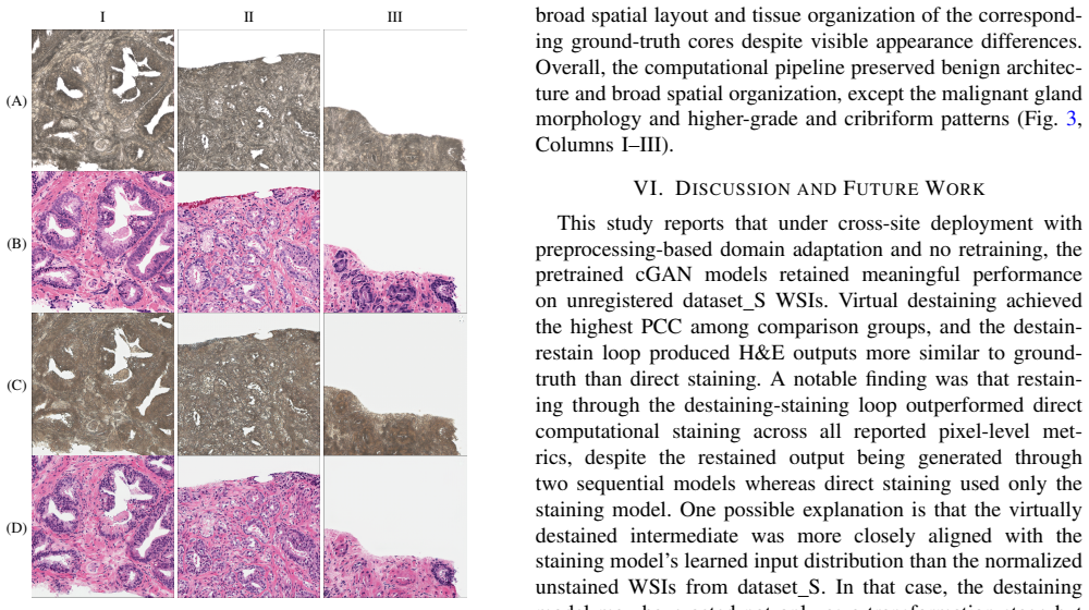
Recognition: no theorem link
Generative Deep Learning for Computational Destaining and Restaining of Unregistered Digital Pathology Images
Pith reviewed 2026-05-15 02:57 UTC · model grok-4.3
The pith
Conditional GANs can destain and restain unregistered pathology slides from external institutions after simple preprocessing
A machine-rendered reading of the paper's core claim, the machinery that carries it, and where it could break.
Core claim
Conditional generative adversarial networks trained on 102 registered prostate core biopsy WSIs from one institution can be deployed on 82 spatially unregistered WSIs from another institution using only a histogram-based stain normalization and channel-wise intensity calibration preprocessing pipeline, yielding destaining performance of PCC 0.854, SSIM 0.699, and PSNR 18.41 dB, with H&E restaining from computationally destained outputs outperforming direct staining from ground-truth unstained inputs (PCC 0.798 vs 0.715, SSIM 0.756 vs 0.718, PSNR 20.08 vs 18.51 dB).
What carries the argument
A conditional generative adversarial network (cGAN) that maps between H&E-stained and unstained pathology images, adapted to new data solely through histogram-based stain normalization and channel-wise intensity calibration without any image registration.
If this is right
- Virtual destaining of external unregistered slides becomes feasible using preprocessing alone.
- Restaining from model-generated destained images produces higher fidelity results than applying the model directly to real unstained slides.
- Preprocessing quality is the dominant performance bottleneck rather than the generative model itself.
- Generative staining models can be shared across institutions without retraining under realistic unregistered conditions.
Where Pith is reading between the lines
- Standardizing the preprocessing steps could enable consistent computational staining across different labs and scanners.
- Targeted improvements to preserve malignant gland morphology would be needed before routine clinical use.
- The same preprocessing approach might transfer to other generative models for different stain types or imaging modalities.
Load-bearing premise
Histogram-based stain normalization and channel-wise intensity calibration are enough to reduce domain shift between unregistered whole-slide images from different institutions without introducing artifacts that change how pathologists read malignant structures.
What would settle it
Side-by-side comparison of model outputs against registered ground-truth images or expert pathologist annotations on the same malignant glands to measure whether vessel-like morphology artifacts disappear when spatial alignment is restored.
Figures


read the original abstract
Conditional generative adversarial networks (cGANs) have enabled high-fidelity computational staining and destaining of hematoxylin and eosin (H&E) in digital pathology whole-slide images (WSI). However, their ability to generalize to out-of-distribution WSI across institutions without retraining remains insufficiently characterized. Previously developed cGAN models trained on 102 registered prostate core biopsy WSIs from Brigham and Women's Hospital were evaluated on 82 spatially unregistered WSIs acquired at Stanford University. To mitigate domain shift without retraining, a preprocessing pipeline consisting of histogram-based stain normalization for H&E-stained WSIs and channel-wise intensity calibration for unstained WSIs was developed. Because image registration was intentionally omitted for real-world deployment conditions, the reported quantitative results are conservative lower bounds reflecting both model performance and limited spatial alignment. Under these conditions, virtual destaining achieved a Pearson correlation coefficient (PCC) of 0.854, structural similarity index measure (SSIM) of 0.699, and peak signal-to-noise ratio (PSNR) of 18.41 dB. H&E restaining from computationally destained outputs outperformed direct staining from ground-truth unstained inputs across all metrics (PCC: 0.798 vs. 0.715; SSIM: 0.756 vs. 0.718; PSNR: 20.08 vs. 18.51 dB), suggesting that preprocessing quality may be more limiting than model capacity. Qualitative pathological review indicated preservation of benign glandular structures while showing that malignant glands were often rendered with vessel-like morphologies. These findings support the feasibility of applying cGAN-based computational H&E staining and destaining generative models to external WSI datasets using preprocessing-based adaptation alone while defining specific morphological targets for future domain adaptation.
Editorial analysis
A structured set of objections, weighed in public.
Referee Report
Summary. The manuscript evaluates pre-trained cGAN models for computational destaining of H&E-stained prostate WSIs and restaining of unstained inputs on an external set of 82 unregistered WSIs from a different institution. A preprocessing pipeline (histogram-based stain normalization for H&E and channel-wise intensity calibration for unstained images) is applied to mitigate domain shift without retraining or registration. Reported metrics include destaining PCC 0.854/SSIM 0.699/PSNR 18.41 dB; restaining from destained outputs outperforms direct staining from ground-truth unstained inputs (PCC 0.798 vs. 0.715, SSIM 0.756 vs. 0.718, PSNR 20.08 vs. 18.51 dB). Qualitative review notes preservation of benign glandular structures but vessel-like morphologies in malignant glands, with all quantitative results acknowledged as conservative lower bounds due to omitted registration.
Significance. If the central claim holds, the work shows that preprocessing alone can support cross-institutional generalization of generative pathology models without retraining, which would lower barriers to deployment. A strength is the empirical evaluation on held-out external unregistered data with explicit caveats on metric interpretation; the outperformance of the destain-then-restain pipeline provides a concrete, falsifiable observation that preprocessing quality may be the primary limiter.
major comments (2)
- [Abstract and qualitative review] Abstract and qualitative pathological review: the observation that malignant glands are often rendered with vessel-like morphologies (while benign structures are preserved) indicates a potential loss of diagnostic glandular architecture. Because all metrics are computed without registration and capture only low-level intensity correlation, this artifact is not quantified and directly challenges the feasibility conclusion for real-world unregistered WSIs.
- [Results (restaining comparison)] Results (restaining comparison): the interpretation that preprocessing quality—not model capacity—is the limiter rests on the metric gains (e.g., PCC 0.798 vs. 0.715). However, the systematic morphological mismatch in malignant glands undermines this without additional evidence such as pathologist diagnostic scoring or a registered subset comparison.
minor comments (1)
- [Abstract] The abstract would benefit from explicitly stating the training set size (102 WSIs) alongside the test set size for immediate context.
Simulated Author's Rebuttal
We thank the referee for their constructive comments, which have helped us improve the clarity and balance of our manuscript. We address each major comment below.
read point-by-point responses
-
Referee: [Abstract and qualitative review] Abstract and qualitative pathological review: the observation that malignant glands are often rendered with vessel-like morphologies (while benign structures are preserved) indicates a potential loss of diagnostic glandular architecture. Because all metrics are computed without registration and capture only low-level intensity correlation, this artifact is not quantified and directly challenges the feasibility conclusion for real-world unregistered WSIs.
Authors: We agree that the vessel-like morphologies in malignant glands represent a significant artifact that could impact diagnostic utility. This observation was noted in our qualitative review, and we have revised the abstract to more explicitly state that while low-level metrics show generalization, high-level morphological fidelity in malignant structures remains a challenge. We have added discussion in the results section emphasizing this as a key limitation and a target for future domain adaptation methods. The lack of registration means we cannot quantify the artifact precisely, but we maintain that the conservative metrics still support the feasibility of preprocessing-based adaptation for initial deployment, with caveats for diagnostic use. revision: yes
-
Referee: [Results (restaining comparison)] Results (restaining comparison): the interpretation that preprocessing quality—not model capacity—is the limiter rests on the metric gains (e.g., PCC 0.798 vs. 0.715). However, the systematic morphological mismatch in malignant glands undermines this without additional evidence such as pathologist diagnostic scoring or a registered subset comparison.
Authors: The metric gains in the destain-then-restain pipeline do provide evidence that preprocessing can improve performance, but we acknowledge that the morphological mismatches undermine claims about overall diagnostic equivalence. We have revised the results and discussion to qualify our interpretation, noting that the improvements are primarily in intensity correlation and that morphological fidelity requires further validation. Unfortunately, pathologist diagnostic scoring was not performed in this study, and the unregistered nature of the external dataset precludes direct registered comparisons. We have included this as an explicit limitation. revision: partial
- Pathologist diagnostic scoring of the generated images
- Quantitative comparison on a registered subset of the external WSIs
Circularity Check
No circularity: empirical evaluation on external unregistered data with no self-referential derivations
full rationale
The paper reports direct empirical metrics (PCC, SSIM, PSNR) computed on held-out unregistered WSIs from a different institution after applying histogram-based stain normalization and intensity calibration. No equations, ansatzes, or uniqueness theorems are invoked that reduce the reported performance numbers to parameters fitted within the same study. The central observation—that destain-then-restain outperforms direct restaining—is presented as a measured outcome on external data rather than a constructed identity. Preprocessing steps are described as practical adaptations, not as part of a closed mathematical loop. Self-citations, if present, are not load-bearing for the quantitative claims. This is a standard empirical ML paper whose results stand or fall on the external test set.
Axiom & Free-Parameter Ledger
axioms (2)
- domain assumption Histogram-based stain normalization sufficiently aligns color distributions across institutions for cGAN input
- domain assumption Channel-wise intensity calibration for unstained WSIs preserves structural information needed for destaining
Reference graph
Works this paper leans on
-
[1]
U.s. cancer statistics prostate cancer stat bite,
Centers for Disease Control and Prevention, “U.s. cancer statistics prostate cancer stat bite,” 2025
work page 2025
-
[2]
The 20-core prostate biopsy protocol–a new gold standard?
V . Raveryet al., “The 20-core prostate biopsy protocol–a new gold standard?”The Journal of Urology, vol. 179, no. 2, pp. 504–507, 2008
work page 2008
-
[3]
Tissue processing and hematoxylin and eosin staining,
A. T. Feldman and D. Wolfe, “Tissue processing and hematoxylin and eosin staining,” inHistopathology, C. E. Day, Ed. Springer, 2014, pp. 31–43
work page 2014
-
[4]
Automated histology analysis: Opportunities for signal processing,
M. T. McCannet al., “Automated histology analysis: Opportunities for signal processing,”IEEE Signal Processing Magazine, vol. 32, pp. 78– 87, 2015
work page 2015
-
[5]
Identification of a cell of origin for human prostate cancer,
A. S. Goldsteinet al., “Identification of a cell of origin for human prostate cancer,”Science, vol. 329, no. 5991, pp. 568–571, 2010
work page 2010
-
[6]
I. J. Goodfellow, J. Pouget-Abadie, M. Mirza, B. Xu, D. Warde-Farley, S. Ozair, A. Courville, and Y . Bengio, “Generative adversarial nets,” in Advances in Neural Information Processing Systems, 2014, pp. 2672– 2680
work page 2014
-
[7]
Image-to-image trans- lation with conditional adversarial networks,
P. Isola, J.-Y . Zhu, T. Zhou, and A. A. Efros, “Image-to-image trans- lation with conditional adversarial networks,” inIEEE Conference on Computer Vision and Pattern Recognition (CVPR), 2017
work page 2017
-
[8]
Deep learning-enabled virtual histological staining of biological samples,
B. Baiet al., “Deep learning-enabled virtual histological staining of biological samples,”Light: Science & Applications, vol. 12, p. 57, 2023
work page 2023
-
[9]
Virtual staining for histology by deep learning,
L. Latonenet al., “Virtual staining for histology by deep learning,” Trends in Biotechnology, vol. 42, no. 9, pp. 1177–1191, 2024
work page 2024
-
[10]
H&E to IHC virtual staining methods in breast cancer: an overview and benchmarking,
P. Kl ¨ockneret al., “H&E to IHC virtual staining methods in breast cancer: an overview and benchmarking,”npj Digital Medicine, vol. 8, no. 1, p. 384, 2025
work page 2025
-
[11]
A. Rana, G. Yauney, A. Lowe, and P. Shah, “Computational histological staining and destaining of prostate core biopsy RGB images with gen- erative adversarial neural networks,” inIEEE International Conference on Machine Learning and Applications (ICMLA), 2018, pp. 828–834
work page 2018
-
[12]
A. Rana, A. Lowe, M. Lithgow, K. Horback, T. Janovitz, A. Da Silva, H. Tsai, V . Shanmugam, A. Bayat, and P. Shah, “Use of deep learning to develop and analyze computational hematoxylin and eosin staining of prostate core biopsy images for tumor diagnosis,”JAMA Network Open, vol. 3, no. 5, p. e205111, 2020
work page 2020
-
[13]
A. Bayat, C. Anderson, and P. Shah, “Automated end-to-end deep learning framework for classification and tumor localization from native non-stained pathology images,” inProc. SPIE 11596, Medical Imaging 2021: Image Processing, 2021, p. 115960A
work page 2021
-
[14]
Digital pathology and artificial intelligence,
M. K. K. Niaziet al., “Digital pathology and artificial intelligence,”The Lancet Oncology, vol. 20, pp. e253–e261, 2019
work page 2019
-
[15]
FLEX Consortiumet al., “Knowledge-guided adaptation of pathology foundation models effectively improves cross-domain generalization,” Nature Communications, vol. 16, p. 11485, 2025
work page 2025
-
[16]
Generative AI for misalignment-resistant virtual staining to accelerate histopathology workflows,
J. Ma, W. Li, J. Liet al., “Generative AI for misalignment-resistant virtual staining to accelerate histopathology workflows,”Nature Com- munications, 2026
work page 2026
-
[17]
QuPath: Open source software for digital pathology image analysis,
P. Bankheadet al., “QuPath: Open source software for digital pathology image analysis,”Scientific Reports, vol. 7, p. 16878, 2017
work page 2017
-
[18]
TIAToolbox as an end-to-end library for advanced tissue image analytics,
J. Pocock, S. Graham, Q. D. Vu, M. Jahanifar, A. Azam, F. Minhas, and N. M. Rajpoot, “TIAToolbox as an end-to-end library for advanced tissue image analytics,”Communications Medicine, vol. 2, no. 1, p. 120, 2022
work page 2022
discussion (0)
Sign in with ORCID, Apple, or X to comment. Anyone can read and Pith papers without signing in.