High resolution large working distance scanning helium microscopy
Pith reviewed 2026-05-20 04:25 UTC · model grok-4.3
The pith
Redesigned pinhole and optimized optics yield 340 nm resolution in large-working-distance scanning helium microscopy.
A machine-rendered reading of the paper's core claim, the machinery that carries it, and where it could break.
Core claim
The central discovery is an intrinsic beamwidth of 340 nm achieved at working distances of 770 μm to 850 μm in a pinhole-based scanning helium microscope, representing a sixfold improvement. This is realized through constrained optimisation of the atom optics together with a redesigned high-resolution pinhole-plate, a reduced pinhole diameter, an increased source--pinhole distance and a larger detector aperture. The beamwidths agree with the modified optimisation model, showing geometric, source-size and diffraction terms now contribute on a similar footing.
What carries the argument
Constrained optimisation of the atom optics combined with the redesigned high-resolution pinhole-plate that enables the sub-micron beam at large distances.
If this is right
- The instrument can now image bacterial specimens and eroded diamond with sub-micron detail at large working distances.
- Large-working-distance pinhole SHeM becomes a viable platform for sub-micron imaging.
- Useful depth of field and practical sample access support topographic imaging and micro-diffraction applications.
- Multiple resolution-limiting factors contribute equally, indicating a near-optimised regime.
Where Pith is reading between the lines
- This design strategy could be applied to improve resolution in other scanning microscopies using neutral probes.
- With the current balance of terms, small further changes to pinhole size or distances might yield additional gains.
- The large working distance allows imaging of samples that require significant clearance, such as in situ experiments.
Load-bearing premise
That the measured beamwidths accurately isolate the intrinsic resolution after subtracting geometric, source-size, and diffraction contributions with no significant unmodeled experimental factors.
What would settle it
Observation of beamwidth measurements at 800 micrometer working distance that deviate substantially from the predicted 340 nm after applying the subtraction of contributions.
Figures


read the original abstract
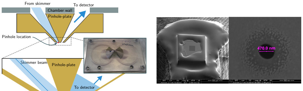
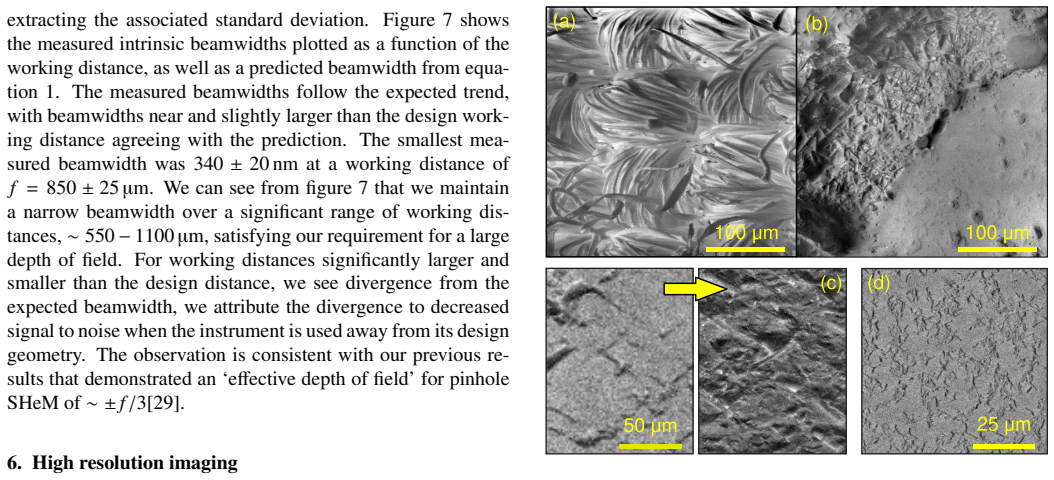
Scanning helium microscopy (SHeM) is attractive for imaging delicate and insulating surfaces because it combines a non-destructive neutral-atom probe with strong surface sensitivity. However, large-working-distance pinhole instruments have so far been limited in spatial resolution. Here we report sub-micron resolution in a large-working-distance pinhole SHeM, with an intrinsic beamwidth of 340nm achieved at working distances of 770 {\mu}m to 850 {\mu}m. This sixfold improvement over our previous long-working-distance configuration is enabled by constrained optimisation of the atom optics together with a redesigned high-resolution pinhole-plate, a reduced pinhole diameter, an increased source--pinhole distance and a larger detector aperture. Beamwidth measurements agree well with the modified optimisation model and show that geometric, source-size and diffraction terms now contribute on a similar footing, placing the instrument in a near-optimised regime. The resulting combination of sub-micron beam size, useful depth of field and practical sample access is demonstrated on bacterial specimens and eroded diamond. The work establishes large-working-distance pinhole SHeM as a viable sub-micron imaging platform and extends its usefulness for topographic imaging and micro-diffraction applications.
Editorial analysis
A structured set of objections, weighed in public.
Referee Report
Summary. The manuscript reports sub-micron resolution in a large-working-distance pinhole scanning helium microscope (SHeM), achieving an intrinsic beamwidth of 340 nm at working distances of 770–850 μm. This sixfold improvement over prior long-working-distance configurations is obtained via constrained optimisation of the atom optics, a redesigned high-resolution pinhole plate, reduced pinhole diameter, increased source–pinhole distance, and enlarged detector aperture. Measured beamwidths are stated to agree with a modified optimisation model in which geometric, source-size, and diffraction contributions are now comparable, placing the instrument near an optimised regime. The combination of resolution, depth of field, and sample access is illustrated on bacterial specimens and eroded diamond.
Significance. If the central experimental result holds, the work establishes large-working-distance pinhole SHeM as a practical sub-micron platform for non-destructive imaging of delicate and insulating surfaces. The balanced contribution of broadening mechanisms and the explicit hardware changes that realise the improvement constitute a clear advance over previous long-working-distance implementations. The demonstrated topographic and micro-diffraction applications further indicate utility beyond pure resolution metrics.
major comments (1)
- Beamwidth measurements and model comparison: the reported 340 nm intrinsic width is obtained after subtracting geometric, source-size, and diffraction terms from the measured profiles. While agreement with the modified optimisation model is claimed, the manuscript does not provide a propagated uncertainty budget for the subtracted components or raw line-scan data; this omission weakens the isolation of the intrinsic resolution and should be addressed with explicit error analysis before the central claim can be considered fully substantiated.
Simulated Author's Rebuttal
We thank the referee for their positive evaluation of the work and for the constructive comment. We address the point below and will revise the manuscript accordingly.
read point-by-point responses
-
Referee: Beamwidth measurements and model comparison: the reported 340 nm intrinsic width is obtained after subtracting geometric, source-size, and diffraction terms from the measured profiles. While agreement with the modified optimisation model is claimed, the manuscript does not provide a propagated uncertainty budget for the subtracted components or raw line-scan data; this omission weakens the isolation of the intrinsic resolution and should be addressed with explicit error analysis before the central claim can be considered fully substantiated.
Authors: We agree that an explicit uncertainty budget and access to the raw data would strengthen the central claim. In the revised manuscript we will add the raw line-scan profiles to the supplementary information and include a propagated uncertainty analysis for the geometric, source-size and diffraction contributions to the measured beamwidth. This will allow readers to assess the isolation of the reported 340 nm intrinsic resolution and the agreement with the optimisation model. revision: yes
Circularity Check
No significant circularity identified
full rationale
The paper reports an experimental result: sub-micron resolution achieved via documented hardware changes (redesigned pinhole-plate, smaller pinhole, increased source-pinhole distance, larger detector aperture) plus constrained optimization of atom optics. Beamwidth measurements at 770-850 μm working distance are presented as direct data that agree with the modified model, with geometric, source-size and diffraction contributions now comparable. No load-bearing step reduces the reported 340 nm intrinsic beamwidth to a fitted parameter, self-citation chain, or input by construction; the central claim rests on physical implementation and measurement that remains independent of the listed circularity patterns.
Axiom & Free-Parameter Ledger
axioms (1)
- standard math Standard atom optics principles govern helium beam focusing and diffraction in pinhole systems
Reference graph
Works this paper leans on
-
[1]
R. F. Egerton, P. Li, M. Malac, Radiation damage in the TEM and SEM, Micron 35 (6) (2004) 399–409.doi: 10.1016/j.micron.2004.02.003
-
[2]
M. T. Postek, A. E. Vladár, Does your SEM really tell the truth?—how would you know? part 1, Scanning 35 (6) (2013) 355–361.doi:https://doi.org/10. 1002/sca.21075
work page 2013
-
[3]
A. S. Palau, S. D. Eder, G. Bracco, B. Holst, Neutral helium atom microscopy, Ultramicroscopy 251 (2023) 113753.doi:10.1016/j.ultramic.2023.113753
-
[4]
S. D. Eder, A. Fahy, M. G. Barr, J. R. Manson, B. Holst, P. C. Dastoor, Sub-resolution contrast in neutral helium microscopy through facet scattering for quantitative imag- ing of nanoscale topographies on macroscopic surfaces, Nature Communications 14 (1) (2023) 904.doi:10. 1038/s41467-023-36578-x
work page 2023
-
[5]
N. A. von Jeinsen, S. M. Lambrick, M. Bergin, A. Radi ´c, B. Liu, D. Seremet, A. P. Jardine, D. J. Ward, 2d he- lium atom diffraction from a microscopic spot, Physical Review Letters 131 (23) (2023) 236202.doi:10.1103/ PhysRevLett.131.236202
work page 2023
-
[6]
B. Holst, G. Alexandrowicz, N. Avidor, G. Benedek, G. Bracco, W. E. Ernst, D. Farías, A. P. Jardine, K. Lef- mann, J. R. Manson, R. Marquardt, S. M. Artés, S. J. Sibener, J. W. Wells, A. Tamtögl, W. Allison, Mate- rial properties particularly suited to be measured with helium scattering: selected examples from 2d materi- als, van der waals heterostructure...
work page 2021
-
[7]
C. J. Hatchwell, M. Bergin, B. Carr, M. G. Barr, A. Fahy, P. C. Dastoor, Measuring scattering distributions in scan- ning helium microscopy, Ultramicroscopy 260 (2024) 113951.doi:10.1016/j.ultramic.2024.113951
-
[8]
R. Flatabø, S. D. Eder, T. Reisinger, G. Bracco, P. Baltzer, B. Samelin, B. Holst, Reflection imaging with a he- lium zone plate microscope, Ultramicroscopy 261 (2024) 113961.doi:10.1016/j.ultramic.2024.113961
-
[9]
M. Barr, A. Fahy, A. Jardine, J. Ellis, D. Ward, D. Ma- cLaren, W. Allison, P. Dastoor, A design for a pinhole scanning helium microscope, Nuclear Instruments and Methods in Physics Research Section B: Beam Interac- tions with Materials and Atoms 340 (2014) 76–80.doi: 10.1016/j.nimb.2014.06.028
-
[10]
G. Bhardwaj, K. R. Sahoo, R. Sharma, P. Nath, P. R. Shirhatti, Neutral-atom-scattering-based mapping of atomically thin layers, Physical Review A 105 (2) (2022) 022828.doi:10.1103/PhysRevA.105.022828
-
[11]
P. Witham, E. Sánchez, A simple approach to neutral atom microscopy, Review of Scientific Instruments 82 (10) (2011) 103705.doi:10.1063/1.3650719
-
[12]
G. Bhardwaj, P. R. Shirhatti, Contrast inversion in neutral- atom microscopy using atomic cluster beams, Physi- cal Review A 107 (6) (2023) 062813.doi:10.1103/ PhysRevA.107.062813
work page 2023
-
[13]
A. Radic, N. v. Jeinsen, V . Perez, K. Wang, M. Lin, B. Liu, Y . Zhu, I. Sami, K. Watanabe, T. Taniguchi, 8 D. Ward, A. Jardine, A. Rao, M. Chhowalla, S. Lam- brick, Measuring vacancy-type defect density in mono- layer MoS$_2$ (2025).arXiv:2409.18637[physics], doi:10.48550/arXiv.2409.18637
-
[14]
T. A. Myles, S. D. Eder, M. G. Barr, A. Fahy, J. Martens, P. C. Dastoor, Taxonomy through the lens of neutral he- lium microscopy, Scientific Reports 9 (1) (2019) 2148. doi:10.1038/s41598-018-36373-5
-
[15]
S. M. Lambrick, L. V ozdecký, M. Bergin, J. E. Halpin, D. A. MacLaren, P. C. Dastoor, S. A. Przyborski, A. P. Jardine, D. J. Ward, Multiple scattering in scanning he- lium microscopy, Applied Physics Letters 116 (6) (2020) 061601.doi:10.1063/1.5143950
-
[16]
M. Bergin, W. Roland-Batty, C. J. Hatchwell, T. A. Myles, J. Martens, A. Fahy, M. Barr, W. J. Belcher, P. C. Das- toor, Standardizing resolution definition in scanning he- lium microscopy, Ultramicroscopy 233 (2022) 113453. doi:10.1016/j.ultramic.2021.113453
-
[17]
S. D. Eder, T. Reisinger, M. M. Greve, G. Bracco, B. Holst, Focusing of a neutral helium beam below one micron, New Journal of Physics 14 (7) (2012) 073014. doi:10.1088/1367-2630/14/7/073014
-
[18]
S. D. Eder, A. Salvador Palau, T. Kaltenbacher, G. Bracco, B. Holst, Velocity distributions in microskimmer super- sonic expansion helium beams: High precision measure- ments and modeling, Review of Scientific Instruments 89 (11) (2018) 113301.doi:10.1063/1.5044203
-
[19]
M. Bergin, D. J. Ward, S. M. Lambrick, N. A. von Jeinsen, B. Holst, J. Ellis, A. P. Jardine, W. Allison, Low-energy electron ionization mass spectrometer for efficient detec- tion of low mass species, Review of Scientific Instruments 92 (7) (2021) 073305.doi:10.1063/5.0050292
-
[20]
P. J. Witham, E. J. Sánchez, Increased resolution in neutral atom microscopy, Journal of Microscopy 248 (3) (2012) 223–227.doi:10.1111/j.1365-2818.2012.03665. x
-
[21]
M. Koch, S. Rehbein, G. Schmahl, T. Reisinger, G. Bracco, W. E. Ernst, B. Holst, Imaging with neu- tral atoms—a new matter-wave microscope, Journal of Microscopy 229 (1) (2008) 1–5.doi:10.1111/j. 1365-2818.2007.01874.x
work page doi:10.1111/j 2008
-
[22]
S. D. Eder, X. Guo, T. Kaltenbacher, M. M. Greve, M. Kalläne, L. Kipp, B. Holst, Focusing of a neutral he- lium beam with a photon-sieve structure, Physical Review A 91 (4) (2015) 043608.doi:10.1103/PhysRevA.91. 043608
-
[23]
R. Flatabø, M. M. Greve, S. D. Eder, M. Kalläne, A. S. Palau, K. K. Berggren, B. Holst, Atom sieve for nanome- ter resolution neutral helium microscopy, Journal of Vac- uum Science & Technology B 35 (6) (2017) 06G502. doi:10.1116/1.4994330
-
[25]
A. Fahy, M. Barr, J. Martens, P. C. Dastoor, A highly con- trasting scanning helium microscope, Review of Scien- tific Instruments 86 (2) (2015) 023704.doi:10.1063/ 1.4907539
work page 2015
-
[26]
M. Bergin, D. J. Ward, J. Ellis, A. P. Jardine, A method for constrained optimisation of the design of a scanning helium microscope, Ultramicroscopy (2019) 112833doi: 10.1016/j.ultramic.2019.112833
-
[27]
A. Salvador Palau, G. Bracco, B. Holst, Theoretical model of the helium zone plate microscope, Physical Review A 95 (1) (2017).doi:10.1103/PhysRevA.95.013611
-
[28]
D. P. DePonte, S. D. Kevan, F. S. Patton, Brightness of micronozzle helium source, Review of Scientific Instru- ments 77 (5) (2006) 055107.doi:10.1063/1.2198813
-
[29]
S. M. Lambrick, M. Bergin, A. P. Jardine, D. J. Ward, A ray tracing method for predicting contrast in neutral atom beam imaging, Micron 113 (2018) 61–68.doi: 10.1016/j.micron.2018.06.014
-
[30]
A. S. Palau, G. Bracco, B. Holst, Theoretical model of the helium pinhole microscope, Physical Review A 94 (6) (2016).doi:10.1103/PhysRevA.94.063624
-
[31]
J. Kelsall, A. Radi ´c, J. Ellis, D. J. Ward, A. P. Jardine, Minimizing interference in low-pressure supersonic beam sources, The Journal of Chemical Physics 162 (9) (2025) 094304.doi:10.1063/5.0247870
-
[32]
Bergin, Instrumentation and contrast mechanisms in scanning helium microscopy, Ph.D
M. Bergin, Instrumentation and contrast mechanisms in scanning helium microscopy, Ph.D. thesis, Fitzwilliam College, University of Cambridge (2018).doi:10. 17863/CAM.37853
work page 2018
-
[33]
Lambrick, The formation of contrast in scanning helium microscopy, Ph.D
Sam M. Lambrick, The formation of contrast in scanning helium microscopy, Ph.D. thesis, Hughes Hall, University of Cambridge (2021).doi:10.17863/CAM.83463
-
[34]
A. Radi ´c, S. M. Lambrick, N. A. von Jeinsen, A. P. Jar- dine, D. J. Ward, 3d surface profilometry using neutral helium atoms, Applied Physics Letters 124 (20) (2024) 204101.doi:10.1063/5.0206374
-
[35]
M. Bergin, T. A. Myles, A. Radi ´c, C. J. Hatchwell, S. M. Lambrick, D. J. Ward, S. D. Eder, A. Fahy, M. Barr, P. C. Dastoor, Complex optical elements for scanning helium microscopy through 3d printing, Journal of Physics D: Applied Physics 55 (9) (2021) 095305.doi:10.1088/ 1361-6463/ac3a3e. 9
work page 2021
-
[36]
A. Radi ´c, S. M. Lambrick, S. Rhodes, D. J. Ward, On the application of components manufactured with stereolitho- graphic 3d printing in high vacuum systems, Vacuum 232 (2025) 113809.doi:10.1016/j.vacuum.2024. 113809
-
[37]
S. M. Lambrick, M. Bergin, D. J. Ward, M. Barr, A. Fahy, T. Myles, A. Radi ´c, P. C. Dastoor, J. Ellis, A. P. Jardine, Observation of diffuse scattering in scanning helium mi- croscopy, Physical Chemistry Chemical Physics 24 (43) (2022) 26539–26546.doi:10.1039/D2CP01951E
-
[38]
M. Bergin, S. M. Lambrick, H. Sleath, D. J. Ward, J. Ellis, A. P. Jardine, Observation of diffraction contrast in scan- ning helium microscopy, Scientific Reports 10 (1) (2020) 1–8.doi:10.1038/s41598-020-58704-1
-
[39]
N. A. von Jeinsen, D. J. Ward, M. Bergin, S. M. Lam- brick, D. M. Williamson, R. M. Langford, L. F. Dawson, V . Rana, S. Shivaswamy, X. Zhou, M. Mikesh, V . D. Gor- don, B. W. Wren, K. A. Brown, P. C. Dastoor, Surface visualisation of bacterial biofilms using neutral atom mi- croscopy, Journal of Microscopy 301 (1) (2026) 107–115. doi:10.1111/jmi.70038
-
[40]
A. J. Henderson, The Solid Particle Erosion of Polycrys- talline Diamond at High and Low Temperatures, Ph.D. thesis, Robinson College, University of Cambridge (May 2020).doi:10.17863/CAM.51694
-
[41]
L. F. Dawson, J. Peltier, C. L. Hall, M. A. Harrison, M. Derakhshan, H. A. Shaw, N. F. Fairweather, B. W. Wren, Extracellular DNA, cell surface proteins and c-di- GMP promote biofilm formation in Clostridioides diffi- cile, Scientific Reports 11 (1) (2021) 3244.doi:10. 1038/s41598-020-78437-5
work page 2021
-
[42]
R. Flatabø, S. D. Eder, T. Reisinger, G. Bracco, P. Baltzer, B. Samelin, B. Holst, Reflection imaging with a helium zone plate microscope (2023).arXiv: 2308.11749[physics],doi:10.48550/arXiv.2308. 11749
-
[43]
A. Radic, N. von Jeinsen, K. Wang, Y . Zhu, I. Sami, V . Perez, D. Ward, A. Jardine, M. Chhowalla, S. Lam- brick, Defect density quantification in monolayer MoS2 using helium atom micro-diffraction (2024).doi:10. 48550/arXiv.2409.18637. 10 Appendix A. Constrained optimisation for non-normal in- cidence The formula for the beam standard deviation,ϕ, derive...
discussion (0)
Sign in with ORCID, Apple, or X to comment. Anyone can read and Pith papers without signing in.