Robustness of breast lesion segmentation under MRI undersampling improves with k-space-aware deep learning
Pith reviewed 2026-05-22 06:51 UTC · model grok-4.3
The pith
K-space-aware deep learning improves robustness of breast lesion segmentation under MRI undersampling and noise while matching standard methods at full sampling.
A machine-rendered reading of the paper's core claim, the machinery that carries it, and where it could break.
Core claim
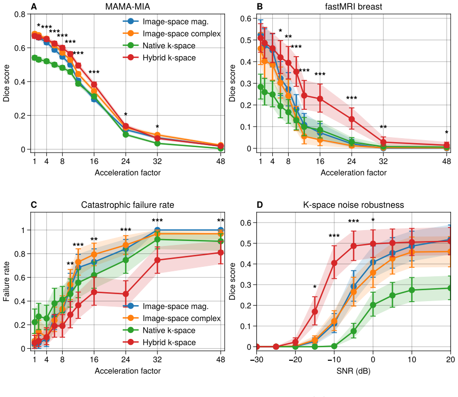
A hybrid k-space-to-image 3D U-Net variant for breast lesion segmentation matches the patient-level Dice similarity coefficient of magnitude image-space baselines at full sampling but significantly outperforms them across moderate to high undersampling levels and when complex Gaussian noise is added directly to k-space, with the advantage reproduced in a within-dataset synthetic control and supported by analysis showing complementary roles for the k-space stage in frequency-domain filtering and the image stage in lesion localization.
What carries the argument
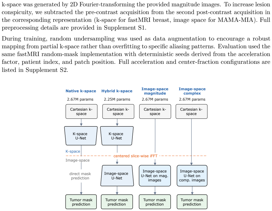
The hybrid k-space-to-image 3D U-Net variant that processes data first in the frequency domain before transitioning to image space, enabling complementary frequency filtering and lesion localization.
If this is right
- The hybrid model retains substantially more segmentation accuracy than image-space baselines as acceleration increases.
- The hybrid model degrades more slowly than image-space baselines when complex Gaussian noise is added to k-space.
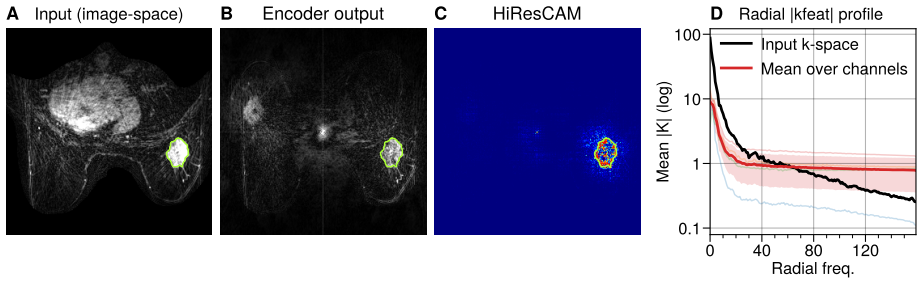
- Feature analysis shows the k-space stage concentrates on frequency-domain filtering while the image stage performs lesion localization.
- The performance advantage is reproduced in a within-dataset synthetic control.
- The hybrid and image-space models perform similarly at full sampling without added noise.
Where Pith is reading between the lines
- The hybrid architecture could be tested on segmentation tasks for other organs commonly imaged with accelerated MRI protocols.
- Prospective validation on real accelerated clinical scans would be required to confirm whether the synthetic undersampling results hold in practice.
- Combining the k-space stage with existing compressed-sensing or parallel-imaging reconstruction methods might produce even more robust end-to-end pipelines.
- The observed complementary roles suggest hybrid k-space-image models may benefit other inverse imaging problems where raw sensor data preserves information lost in standard reconstructions.
Load-bearing premise
Synthetic undersampling and added complex Gaussian noise applied to retrospective public datasets accurately represent the characteristics of real prospectively accelerated clinical MRI acquisitions and their noise.
What would settle it
A direct comparison on prospectively undersampled breast DCE-MRI data acquired on clinical scanners showing that the hybrid model's Dice advantage over image-space baselines disappears or reverses.
Figures


read the original abstract
Purpose: To assess whether breast lesion segmentation can be learned directly from acquired MRI k-space, and whether doing so improves robustness when data are accelerated or noisy. Materials and Methods: This retrospective study used public breast dynamic contrast-enhanced MRI (DCE-MRI) datasets with acquired and synthetic k-space, together with a within-dataset synthetic control. We compared four 3D U-Net variants: a hybrid k-space-to-image model, a native k-space model, and magnitude and complex image-space baselines. Models were evaluated under increasing undersampling and added complex Gaussian k-space noise. The primary outcome was patient-level Dice similarity coefficient under cross-validation, with the hybrid model prespecified as the main comparison against the magnitude image-space baseline. Results: At full sampling, the hybrid and image-space models performed similarly. As acceleration increased, the hybrid model retained substantially more segmentation accuracy and significantly outperformed the magnitude image-space baseline across moderate to high undersampling levels. The same pattern was observed when noise was added directly to k-space: the hybrid model degraded more slowly, whereas the image-space baseline failed under heavier noise. This advantage was reproduced in the within-dataset synthetic control. Feature analysis suggested that the k-space stage and image-space stage played complementary roles, with frequency-domain filtering concentrated before image-domain lesion localization. Conclusion: K-space-aware deep learning improves the robustness of breast lesion segmentation under MRI undersampling and k-space noise, while matching image-space methods at full sampling.
Editorial analysis
A structured set of objections, weighed in public.
Referee Report
Summary. The paper evaluates whether breast lesion segmentation from DCE-MRI can be performed directly in k-space or via hybrid k-space-to-image models using 3D U-Net variants. It claims that a hybrid model matches image-space baselines at full sampling but retains substantially higher patient-level Dice scores under retrospective undersampling and added complex Gaussian k-space noise, with the advantage reproduced in a within-dataset synthetic control. The primary prespecified comparison is the hybrid model versus the magnitude image-space baseline, evaluated via cross-validation on public datasets.
Significance. If the central empirical findings hold, the work provides evidence that incorporating k-space processing stages can improve robustness of segmentation models to common MRI acquisition degradations without sacrificing performance at full sampling. Strengths include the use of public datasets, cross-validation, a prespecified primary endpoint, and consistent patterns across acceleration factors and noise levels. However, the significance is tempered by reliance on synthetic retrospective degradations whose fidelity to prospective clinical scans remains unproven.
major comments (2)
- [§3] §3 (Methods, simulation pipeline): The central robustness claim depends on retrospective undersampling masks plus i.i.d. complex Gaussian noise added to k-space from public DCE-MRI data being representative of prospective accelerated acquisitions. Real scans introduce trajectory-dependent aliasing, coil-sensitivity effects, B0/B1 inhomogeneities, and non-i.i.d. noise components that are not reproduced; without additional validation (e.g., on prospectively undersampled data or more realistic noise models), the observed Dice advantage may be an artifact of the simulation rather than an intrinsic property of k-space-aware architectures.
- [Results] Results (primary outcome reporting): While the abstract states that the hybrid model 'significantly outperformed' the magnitude baseline across moderate-to-high undersampling, exact effect sizes, confidence intervals, p-values, and the precise statistical test used for the prespecified comparison are not detailed. This information is load-bearing for interpreting whether the retained accuracy constitutes a clinically meaningful improvement.
minor comments (2)
- [Abstract / Results] The abstract mentions 'feature analysis suggested that the k-space stage and image-space stage played complementary roles' but does not specify the analysis method (e.g., activation visualization, ablation, or frequency-domain filtering quantification). Adding a brief methods paragraph or supplementary figure would improve reproducibility.
- [Methods] Notation for the four U-Net variants (hybrid, native k-space, magnitude image-space, complex image-space) should be defined consistently with a table or equation early in the methods to avoid ambiguity when comparing performance curves.
Simulated Author's Rebuttal
We thank the referee for the constructive and detailed review. The comments have prompted us to improve the statistical reporting and to more explicitly discuss the limitations of our simulation approach. We respond to each major comment below.
read point-by-point responses
-
Referee: [§3] §3 (Methods, simulation pipeline): The central robustness claim depends on retrospective undersampling masks plus i.i.d. complex Gaussian noise added to k-space from public DCE-MRI data being representative of prospective accelerated acquisitions. Real scans introduce trajectory-dependent aliasing, coil-sensitivity effects, B0/B1 inhomogeneities, and non-i.i.d. noise components that are not reproduced; without additional validation (e.g., on prospectively undersampled data or more realistic noise models), the observed Dice advantage may be an artifact of the simulation rather than an intrinsic property of k-space-aware architectures.
Authors: We agree that retrospective undersampling combined with i.i.d. complex Gaussian noise does not fully reproduce the artifacts present in prospective accelerated acquisitions, including trajectory-specific aliasing, coil-sensitivity maps, B0/B1 inhomogeneities, and non-i.i.d. noise. Because the study relies on public fully sampled retrospective datasets, prospectively undersampled data were not available. We have added a dedicated limitations paragraph in the Discussion that acknowledges these gaps and recommends prospective validation in future work. At the same time, the advantage of the hybrid model remained consistent across multiple acceleration factors and noise amplitudes and was reproduced in the within-dataset synthetic control, suggesting that the architectural difference confers some robustness even under the controlled degradations we could simulate. revision: partial
-
Referee: [Results] Results (primary outcome reporting): While the abstract states that the hybrid model 'significantly outperformed' the magnitude baseline across moderate-to-high undersampling, exact effect sizes, confidence intervals, p-values, and the precise statistical test used for the prespecified comparison are not detailed. This information is load-bearing for interpreting whether the retained accuracy constitutes a clinically meaningful improvement.
Authors: We appreciate this observation. In the revised manuscript we have expanded the Results section to report, for the prespecified hybrid-versus-magnitude comparison at each acceleration factor: mean patient-level Dice scores with standard deviations, 95 % confidence intervals, p-values from paired t-tests (or Wilcoxon signed-rank tests where normality assumptions were not met), and Cohen’s d effect sizes. These quantities are now presented both in the text and in an updated supplementary table so that readers can directly evaluate the magnitude and statistical significance of the observed differences. revision: yes
- Prospective validation on real accelerated clinical acquisitions is not possible with the public retrospective datasets used in this study.
Circularity Check
No circularity: empirical ML comparison on held-out data
full rationale
The manuscript describes a retrospective empirical study that trains and evaluates four 3D U-Net variants (hybrid k-space-to-image, native k-space, magnitude and complex image-space baselines) on public breast DCE-MRI datasets using cross-validation. Performance is measured by patient-level Dice under synthetic undersampling and added complex Gaussian noise, with direct numerical comparisons reported. No equations, derivations, or predictions are presented that reduce to fitted parameters by construction, and no load-bearing self-citations or uniqueness theorems are invoked to justify the central claims. The results rest on standard held-out evaluation rather than self-referential definitions or ansatz smuggling.
Axiom & Free-Parameter Ledger
free parameters (2)
- U-Net architecture and training hyperparameters
- Undersampling acceleration factors and noise levels
axioms (1)
- domain assumption Synthetic k-space undersampling and noise addition faithfully model real accelerated MRI data characteristics
Lean theorems connected to this paper
-
IndisputableMonolith/Foundation/RealityFromDistinction.leanreality_from_one_distinction unclear?
unclearRelation between the paper passage and the cited Recognition theorem.
hybrid k-space-to-image model chains a trainable k-space stage, a non-trainable inverse fast Fourier transform (FFT), and a trainable image-space stage
-
IndisputableMonolith/Cost/FunctionalEquation.leanwashburn_uniqueness_aczel unclear?
unclearRelation between the paper passage and the cited Recognition theorem.
Models were evaluated under increasing undersampling and added complex Gaussian k-space noise
What do these tags mean?
- matches
- The paper's claim is directly supported by a theorem in the formal canon.
- supports
- The theorem supports part of the paper's argument, but the paper may add assumptions or extra steps.
- extends
- The paper goes beyond the formal theorem; the theorem is a base layer rather than the whole result.
- uses
- The paper appears to rely on the theorem as machinery.
- contradicts
- The paper's claim conflicts with a theorem or certificate in the canon.
- unclear
- Pith found a possible connection, but the passage is too broad, indirect, or ambiguous to say the theorem truly supports the claim.
Reference graph
Works this paper leans on
-
[1]
C. K. Kuhl, S. Schrading, K. Strobel, H. H. Schild, R.-D. Hilgers, and H. B. Bieling, “Abbreviated Breast Magnetic Resonance Imaging (MRI): First Postcontrast Subtracted Images and Maximum-Intensity Projection—A Novel Approach to Breast Cancer Screening With MRI,”Journal of Clinical Oncology, vol. 32, pp. 2304–2310, Aug. 2014
work page 2014
-
[2]
Generalized autocalibrating partially parallel acquisitions (GRAPPA),
M. A. Griswold, P. M. Jakob, R. M. Heidemann, M. Nittka, V. Jellus, J. Wang, B. Kiefer, and A. Haase, “Generalized autocalibrating partially parallel acquisitions (GRAPPA),”Magnetic Resonance in Medicine, vol. 47, pp. 1202–1210, June 2002. 10
work page 2002
-
[3]
Sparse MRI: The application of compressed sensing for rapid MR imaging,
M. Lustig, D. Donoho, and J. M. Pauly, “Sparse MRI: The application of compressed sensing for rapid MR imaging,”Magnetic Resonance in Medicine, vol. 58, pp. 1182–1195, Dec. 2007
work page 2007
-
[4]
Learning a variational network for reconstruction of accelerated MRI data,
K. Hammernik, T. Klatzer, E. Kobler, M. P. Recht, D. K. Sodickson, T. Pock, and F. Knoll, “Learning a variational network for reconstruction of accelerated MRI data,”Magnetic Resonance in Medicine, vol. 79, pp. 3055–3071, June 2018
work page 2018
-
[5]
fastMRI: An Open Dataset and Benchmarks for Accelerated MRI,
J. Zbontar, F. Knoll, A. Sriram, T. Murrell, Z. Huang, M. J. Muckley, A. Defazio, R. Stern, P. Johnson, M. Bruno, M. Parente, K. J. Geras, J. Katsnelson, H. Chandarana, Z. Zhang, M. Drozdzal, A. Romero, M. Rabbat, P. Vincent, N. Yakubova, J. Pinkerton, D. Wang, E. Owens, C. L. Zitnick, M. P. Recht, D. K. Sodickson, and Y. W. Lui, “fastMRI: An Open Dataset...
-
[6]
L. Hirsch, Y. Huang, S. Luo, C. Rossi Saccarelli, R. Lo Gullo, I. Daimiel Naranjo, A. G. V. Bitencourt, N. Onishi, E. S. Ko, D. Leithner, D. Avendano, S. Eskreis-Winkler, M. Hughes, D. F. Martinez, K. Pinker, K. Juluru, A. E. El-Rowmeim, P. Elnajjar, E. A. Morris, H. A. Makse, L. C. Parra, and E. J. Sutton, “Radiologist-Level Performance by Using Deep Lea...
work page 2022
-
[7]
L. C. Adams, K. K. Bressem, S. M. Böker, Y.-N. Y. Bender, D. Nörenberg, B. Hamm, and M. R. Makowski, “Diagnostic performance of susceptibility-weighted magnetic resonance imaging for the detection of calcifications: A systematic review and meta-analysis,”Scientific Reports, vol. 7, p. 15506, 2017
work page 2017
-
[8]
Brain Segmentation from k-Space with End-to-End Recurrent Attention Network,
Q. Huang, X. Chen, D. Metaxas, and M. S. Nadar, “Brain Segmentation from k-Space with End-to-End Recurrent Attention Network,” inMedical Image Computing and Computer Assisted Intervention – MICCAI 2019(D. Shen, T. Liu, T. M. Peters, L. H. Staib, C. Essert, S. Zhou, P.-T. Yap, and A. Khan, eds.), vol. 11766, pp. 275–283, Cham: Springer International Publis...
work page 2019
-
[9]
Direct MRI Segmentation from k-Space Data by Iterative Potts Minimization,
L. Kiefer, S. Petra, M. Storath, and A. Weinmann, “Direct MRI Segmentation from k-Space Data by Iterative Potts Minimization,” inScale Space and Variational Methods in Computer Vision(J. Lellmann, M. Burger, and J. Modersitzki, eds.), vol. 11603, pp. 406–418, Cham: Springer International Publishing,
-
[11]
k-strip: A novel segmentation algorithm in k-space for the application of skull stripping,
M. Rempe, F. Mentzel, K. L. Pomykala, J. Haubold, F. Nensa, K. Kroeninger, J. Egger, and J. Kleesiek, “k-strip: A novel segmentation algorithm in k-space for the application of skull stripping,”Computer Methods and Programs in Biomedicine, vol. 243, p. 107912, Jan. 2024
work page 2024
-
[12]
Cardiac MR Segmentation from Undersampled k-space Using Deep Latent Representation Learning,
J. Schlemper, O. Oktay, W. Bai, D. C. Castro, J. Duan, C. Qin, J. V. Hajnal, and D. Rueckert, “Cardiac MR Segmentation from Undersampled k-space Using Deep Latent Representation Learning,” inMedical Image Computing and Computer Assisted Intervention – MICCAI 2018(A. F. Frangi, J. A. Schnabel, C. Davatzikos, C. Alberola-López, and G. Fichtinger, eds.), vol...
work page 2018
-
[13]
Direct Cardiac Segmentation from Undersampled K-Space using Transformers,
Y. Zhang, N. Stolt-Ansó, J. Pan, W. Huang, K. Hammernik, and D. Rueckert, “Direct Cardiac Segmentation from Undersampled K-Space using Transformers,” in2024 IEEE International Symposium on Biomedical Imaging (ISBI), (Athens, Greece), pp. 1–4, IEEE, May 2024
work page 2024
-
[14]
Classification, Regression and Segmentation Directly from K-Space in Cardiac MRI,
R. Li, J. Pan, Y. Zhu, J. Ni, and D. Rueckert, “Classification, Regression and Segmentation Directly from K-Space in Cardiac MRI,” inMachine Learning in Medical Imaging(X. Xu, Z. Cui, I. Rekik, X. Ouyang, and K. Sun, eds.), vol. 15241, pp. 31–41, Cham: Springer Nature Switzerland, 2025. Series Title: Lecture Notes in Computer Science
work page 2025
-
[15]
K2S Challenge: From Undersampled K-Space to Automatic Segmentation,
A. A. Tolpadi, U. Bharadwaj, K. T. Gao, R. Bhattacharjee, F. G. Gassert, J. Luitjens, P. Giesler, J. N. Morshuis, P. Fischer, M. Hein, C. F. Baumgartner, A. Razumov, D. Dylov, Q. V. Lohuizen, S. J. Fransen, X. Zhang, R. Tibrewala, H. L. De Moura, K. Liu, M. V. W. Zibetti, R. Regatte, S. Majumdar, and 11 V. Pedoia, “K2S Challenge: From Undersampled K-Space...
work page 2023
-
[16]
A. D. Desai, A. M. Schmidt, E. B. Rubin, C. M. Sandino, M. S. Black, V. Mazzoli, K. J. Stevens, R. Boutin, C. Ré, G. E. Gold, B. A. Hargreaves, and A. S. Chaudhari, “SKM-TEA: A Dataset for Accelerated MRI Reconstruction with Dense Image Labels for Quantitative Clinical Evaluation,” 2022. Version Number: 1
work page 2022
-
[17]
Domain Influence in MRI Medical Image Segmentation: Spatial Versus k-Space Inputs,
E. Gösche, R. Eghbali, F. Knoll, and A. M. Rauschecker, “Domain Influence in MRI Medical Image Segmentation: Spatial Versus k-Space Inputs,” inMachine Learning in Medical Imaging(X. Xu, Z. Cui, I. Rekik, X. Ouyang, and K. Sun, eds.), vol. 15241, pp. 310–319, Cham: Springer Nature Switzerland,
-
[18]
Series Title: Lecture Notes in Computer Science
-
[19]
Encoding and reconstruction in parallel MRI,
K. P. Pruessmann, “Encoding and reconstruction in parallel MRI,”NMR in Biomedicine, vol. 19, pp. 288–299, May 2006
work page 2006
-
[20]
A. Deshmane, V. Gulani, M. A. Griswold, and N. Seiberlich, “Parallel MR imaging,”Journal of Magnetic Resonance Imaging, vol. 36, pp. 55–72, July 2012
work page 2012
-
[21]
R. M. Mann, N. Cho, and L. Moy, “Breast MRI: State of the Art,”Radiology, vol. 292, pp. 520–536, Sept. 2019
work page 2019
-
[22]
FastMRI Breast: A Publicly Available Radial k-Space Dataset of Breast Dynamic Contrast-enhanced MRI,
E. Solomon, P. M. Johnson, Z. Tan, R. Tibrewala, Y. W. Lui, F. Knoll, L. Moy, S. G. Kim, and L. Heacock, “FastMRI Breast: A Publicly Available Radial k-Space Dataset of Breast Dynamic Contrast-enhanced MRI,”Radiology: Artificial Intelligence, vol. 7, p. e240345, Jan. 2025
work page 2025
-
[23]
A large-scale multicenter breast cancer DCE-MRI benchmark dataset with expert segmentations,
L. Garrucho, K. Kushibar, C.-A. Reidel, S. Joshi, R. Osuala, A. Tsirikoglou, M. Bobowicz, J. Del Riego, A. Catanese, K. Gwoździewicz, M.-L. Cosaka, P. M. Abo-Elhoda, S. W. Tantawy, S. S. Sakrana, N. O. Shawky-Abdelfatah, A. M. A. Salem, A. Kozana, E. Divjak, G. Ivanac, K. Nikiforaki, M. E. Klontzas, R. García-Dosdá, M. Gulsun-Akpinar, O. Lafcı, R. Mann, C...
work page 2025
-
[24]
Checklist for Artificial Intelligence in Medical Imaging (CLAIM): A Guide for Authors and Reviewers,
J. Mongan, L. Moy, and C. E. Kahn, “Checklist for Artificial Intelligence in Medical Imaging (CLAIM): A Guide for Authors and Reviewers,”Radiol. Artif. Intell., vol. 2, p. e200029, Mar. 2020
work page 2020
-
[25]
F. Knoll, J. Zbontar, A. Sriram, M. J. Muckley, M. Bruno, A. Defazio, M. Parente, K. J. Geras, J. Katsnelson, H. Chandarana, Z. Zhang, M. Drozdzalv, A. Romero, M. Rabbat, P. Vincent, J. Pinkerton, D. Wang, N. Yakubova, E. Owens, C. L. Zitnick, M. P. Recht, D. K. Sodickson, and Y. W. Lui, “fastMRI: A Publicly Available Raw k-Space and DICOM Dataset of Knee...
work page 2020
-
[26]
ESPIRiT–an eigenvalue approach to autocalibrating parallel MRI: where SENSE meets GRAPPA,
M. Uecker, P. Lai, M. J. Murphy, P. Virtue, M. Elad, J. M. Pauly, S. S. Vasanawala, and M. Lustig, “ESPIRiT–an eigenvalue approach to autocalibrating parallel MRI: where SENSE meets GRAPPA,” Magn. Reson. Med., vol. 71, pp. 990–1001, Mar. 2014
work page 2014
-
[27]
MONAI: Medical open network for AI,
MONAI Consortium, “MONAI: Medical open network for AI,” July 2022
work page 2022
-
[28]
Y. Li, Y. Qi, Z. Hu, K. Zhang, S. Jia, L. Zhang, W. Xu, S. Shen, Y. X. J. Wáng, Z. Li, D. Liang, X. Liu, H. Zheng, G. Cheng, and N. Zhang, “A novel automatic segmentation method directly based on magnetic resonance imaging K-space data for auxiliary diagnosis of glioma,”Quant. Imaging Med. Surg., vol. 14, pp. 2008–2020, Feb. 2024
work page 2008
-
[29]
Implicit data crimes: Machine learning bias arising from misuse of public data,
E. Shimron, J. I. Tamir, K. Wang, and M. Lustig, “Implicit data crimes: Machine learning bias arising from misuse of public data,”Proceedings of the National Academy of Sciences, vol. 119, p. e2117203119, Mar. 2022. 12 Supplement S1. Dataset preprocessing fastMRI breast The fastMRI breast dataset provides raw multi-coil radial k-space from contrast-enhanc...
work page 2022
discussion (0)
Sign in with ORCID, Apple, or X to comment. Anyone can read and Pith papers without signing in.