VesselSim: learning 3D blood vessel segmentation without expert annotations
Pith reviewed 2026-06-29 22:41 UTC · model grok-4.3
The pith
A neural network trained only on simulated blood vessel volumes can segment real MR and CT scans at levels competitive with models that use expert annotations.
A machine-rendered reading of the paper's core claim, the machinery that carries it, and where it could break.
Core claim
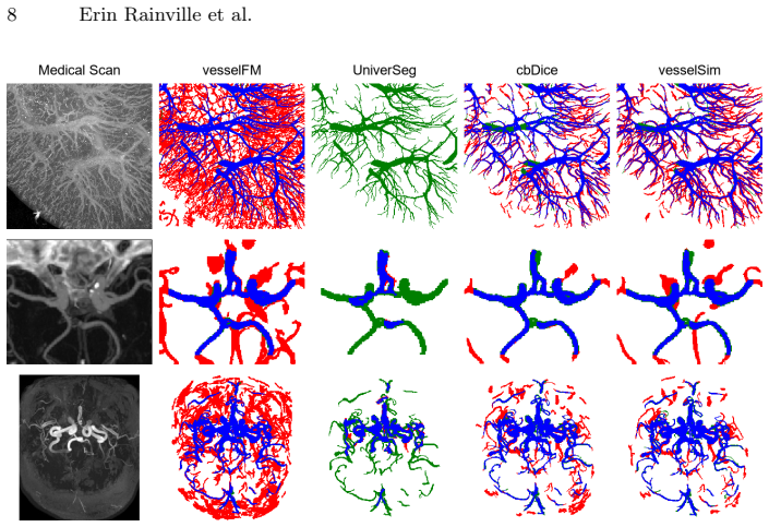
VesselSim first builds a stochastic geometry-driven vascular simulation that produces anatomically plausible 3D angiographic volumes by modeling recursive branching, curvature-controlled growth, and collision-aware topology, then applies domain-randomized intensity synthesis to create 16,500 training examples. A 3D U-Net is trained exclusively on this synthetic set. At test time on real clinical volumes, a self-supervised mask reconstruction decoder adapts the network to the new domain. When evaluated in a zero-shot setting on multiple MR and CT datasets covering brain and kidneys, the adapted model reaches accuracy levels competitive with state-of-the-art vascular segmentation foundation mo
What carries the argument
The stochastic geometry-driven vascular simulation that generates recursive branching, curvature-controlled growth, and collision-aware topology, combined with domain-randomized intensity synthesis to produce training volumes and a self-supervised mask reconstruction decoder for test-time adaptation.
If this is right
- Training relies only on synthetic data, eliminating the collection of real annotated medical volumes for this task.
- The same trained network can be applied directly to new MR and CT scans from different anatomical sites without retraining or additional labels.
- Cross-domain generalization arises from learning vessel geometry rather than scanner-specific intensity patterns.
- Self-supervised test-time adaptation allows the model to handle distribution shifts at inference without domain-specific knowledge.
Where Pith is reading between the lines
- The simulation parameters could be varied to generate training sets matched to specific disease states such as aneurysms or stenoses.
- Similar geometry-driven synthesis might reduce annotation needs for segmentation of other branching anatomical structures like airways or nerves.
- If the adaptation decoder proves stable across modalities, the overall pipeline could be tested on additional imaging types such as ultrasound without new labeled data.
Load-bearing premise
The simulated vessel structures and image appearances are close enough to real clinical angiograms that training on them plus self-supervised adaptation produces a model that works on unseen real scans.
What would settle it
If the adapted model shows substantially lower Dice scores or higher false-positive rates than expert-annotated models when tested on a new collection of real MR or CT angiograms, the claim that synthetic data suffices would be refuted.
Figures

read the original abstract
Blood vessel segmentation is a core task in medical image analysis for the care of vascular diseases and surgical planning, yet the challenges of providing expert vascular annotations pose a major obstacle for the progress of related deep learning techniques. To address this, we propose VesselSim, a two-stage framework for universal 3D blood vessel segmentation that eliminates the need for real annotated data during training. First, we introduce a stochastic, geometry-driven vascular simulation framework that models recursive branching, curvature-controlled growth, and collision-aware topology, followed by domain-randomized intensity synthesis to generate 16,500 anatomically plausible 3D angiographic volumes. Second, a 3D U-Net is trained solely on this synthetic data. To bridge the domain gap from synthetic to real images at inference time, we introduce a test-time adaptation strategy via a self-supervised mask reconstruction decoder, enabling adaptation to unseen clinical scans without prior domain knowledge. We evaluate VesselSim in a zero-shot setting on multiple real-world datasets spanning MR and CT across several anatomical regions, including the brain and kidneys. Despite being trained exclusively on synthetic data, VesselSim achieves performance competitive with state-of-the-art vascular segmentation foundation models. These findings suggest that learning vessel geometry from synthetic tubular structures is effective for robust cross-domain generalization, substantially reducing the reliance on acquired medical imaging data and more importantly, expert annotations.
Editorial analysis
A structured set of objections, weighed in public.
Referee Report
Summary. The manuscript proposes VesselSim, a two-stage framework for universal 3D blood vessel segmentation without expert annotations. Stage one generates 16,500 synthetic 3D angiographic volumes via a stochastic geometry-driven simulation (recursive branching, curvature-controlled growth, collision-aware topology) plus domain-randomized intensity synthesis. A 3D U-Net is trained exclusively on these synthetic volumes. Stage two introduces self-supervised test-time adaptation via a mask-reconstruction decoder to adapt to unseen real MR and CT volumes. The central claim is that this synthetic-only training plus TTA yields performance competitive with state-of-the-art vascular segmentation foundation models on real-world datasets spanning brain and kidney anatomy in a zero-shot setting.
Significance. If the empirical results hold, the contribution would be significant: it demonstrates that geometry-driven synthetic data generation combined with self-supervised TTA can produce robust cross-domain generalization without any real annotations or domain-specific knowledge, directly addressing the annotation bottleneck in vascular segmentation. The approach is internally consistent, avoids circular fitting to real data, and rests on explicit simulation rather than ad-hoc parameter tuning to target datasets.
major comments (1)
- [Abstract] Abstract: the claim that VesselSim 'achieves performance competitive with state-of-the-art vascular segmentation foundation models' is asserted without any accompanying quantitative metrics (e.g., Dice, sensitivity, Hausdorff distance), baseline comparisons, statistical tests, ablation results, or tables/figures. This information is load-bearing for the central claim and prevents evaluation of whether the synthetic distribution plus TTA actually suffices for competitive zero-shot performance on real MR/CT data.
Simulated Author's Rebuttal
We thank the referee for the positive assessment of the work's significance and for the constructive feedback on the abstract. We address the single major comment below.
read point-by-point responses
-
Referee: [Abstract] Abstract: the claim that VesselSim 'achieves performance competitive with state-of-the-art vascular segmentation foundation models' is asserted without any accompanying quantitative metrics (e.g., Dice, sensitivity, Hausdorff distance), baseline comparisons, statistical tests, ablation results, or tables/figures. This information is load-bearing for the central claim and prevents evaluation of whether the synthetic distribution plus TTA actually suffices for competitive zero-shot performance on real MR/CT data.
Authors: We agree that the abstract should include key quantitative support for the central claim to enable immediate evaluation. The results section of the manuscript already contains the requested elements: tables reporting Dice, sensitivity, and Hausdorff distances across multiple real MR and CT datasets, direct comparisons against vascular segmentation foundation models, and ablation studies on the simulation and TTA components. In the revised version we will condense the most salient metrics and comparisons into the abstract (while preserving its length constraints) so that the claim is no longer unsupported at the point of first reading. revision: yes
Circularity Check
No significant circularity in derivation chain
full rationale
The paper's central pipeline—stochastic geometry simulation to generate 16,500 synthetic volumes, domain-randomized synthesis, 3D U-Net training exclusively on synthetics, and self-supervised mask-reconstruction TTA—contains no equations, fitted parameters, or self-citations that reduce any claimed result to its own inputs by construction. The zero-shot evaluation on real MR/CT datasets is presented as an external test of generalization rather than a fitted or renamed quantity. No load-bearing uniqueness theorems, ansatzes smuggled via prior work, or self-definitional steps appear in the described methods or abstract. This is the expected outcome for a simulation-driven approach that does not rely on real-data fitting loops.
Axiom & Free-Parameter Ledger
free parameters (1)
- simulation hyperparameters (branching probability, curvature limits, collision thresholds)
axioms (1)
- domain assumption Synthetic tubular structures generated by recursive branching and curvature rules sufficiently approximate real vessel geometry and topology for downstream deep learning.
Reference graph
Works this paper leans on
-
[1]
In: International Workshop on Machine Learning in Clinical Neuroimaging
Aktar, M., Rivaz, H., Kersten-Oertel, M., Xiao, Y.: Vesselshot: Few-shot learning for cerebral blood vessel segmentation. In: International Workshop on Machine Learning in Clinical Neuroimaging. pp. 46–55 (2023)
2023
-
[2]
International Conference on Com- puter Vision (2023)
Butoi*, V.I., Ortiz*, J.J.G., Ma, T., Sabuncu, M.R., Guttag, J., Dalca, A.V.: Uni- verseg: Universal medical image segmentation. International Conference on Com- puter Vision (2023)
2023
-
[3]
IEEE Transactions on Medical Imaging 34(12), 2478–2491 (2015)
Bériault, S., Xiao, Y., Collins, D.L., Pike, G.B.: Automatic swi venography seg- mentation using conditional random fields. IEEE Transactions on Medical Imaging 34(12), 2478–2491 (2015)
2015
-
[4]
In: 2025 IEEE/CVF Winter Conference on Applications of Computer Vision (WACV)
Colussi, M., Mascetti, S., Dolz, J., Desrosiers, C.: Rec-ttt: Contrastive feature reconstruction for test-time training. In: 2025 IEEE/CVF Winter Conference on Applications of Computer Vision (WACV). pp. 6699–6708 (2025)
2025
-
[5]
DeVries, T., Taylor, G.W.: Improved regularization of convolutional neural net- works with cutout (2017)
2017
-
[6]
In: Medical Image Com- puting and Computer Assisted Intervention – MICCAI 2025
Falcetta, D., Marciano, V., Yang, K., Cleary, J., Legris, L., Rizzaro, M.D., Pitsior- las, I., Chaptoukaev, H., Lemasson, B., Menze, B., Zuluaga, M.A.: Vesselverse: A dataset and collaborative framework for vessel annotation. In: Medical Image Com- puting and Computer Assisted Intervention – MICCAI 2025. vol. LNCS 15972, pp. 656 – 666. Springer Nature Swi...
2025
-
[7]
In: International Con- ference on Medical Image Computing and Computer-Assisted Intervention
Feldman, P., Fainstein, M., Siless, V., Delrieux, C., Iarussi, E.: Vesselvae: Recur- sive variational autoencoders for 3d blood vessel synthesis. In: International Con- ference on Medical Image Computing and Computer-Assisted Intervention. pp. 67–76 (2023)
2023
-
[8]
In: International Conference on Medical Image Computing and Computer-Assisted Intervention
Feldman, P., Sinnona, M., Delrieux, C., Siless, V., Iarussi, E.: Vesselgpt: Autore- gressive modeling of vascular geometry. In: International Conference on Medical Image Computing and Computer-Assisted Intervention. pp. 662–672 (2025)
2025
-
[9]
Galarreta Valverde, M., Macedo, M., Mekkaoui, C.: Three-dimensional synthetic bloodvesselgenerationusingstochasticl-systems.In:MedicalImaging2013:Image Processing. vol. 8669 (03 2013). https://doi.org/10.1117/12.2007532 10 Erin Rainville et al
-
[10]
In: International Conference on Medical Image Computing and Computer-Assisted Intervention
Gu, Y., Sun, Z., Liu, Z., Xu, Y.: Test-time training with local contrast-preserving copy-pasted image for domain generalization in retinal vessel segmentation. In: International Conference on Medical Image Computing and Computer-Assisted Intervention. pp. 614–624 (2025)
2025
-
[11]
IEEE Transactions on Medical Imaging (2025)
Guo, Z., Tan, Z., Feng, J., Zhou, J.: Vesseldiffusion: 3d vascular structure genera- tion based on diffusion model. IEEE Transactions on Medical Imaging (2025)
2025
-
[12]
Proceedings of the National Academy of Sciences121(11), e2310044121 (2024)
Haast, R.A., Kashyap, S., Ivanov, D., Yousif, M.D., DeKraker, J., Poser, B.A., Khan, A.R.: Insights into hippocampal perfusion using high-resolution, multi- modal 7t mri. Proceedings of the National Academy of Sciences121(11), e2310044121 (2024)
2024
-
[13]
Computer- ized medical imaging and graphics34(8), 605–616 (2010)
Hamarneh, G., Jassi, P.: Vascusynth: Simulating vascular trees for generating vol- umetric image data with ground-truth segmentation and tree analysis. Computer- ized medical imaging and graphics34(8), 605–616 (2010)
2010
-
[14]
https://kaggle.com/competitions/blood-vessel-segmentation (2023), kaggle com- petition
Jain, Y., Borner, K., Walsh, C., Ruschman, N., Lee, P.D., Weber, G.M., Hol- brook, R., Howard, A.: Sennet + hoa - hacking the human vasculature in 3d. https://kaggle.com/competitions/blood-vessel-segmentation (2023), kaggle com- petition
2023
-
[15]
IEEE transactions on bio-medical engineeringPP(11 2023)
Jessen, E., Steinbach, M., Debbaut, C., Schillinger, D.: Branching ex- ponents of synthetic vascular trees under different optimality prin- ciples. IEEE transactions on bio-medical engineeringPP(11 2023). https://doi.org/10.1109/TBME.2023.3334758
-
[16]
Computer Methods and Programs in Biomedicine158, 71–91 (2018)
Moccia,S.,DeMomi,E.,ElHadji,S.,Mattos,L.S.:Bloodvesselsegmentationalgo- rithms — review of methods, datasets and evaluation metrics. Computer Methods and Programs in Biomedicine158, 71–91 (2018)
2018
-
[17]
the vascular system and the cost of blood volume
Murray, C.D.: The physiological principle of minimum work: I. the vascular system and the cost of blood volume. Proceedings of the National Academy of Sciences 12(3), 207–214 (1926)
1926
-
[18]
In: Proceedings of the IEEE Conference on Computer Vision and Pattern Recognition (CVPR)
Pathak, D., Krahenbuhl, P., Donahue, J., Darrell, T., Efros, A.: Context en- coders: Feature learning by inpainting. In: Proceedings of the IEEE Conference on Computer Vision and Pattern Recognition (CVPR). pp. 2536–2544 (06 2016). https://doi.org/10.1109/CVPR.2016.278
-
[19]
PyTorch MONAI Team: Medical open network for ai (MONAI), https://github.com/Project-MONAI/MONAI
-
[20]
In: International Conference on Medical Image Computing and Computer-Assisted Intervention
Shi,P.,Hu,J.,Yang,Y.,Gao,Z.,Liu,W.,Ma,T.:Centerlineboundarydicelossfor vascular segmentation. In: International Conference on Medical Image Computing and Computer-Assisted Intervention. pp. 46–56. Springer (2024)
2024
-
[21]
Advanced Science11(32), 2402195 (2024)
Sweeney, P.W., Hacker, L., Lefebvre, T.L., Brown, E.L., Gröhl, J., Bohndiek, S.E.: Unsupervised segmentation of 3d microvascular photoacoustic images using deep generative learning. Advanced Science11(32), 2402195 (2024)
2024
- [22]
-
[23]
In: Proceedings of the IEEE/CVF Conference on Computer Vision and Pattern Recognition
Wittmann, B., Wattenberg, Y., Amiranashvili, T., Shit, S., Menze, B.: vesselfm: A foundation model for universal 3d blood vessel segmentation. In: Proceedings of the IEEE/CVF Conference on Computer Vision and Pattern Recognition. pp. 20874–20884 (2025)
2025
-
[24]
International Journal for Numerical Methods in Biomedical Engineering29(4), 515–529 (2013) VesselSim: learning 3D blood vessel segmentation without expert annotations 11
Yang, J., Wang, Y.: Design of vascular networks: A mathematical model approach. International Journal for Numerical Methods in Biomedical Engineering29(4), 515–529 (2013) VesselSim: learning 3D blood vessel segmentation without expert annotations 11
2013
- [25]
-
[26]
Information Fusion123, 103281 (2025)
Yang, Y., Xu, J., Xu, M., Tang, X., Wang, B., Shu, K., You, Z.: Fsvs-net: A few-shot semi-supervised vessel segmentation network for multiple organs based on feature distillation and bidirectional weighted fusion. Information Fusion123, 103281 (2025)
2025
-
[27]
arXiv preprint arXiv:2512.02497 (2025)
Yu, W., Jiang, S., Chen, Y., Chang, S., Wang, Y., Wu, B., Dong, J., Liu, M., Zhu, S., Qin, F., Wang, C., Tian, Q.: A large scale benchmark for test time adaptation methods in medical image segmentation. ArXivabs/2512.02497(2025)
-
[28]
In: Proceedings of the IEEE/CVF International Conference on Computer Vision (ICCV) (2025)
Zhou, J., Wang, W., Li, S., Qu, X., Guo, X., Liu, Y., Tang, W., Lin, X., Zheng, Y.: Topotta: Topology-enhanced test-time adaptation for tubular structure segmen- tation. In: Proceedings of the IEEE/CVF International Conference on Computer Vision (ICCV) (2025)
2025
discussion (0)
Sign in with ORCID, Apple, or X to comment. Anyone can read and Pith papers without signing in.