DualGate-Net: A Prior-Gated Dual-Encoder Framework for Histopathology Cell Detection
Pith reviewed 2026-06-27 22:38 UTC · model grok-4.3
The pith
DualGate-Net combines local and global encoders with learnable prior-gated fusion to adaptively incorporate tissue context for cell detection in histopathology images.
A machine-rendered reading of the paper's core claim, the machinery that carries it, and where it could break.
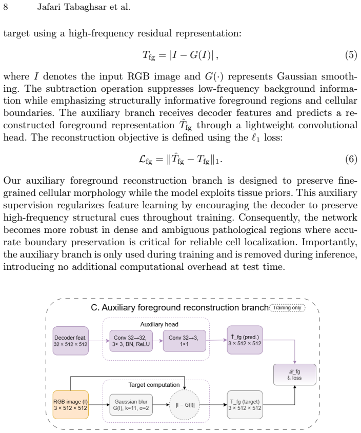
Core claim
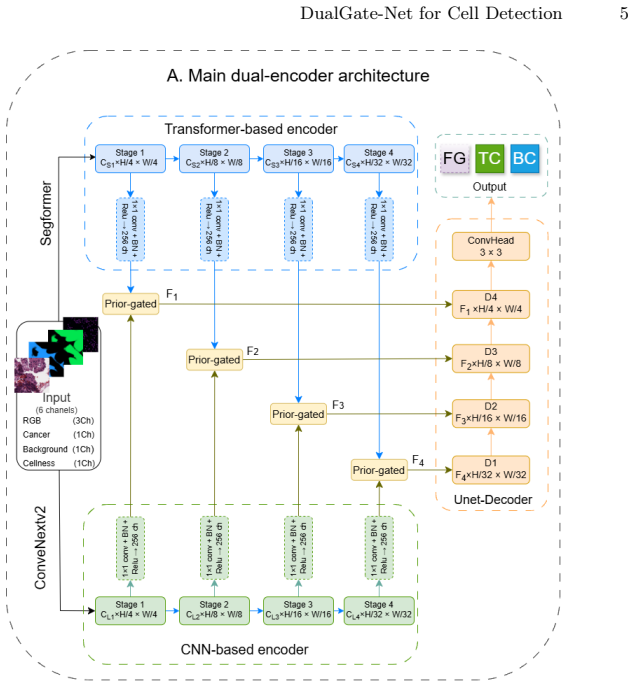
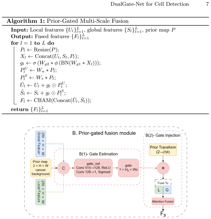
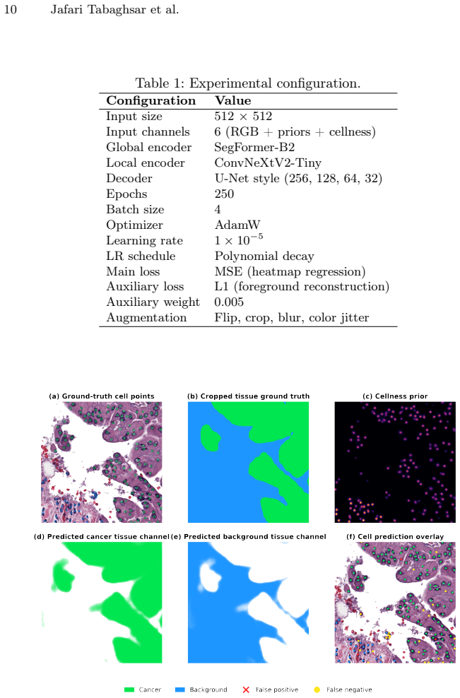
DualGate-Net combines a ConvNeXtV2-based local encoder and a SegFormer-based global encoder through a learnable prior-gated fusion mechanism that adaptively regulates the influence of tissue priors across spatial locations. An auxiliary foreground reconstruction branch preserves high-frequency cellular structures during training, and auxiliary cellness-guided cues further improve localization robustness. Experiments on the OCELOT benchmark demonstrate consistent improvements, achieving macro F1-scores of 0.7722 on the validation set and 0.7345 on the test set.
What carries the argument
The learnable prior-gated fusion mechanism that adaptively regulates the influence of tissue priors across spatial locations.
If this is right
- Adaptive per-location regulation of priors reduces noise propagation relative to static fusion strategies.
- The auxiliary foreground reconstruction branch maintains high-frequency cellular structures that would otherwise be lost.
- Cellness-guided cues add localization robustness on top of the gated fusion.
- The reported macro F1 scores of 0.7722 validation and 0.7345 test represent measurable gains on the OCELOT benchmark.
Where Pith is reading between the lines
- The same learnable gating pattern could be inserted into other dual-stream medical imaging models that currently use fixed context fusion.
- Evaluating the framework on additional histopathology cohorts with different staining protocols would test whether the adaptive regulation generalizes beyond OCELOT.
- The spatial maps produced by the gate itself could be inspected to identify which tissue microenvironments most strongly influence particular cell classes.
Load-bearing premise
The learnable prior-gated fusion module will adaptively regulate tissue-prior influence across locations without propagating noise, and the auxiliary foreground reconstruction branch will reliably preserve high-frequency cellular structures.
What would settle it
Running an ablation on the OCELOT test set that removes the learnable gate or the auxiliary reconstruction branch and shows no drop in macro F1, or visual inspection of fused feature maps that reveals increased noise, would falsify the central claim.
Figures
read the original abstract
Cell detection in histopathology images strongly depends on surrounding tissue context, where visually similar cells may belong to different classes under different microenvironments. Recent tissue-aware methods incorporate contextual priors, but often rely on static fusion strategies that may propagate noisy information. In this work, we propose DualGate-Net, a prior-aware dual-encoder framework that combines a ConvNeXtV2-based local encoder and a SegFormer-based global encoder through a learnable prior-gated fusion mechanism. The proposed module adaptively regulates the influence of tissue priors across spatial locations, while an auxiliary foreground reconstruction branch preserves high-frequency cellular structures during training. In addition, auxiliary cellness-guided cues are incorporated to further improve localization robustness. Experiments on the OCELOT benchmark demonstrate consistent improvements, achieving macro F1-scores of 0.7722 on the validation set and 0.7345 on the test set, highlighting the effectiveness of adaptive prior integration for robust histopathology cell detection.
Editorial analysis
A structured set of objections, weighed in public.
Referee Report
Summary. The paper proposes DualGate-Net, a dual-encoder architecture combining a ConvNeXtV2 local encoder and SegFormer global encoder via a learnable prior-gated fusion module, plus an auxiliary foreground reconstruction branch and cellness-guided cues, for context-aware cell detection in histopathology images. It reports macro F1 scores of 0.7722 on the OCELOT validation set and 0.7345 on the test set, attributing gains to adaptive regulation of tissue priors and preservation of high-frequency structures.
Significance. If the empirical gains can be rigorously attributed to the proposed mechanisms rather than training choices or backbone, the work addresses a practical need for robust tissue-context integration in computational pathology. The absence of ablations, baselines, and mechanism-specific diagnostics in the current presentation prevents assessing whether the adaptive fusion and auxiliary branch deliver the claimed benefits.
major comments (3)
- [Abstract] Abstract: The reported macro F1 scores of 0.7722/0.7345 are presented without baseline comparisons, statistical tests, error bars, ablation studies, or details on dataset splits and training protocol. This makes it impossible to determine whether improvements stem from the learnable prior-gated fusion or auxiliary branch rather than other factors.
- [Abstract] Abstract (framework description): The central claim that the learnable prior-gated fusion 'adaptively regulates the influence of tissue priors across spatial locations' without propagating noise lacks supporting evidence such as gating visualizations, attention maps, or noise-injection ablations. Without these, attribution of the F1 gains to this module cannot be verified.
- [Abstract] Abstract (framework description): The auxiliary foreground reconstruction branch is asserted to 'preserve high-frequency cellular structures,' yet no frequency-domain metrics, reconstruction error analysis, or targeted ablations are reported to confirm this behavior or its contribution to detection performance.
Simulated Author's Rebuttal
We thank the referee for the constructive feedback. We address each major comment below and will revise the manuscript to strengthen the evidence supporting our claims.
read point-by-point responses
-
Referee: [Abstract] Abstract: The reported macro F1 scores of 0.7722/0.7345 are presented without baseline comparisons, statistical tests, error bars, ablation studies, or details on dataset splits and training protocol. This makes it impossible to determine whether improvements stem from the learnable prior-gated fusion or auxiliary branch rather than other factors.
Authors: We agree that the abstract is too concise to fully contextualize the results. The full manuscript contains baseline comparisons, dataset details, and training protocol in the experiments section. To address the concern directly, we will revise the abstract to briefly reference these elements and add error bars plus statistical significance testing to the reported F1 scores in the results. revision: yes
-
Referee: [Abstract] Abstract (framework description): The central claim that the learnable prior-gated fusion 'adaptively regulates the influence of tissue priors across spatial locations' without propagating noise lacks supporting evidence such as gating visualizations, attention maps, or noise-injection ablations. Without these, attribution of the F1 gains to this module cannot be verified.
Authors: We acknowledge that the current manuscript lacks mechanism-specific diagnostics for the prior-gated fusion. We will add gating visualizations, attention maps, and noise-injection ablations in the revised version to provide direct evidence for the adaptive regulation claim and its contribution to the observed performance. revision: yes
-
Referee: [Abstract] Abstract (framework description): The auxiliary foreground reconstruction branch is asserted to 'preserve high-frequency cellular structures,' yet no frequency-domain metrics, reconstruction error analysis, or targeted ablations are reported to confirm this behavior or its contribution to detection performance.
Authors: We agree that additional targeted analysis is needed to substantiate the auxiliary branch claim. In the revision, we will incorporate frequency-domain metrics, reconstruction error analysis, and ablations isolating the branch to confirm its effect on high-frequency structures and detection performance. revision: yes
Circularity Check
No circularity: empirical results on external benchmark with no self-referential derivations
full rationale
The paper reports macro F1 scores of 0.7722/0.7345 on the OCELOT benchmark after describing a dual-encoder architecture with learnable gated fusion and auxiliary branches. No equations, fitted parameters, or derivation steps appear in the abstract or described framework that reduce any claimed output to an input by construction. No self-citations are invoked as load-bearing uniqueness theorems, no ansatzes are smuggled, and no predictions are statistically forced from subsets of the same data. The performance numbers are standard empirical evaluations on an independent external dataset, making the derivation chain self-contained against external benchmarks.
Axiom & Free-Parameter Ledger
Reference graph
Works this paper leans on
-
[1]
The Journal of pathol- ogy249(3), 286–294 (2019)
Abels, E., Pantanowitz, L., Aeffner, F., Zarella, M.D., Van der Laak, J., Bui, M.M., Vemuri, V.N., Parwani, A.V., Gibbs, J., Agosto-Arroyo, E., et al.: Computational pathology definitions, best practices, and recommendations for regulatory guid- ance: a white paper from the digital pathology association. The Journal of pathol- ogy249(3), 286–294 (2019)
2019
-
[2]
In: Proceedings of the IEEE/CVF International Conference on Computer Vision
Abousamra, S., Belinsky, D., Van Arnam, J., Allard, F., Yee, E., Gupta, R., Kurc, T., Samaras, D., Saltz, J., Chen, C.: Multi-class cell detection using spatial con- text representation. In: Proceedings of the IEEE/CVF International Conference on Computer Vision. pp. 4005–4014 (2021)
2021
-
[3]
arXiv preprint arXiv:2003.10778 (2020)
Gamper, J., Koohbanani, N.A., Benes, K., Graham, S., Jahanifar, M., Khurram, S.A., Azam, A., Hewitt, K., Rajpoot, N.: Pannuke dataset extension, insights and baselines. arXiv preprint arXiv:2003.10778 (2020)
-
[4]
Medical image analysis58, 101563 (2019)
Graham, S., Vu, Q.D., Raza, S.E.A., Azam, A., Tsang, Y.W., Kwak, J.T., Rajpoot, N.: Hover-net: Simultaneous segmentation and classification of nuclei in multi- tissue histology images. Medical image analysis58, 101563 (2019)
2019
-
[5]
In: International Conference on Medical Image Comput- ing and Computer-Assisted Intervention
Ha, S.M., Ko, Y.S., Park, Y.: Generating blobcell label from weak annotations for precise cell segmentation. In: International Conference on Medical Image Comput- ing and Computer-Assisted Intervention. pp. 161–170. Springer (2023)
2023
-
[6]
Medical image analysis94, 103143 (2024)
Hörst, F., Rempe, M., Heine, L., Seibold, C., Keyl, J., Baldini, G., Ugurel, S., Siveke, J., Grünwald, B., Egger, J., et al.: Cellvit: Vision transformers for precise cell segmentation and classification. Medical image analysis94, 103143 (2024)
2024
-
[7]
IEEE transactions on medical imaging39(5), 1380–1391 (2019)
Kumar, N., Verma, R., Anand, D., Zhou, Y., Onder, O.F., Tsougenis, E., Chen, H., Heng, P.A., Li, J., Hu, Z., et al.: A multi-organ nucleus segmentation challenge. IEEE transactions on medical imaging39(5), 1380–1391 (2019)
2019
-
[8]
In: International Conference on Medical Image Computing and Computer-Assisted Intervention
Lafarge, M.W., Koelzer, V.H.: Detecting cells in histopathology images with a resnet ensemble model. In: International Conference on Medical Image Computing and Computer-Assisted Intervention. pp. 123–129. Springer (2023)
2023
-
[9]
In: International Conference on Medi- cal Image Computing and Computer-Assisted Intervention
Lo, Y.W., Yang, C.H.: Enhancing cell detection via fc-hardnet and tissue seg- mentation: Ocelot 2023 challenge approach. In: International Conference on Medi- cal Image Computing and Computer-Assisted Intervention. pp. 130–137. Springer (2023)
2023
-
[10]
Millward, J., He, Z., Nibali, A.: Dense prediction of cell centroids using tissue con- textandcellrefinement.In:InternationalConferenceonMedicalImageComputing and Computer-Assisted Intervention. pp. 138–149. Springer (2023) DualGate-Net for Cell Detection 15
2023
-
[11]
In: Proceedings of the IEEE/CVF Conference on Computer Vision and Pattern Recognition
Ryu, J., Puche, A.V., Shin, J., Park, S., Brattoli, B., Lee, J., Jung, W., Cho, S.I., Paeng, K., Ock, C.Y., et al.: Ocelot: Overlapped cell on tissue dataset for histopathology. In: Proceedings of the IEEE/CVF Conference on Computer Vision and Pattern Recognition. pp. 23902–23912 (2023)
2023
-
[12]
In: International Conference on Medical Image Computing and Computer-Assisted Intervention
Schoenpflug, L.A., Koelzer, V.H.: Softctm: cell detection by soft instance segmen- tation and consideration of cell-tissue interaction. In: International Conference on Medical Image Computing and Computer-Assisted Intervention. pp. 109–122. Springer (2023)
2023
-
[13]
GigaScience 14, giaf011 (2025)
Schuiveling, M., Liu, H., Eek, D., Breimer, G.E., Suijkerbuijk, K.P., Blokx, W.A., Veta, M.: A novel dataset for nuclei and tissue segmentation in melanoma with baseline nuclei segmentation and tissue segmentation benchmarks. GigaScience 14, giaf011 (2025)
2025
-
[14]
In: Proceedings of the AAAI Conference on Artificial Intelligence
Shui, Z., Li, H., Zhang, Y., Sun, Y., Ye, Y., Chen, P., Guo, R., Cui, L., Zhu, C., Yang, L.: Towards effective and efficient context-aware nucleus detection in histopathology whole slide images. In: Proceedings of the AAAI Conference on Artificial Intelligence. vol. 40, pp. 9042–9050 (2026)
2026
-
[15]
arXiv preprint arXiv:2510.20754 (2025)
Torbati, N., Meshcheryakova, A., Woitek, R., Mechtcheriakova, D., Mahbod, A.: Acs-segnet: An attention-based cnn-segformer segmentation network for tissue seg- mentation in histopathology. arXiv preprint arXiv:2510.20754 (2025)
-
[16]
In: Proceedings of the IEEE/CVF conference on computer vision and pattern recognition
Woo, S., Debnath, S., Hu, R., Chen, X., Liu, Z., Kweon, I.S., Xie, S.: Convnext v2: Co-designing and scaling convnets with masked autoencoders. In: Proceedings of the IEEE/CVF conference on computer vision and pattern recognition. pp. 16133– 16142 (2023)
2023
-
[17]
PeerJ11, e15408 (2023)
Wu, Y., Liu, X., Liu, F., Li, Y., Xiong, X., Sun, H., Lin, B., Li, Y., Xu, B.: A multi- class classification algorithm based on hematoxylin-eosin staining for neoadjuvant therapy in rectal cancer: a retrospective study. PeerJ11, e15408 (2023)
2023
-
[18]
Advances in neural information processing systems34, 12077–12090 (2021)
Xie, E., Wang, W., Yu, Z., Anandkumar, A., Alvarez, J.M., Luo, P.: Segformer: Simple and efficient design for semantic segmentation with transformers. Advances in neural information processing systems34, 12077–12090 (2021)
2021
discussion (0)
Sign in with ORCID, Apple, or X to comment. Anyone can read and Pith papers without signing in.