Mitosis Detection in the Wild: Multi-Tumor and Context-Aware Generalization in the MIDOG 2025 Challenge
Pith reviewed 2026-06-27 22:11 UTC · model grok-4.3
The pith
Mitosis detection models degrade sharply outside hotspots and across diverse tumor types in MIDOG 2025.
A machine-rendered reading of the paper's core claim, the machinery that carries it, and where it could break.
Core claim
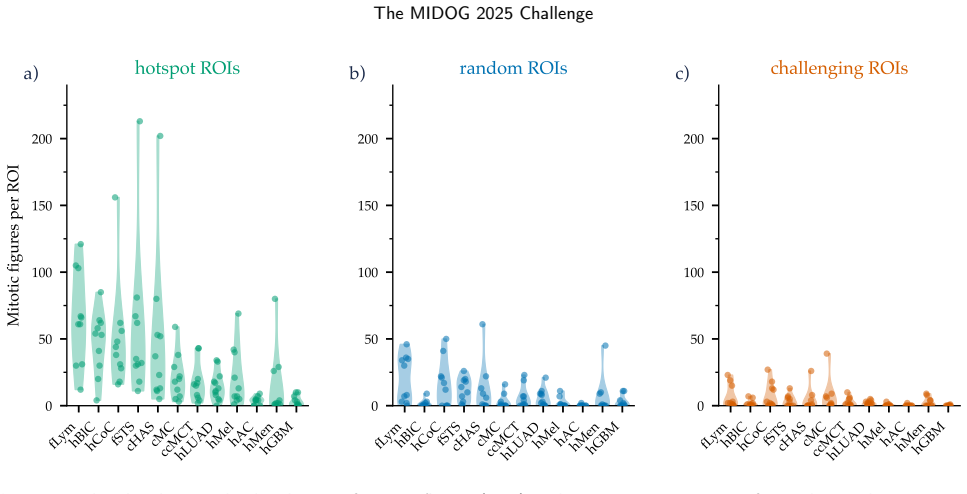
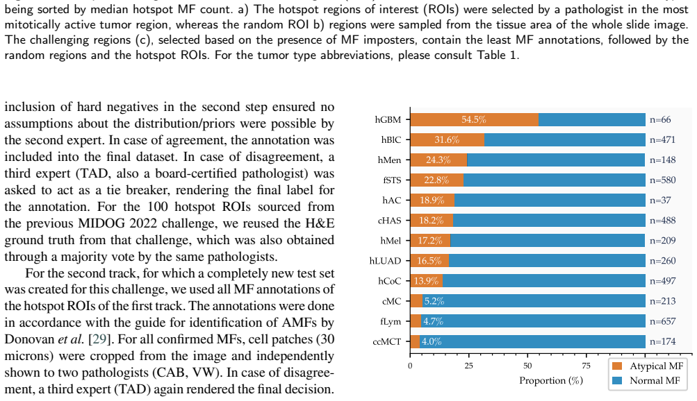
MIDOG 2025 establishes that current mitosis detectors remain unreliable in the wild: performance holds in conventional hotspots but declines substantially in random and hard-negative regions and varies across the 12 tumor types, with the curated multi-tumor, multi-scanner test set intended to expose these limitations more realistically than hotspot-only benchmarks.
What carries the argument
The multi-contextual evaluation protocol that mandates mitosis detection across hotspots, random tissue areas, and hard-negative regions together with atypical mitotic figure classification on a 365-case test set spanning 12 tumor types.
If this is right
- False-positive rates triple in regions rich in hard negatives compared with hotspot evaluation.
- Detection accuracy differs substantially across tumor types, exposing blind spots for rare or highly pleomorphic cases.
- Simple ensembling raises average F1 by 1.5 points and balanced accuracy by 1.3 points; test-time augmentation yields no meaningful change.
Where Pith is reading between the lines
- Models may require targeted handling of pleomorphic mitotic figures to close the observed tumor-type gaps.
- Clinical pipelines might still need per-tumor or per-scanner calibration even after training on this diverse set.
- The absence of benefit from test-time augmentation suggests that certain standard robustness tricks transfer poorly to this histology task.
Load-bearing premise
The 365-case collection across 12 tumor types and multiple scanners captures enough of the biological and contextual variation that occurs in routine clinical whole-slide analysis.
What would settle it
A submitted model that maintains its hotspot F1 score without increase in false positives when tested on the challenging ROIs and shows no performance gaps across all 12 tumor types.
Figures

read the original abstract
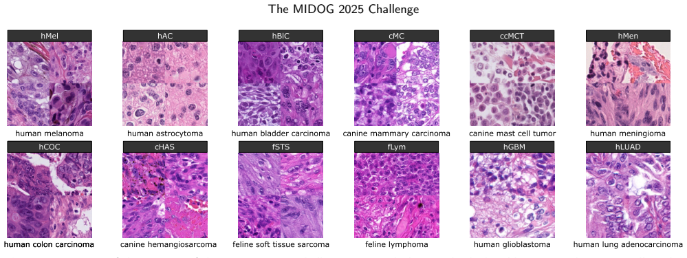
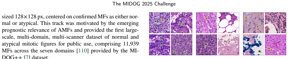
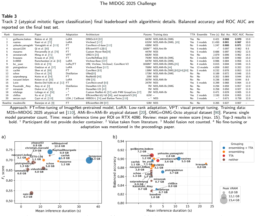
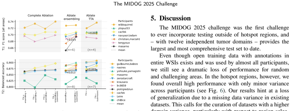
Automated mitosis detection is a well-established task in computational pathology. While previous benchmarks focused on scanner-induced domain shift, clinical "real-world" application requires models to be robust across the vast variance to be expected in the histological landscape. The MItosis DOmain Generalization (MIDOG) 2025 challenge was designed to evaluate algorithmic performance across unprecedented biological and contextual diversity. We curated a test dataset of 365 cases, encompassing 12 distinct human, canine and feline tumor types, digitized across multiple scanning platforms. Moving beyond hand-selected hotspots, the challenge required detection also in random tissue areas (representative of the whole slide detection situation) and challenging areas (areas rich in hard negatives). In the second track, we introduced the classification of atypical mitotic figures (AMFs). There were 18 teams submitting to the detection track, with F1 scores ranging up to 0.740. In the AMF detection track, we had 21 submissions with balanced accuracy values up to 0.908. Our analysis reveals that while most models perform reliably in traditional hotspots, significant performance degradation occurs in challenging ROIs, where false positive rates tripled. Furthermore, performance varied significantly across the 12 tumor types, highlighting "blind spots" in current state-of-the-art architectures when encountering rare or highly pleomorphic malignancies. Moreover, we evaluated the effectiveness of ensembling and found a mean increases of 1.5 and 1.3 percentage points in F1 score and balanced accuracy, respectively. In contrast, TTA showed no relevant improvement. MIDOG 2025 demonstrates that "in the wild" mitosis detection remains a significant hurdle. The transition from hotspot-only evaluation to a multi-contextual framework provides a more realistic proxy for clinical reliability.
Editorial analysis
A structured set of objections, weighed in public.
Referee Report
Summary. The manuscript reports results from the MIDOG 2025 challenge on mitosis detection. It describes curation of a 365-case test set spanning 12 tumor types (human, canine, feline) and multiple scanners, with evaluation in hotspots, random tissue areas, and hard-negative ROIs. A second track addresses atypical mitotic figure classification. Aggregated results from 18 detection-track and 21 AMF-track submissions show peak F1 of 0.740 and balanced accuracy of 0.908; performance degrades markedly in challenging ROIs (FP rates triple) and varies across tumor types. Ensembling yields modest gains (+1.5 pp F1, +1.3 pp balanced accuracy) while test-time augmentation does not. The central claim is that in-the-wild mitosis detection remains a significant hurdle and that the multi-contextual protocol supplies a more realistic clinical proxy.
Significance. If the test-set representativeness holds, the work is significant for shifting mitosis-detection benchmarks from scanner-only domain shift to biological and contextual diversity, exposing model blind spots on rare or pleomorphic tumors. The scale of participation, explicit comparison of ensembling versus TTA, and move beyond hotspot-only evaluation constitute concrete strengths that can guide future architecture design.
major comments (2)
- [Abstract] Abstract (dataset curation paragraph): The headline claim that performance degradation demonstrates a general clinical barrier rests on the 365-case set being representative of clinical whole-slide variance in mitotic density, pleomorphism, and hard-negative prevalence; however, no quantitative anchor (Wasserstein distance, KS tests, or prevalence statistics) against a broader clinical reference cohort is supplied.
- [Abstract] Abstract (results paragraph): The reported FP-rate tripling and tumor-type gaps are presented as evidence of generalization failure, yet the manuscript supplies neither per-tumor-type case counts nor per-ROI mitotic-density statistics, preventing assessment of whether the observed drops are driven by a few atypical cases or reflect systematic limitations.
minor comments (2)
- The manuscript would benefit from reporting mean and standard deviation of F1 and balanced-accuracy scores across all submissions rather than only the maximum values, to contextualize the range 0–0.740.
- Clarify whether the 365 cases include any overlap with prior MIDOG editions or whether all material is newly acquired for this challenge.
Simulated Author's Rebuttal
We thank the referee for the constructive comments. We address each major comment below.
read point-by-point responses
-
Referee: [Abstract] Abstract (dataset curation paragraph): The headline claim that performance degradation demonstrates a general clinical barrier rests on the 365-case set being representative of clinical whole-slide variance in mitotic density, pleomorphism, and hard-negative prevalence; however, no quantitative anchor (Wasserstein distance, KS tests, or prevalence statistics) against a broader clinical reference cohort is supplied.
Authors: The test set was curated to span 12 tumor types across species and multiple scanners to capture substantial biological and contextual diversity, based on expert selection rather than formal distributional matching to an external reference cohort. No such large-scale annotated reference cohort was available for computing Wasserstein distances or KS tests. We will revise the abstract to replace the phrasing 'general clinical barrier' with 'significant challenge under diverse conditions' and add a clarifying sentence on the diversity-driven curation approach. revision: partial
-
Referee: [Abstract] Abstract (results paragraph): The reported FP-rate tripling and tumor-type gaps are presented as evidence of generalization failure, yet the manuscript supplies neither per-tumor-type case counts nor per-ROI mitotic-density statistics, preventing assessment of whether the observed drops are driven by a few atypical cases or reflect systematic limitations.
Authors: The full manuscript (Section 3.1 and Table 1) reports the number of cases per tumor type. Per-ROI mitotic density statistics are not provided because ROIs were expert-selected into qualitative categories (hotspots, random tissue, hard-negative) without per-ROI density quantification. The performance degradation and tumor-type variation are observed consistently across the set rather than driven by outliers. We will add a short reference to the case distribution in the abstract if space allows. revision: partial
Circularity Check
Empirical challenge report with no derivations or self-referential predictions
full rationale
The paper is a challenge summary reporting F1 scores and balanced accuracies from 18-21 external team submissions on a fixed test set of 365 cases. No equations, fitted parameters, or predictions are derived within the manuscript; all quantitative results originate from participant algorithms evaluated on the provided data. The central claim (performance degradation in non-hotspot ROIs and across tumor types) is a direct empirical observation, not a reduction to any self-defined input or self-citation chain. No load-bearing steps match the enumerated circularity patterns.
Axiom & Free-Parameter Ledger
axioms (1)
- domain assumption The curated 365-case test set across 12 tumor types and multiple scanners captures the biological and contextual diversity of real-world histology slides.
Reference graph
Works this paper leans on
-
[1]
Ammeling, J., Aubreville, M., Banerjee, S., Bertram, C.A., Breininger, K., Hirling, D., Horvath, P., Stathonikos, N., Veta, M.,
-
[2]
URL:https: //doi.org/10.5281/zenodo.15077361, doi:10.5281/zenodo.15077361
Mitosis domain generalization challenge 2025. URL:https: //doi.org/10.5281/zenodo.15077361, doi:10.5281/zenodo.15077361
-
[3]
Atey, K., Jha, S.A., Bala, G., Sethi, A., 2026. Mix, Align, Distil: Reliable Cross-Domain Atypical Mitosis Classification, in: Aubre- ville,M.,Bertram,C.A.(Eds.),MitoticFigureDetectionandAtypia Classification in Whole Slide Images, Springer. pp. 137–143
2026
-
[4]
Aubreville, M., Bertram, C.A., Donovan, T.A., Marzahl, C., Maier, A., Klopfleisch, R., 2020a. A completely annotated whole slide imagedatasetofcaninebreastcancertoaidhumanbreastcancerre- search.Scientificdata7:417,1–10.doi:10.1038/s41597-020-00756-z
-
[5]
Aubreville, M., Bertram, C.A., Marzahl, C., Gurtner, C., Dettwiler, M.,Schmidt,A.,Bartenschlager,F.,Merz,S.,Fragoso,M.,Kershaw, O., et al., 2020b. Deep learning algorithms out-perform veterinary pathologists in detecting the mitotically most active tumor region. ScientificReports10:16447,1–11.doi:10.1038/s41598-020-73246-2
-
[6]
Mitosis domain generalization in histopathology images—the MIDOG challenge
Aubreville, M., Stathonikos, N., Bertram, C.A., Klopfleisch, R., Ter Hoeve, N., Ciompi, F., Wilm, F., Marzahl, C., Donovan, T.A., Maier, A., et al., 2023a. Mitosis domain generalization in histopathology images—the MIDOG challenge. Medical Image Analysis 84, 102699
-
[7]
Domaingeneralizationacrosstumortypes,laboratories, and species—insights from the 2022 edition of the mitosis domain generalization challenge
Aubreville, M., Stathonikos, N., Donovan, T.A., Klopfleisch, R., Ammeling, J., Ganz, J., Wilm, F., Veta, M., Jabari, S., Eckstein, M., etal.,2024. Domaingeneralizationacrosstumortypes,laboratories, and species—insights from the 2022 edition of the mitosis domain generalization challenge. Medical Image Analysis 94, 103155
2024
-
[8]
A comprehensive multi-domain dataset for mitotic figure detection
Aubreville,M.,Wilm,F.,Stathonikos,N.,Breininger,K.,Donovan, T.A., Jabari, S., Veta, M., Ganz, J., Ammeling, J., Van Diest, P.J., et al., 2023b. A comprehensive multi-domain dataset for mitotic figure detection. Scientific data 10, 484
-
[9]
Balezo,G.,Bourgade,R.,Feki,H.,Monnier,L.,Blons,M.,Blondel, A., Decencière, E., Planas, A.P., Walter, T., 2026. Efficient Fine- Tuning of DINOv3 Pretrained on Natural Images for Atypical Mi- toticFigureClassification,in:Aubreville,M.,Bertram,C.A.(Eds.), Mitotic Figure Detection and Atypia Classification in Whole Slide Images, Springer. pp. 15–25
2026
-
[10]
Mitosis Domain Generalization (MIDOG) Challenge 2025 Baselines, in: Aubreville, M., Bertram, C.A
Banerjee, S., Ammeling, J., Weiss, V., Donovan, T.A., Klopfleisch, R., Bertram, C.A., Breininger, K., Aubreville, M., 2026a. Mitosis Domain Generalization (MIDOG) Challenge 2025 Baselines, in: Aubreville, M., Bertram, C.A. (Eds.), Mitosis Domain Generaliza- tion (MIDOG) Challenge 2025 Baselines, Springer. pp. 1–14
2025
-
[12]
Bench- marking deep learning and vision foundation models for atyp- ical vs
Banerjee, S., Weiss, V., Donovan, T.A., Fick, R.H., Conrad, T., Ammeling, J., Porsche, N., Klopfleisch, R., Kaltenecker, C.C., Breininger, K., Aubreville, M., Bertram, C.A., 2026c. Bench- marking deep learning and vision foundation models for atyp- ical vs. normal mitosis classification with cross-dataset evalua- tion. Machine Learning for Biomedical Imag...
2026
-
[13]
org/10.59275/j.melba.2026-6c1g
URL:https://melba-journal.org/2026:006, doi:https://doi. org/10.59275/j.melba.2026-6c1g
-
[14]
Is Syn- thetic Image Augmentation Useful for Imbalanced Classification Problems? Case-Study on the MIDOG2025 Atypical Cell Detec- tion Competition, in: Aubreville, M., Bertram, C.A
Benito-Del-Valle, L., Moreno-Sánchez, P.A., Eguskiza, I., Vitoria, I., Picón, A., López-Saratxaga, C., Galdran, A., 2026. Is Syn- thetic Image Augmentation Useful for Imbalanced Classification Problems? Case-Study on the MIDOG2025 Atypical Cell Detec- tion Competition, in: Aubreville, M., Bertram, C.A. (Eds.), Mitotic Figure Detection and Atypia Classific...
2026
-
[15]
Computer-assisted mitotic count using a deep learning–based algorithm improves interobserver reproducibility and accuracy
Bertram,C.A.,Aubreville,M.,Donovan,T.A.,Bartel,A.,Wilm,F., Marzahl, C., Assenmacher, C.A., Becker, K., Bennett, M., Corner, S., et al., 2022. Computer-assisted mitotic count using a deep learning–based algorithm improves interobserver reproducibility and accuracy. Veterinary pathology 59, 211–226. doi:10.1177/ 03009858211067478
2022
-
[16]
Computerized calculation of mitotic count distribution in canine cutaneous mast cell tumor sections: mitotic countisareadependent
Bertram,C.A.,Aubreville,M.,Gurtner,C.,Bartel,A.,Corner,S.M., Dettwiler, M., Kershaw, O., Noland, E.L., Schmidt, A., Sledge, D.G., et al., 2020a. Computerized calculation of mitotic count distribution in canine cutaneous mast cell tumor sections: mitotic countisareadependent. Veterinarypathology57,214–226. doi:10. 1177/0300985819890686
-
[17]
Bertram, C.A., Aubreville, M., Marzahl, C., Maier, A., Klopfleisch, R., 2019. A large-scale dataset for mitotic figure assessment on whole slide images of canine cutaneous mast cell tumor. Scientific data 6, 1–9. doi:10.1038/s41597-019-0290-4
-
[18]
Atyp- ical mitotic figures are prognostically meaningful for canine cuta- neous mast cell tumors
Bertram, C.A., Bartel, A., Donovan, T.A., Kiupel, M., 2023. Atyp- ical mitotic figures are prognostically meaningful for canine cuta- neous mast cell tumors. Veterinary Sciences 11, 5
2023
-
[19]
Mitotic activity: A systematic literature review of the assessment methodology and prognostic value in canine tumors
Bertram, C.A., Donovan, T.A., Bartel, A., 2024a. Mitotic activity: A systematic literature review of the assessment methodology and prognostic value in canine tumors. Veterinary pathology 61, 752–
-
[20]
doi:10.1177/03009858241239565
-
[21]
Mitotic activity: A systematic literature review of the assessment methodology and prognostic value in feline tumors
Bertram, C.A., Donovan, T.A., Bartel, A., 2024b. Mitotic activity: A systematic literature review of the assessment methodology and prognostic value in feline tumors. Veterinary Pathology 61, 743–
-
[22]
doi:10.1177/03009858241239566. M. Aubreville et al.:Preprint submitted to ElsevierPage 14 of 18 The MIDOG 2025 Challenge
-
[23]
Bertram, C.A., Veta, M., Marzahl, C., Stathonikos, N., Maier, A., Klopfleisch, R., Aubreville, M., 2020b. Are pathologist-defined la- bels reproducible? comparison of the tupac16 mitotic figure dataset with an alternative set of labels, in: Interpretable and Annotation- EfficientLearningforMedicalImageComputing.Springer,pp.204–
-
[24]
doi:10.1007/978-3-030-61166-8_22
-
[25]
Bertram, C.A., Weiss, V., Donovan, T.A., Banerjee, S., Conrad, T., Ammeling, J., Klopfleisch, R., Kaltenecker, C., Aubreville, M.,
-
[26]
Histologic dataset of normal and atypical mitotic figures on human breast cancer (ami-br), in: BVM Workshop, Springer. pp. 113–118
-
[27]
Robust Pan-Cancer Mitotic Figure Detection with YOLOv12, in: Aubre- ville,M.,Bertram,C.A.(Eds.),MitoticFigureDetectionandAtypia Classification in Whole Slide Images, Springer
Bourgade, R., Balezo, G., Monier, L., Feki, H., Blons, M., Blondel, A., Loussouarn, D., Vincent-Salomon, A., Walter, T., 2026. Robust Pan-Cancer Mitotic Figure Detection with YOLOv12, in: Aubre- ville,M.,Bertram,C.A.(Eds.),MitoticFigureDetectionandAtypia Classification in Whole Slide Images, Springer. pp. 26–36
2026
-
[28]
MIDOG 2025: Mitotic Figure Detection with Attention-Guided False Pos- itive Correction, in: Aubreville, M., Bertram, C.A
Broad, A., Keighley, J., Godson, L., Wright, A., 2026. MIDOG 2025: Mitotic Figure Detection with Attention-Guided False Pos- itive Correction, in: Aubreville, M., Bertram, C.A. (Eds.), Mitotic Figure Detection and Atypia Classification in Whole Slide Images, Springer. pp. 87–90
2026
-
[29]
End-to-end object detection with transformers
Carion, N., Massa, F., Synnaeve, G., Usunier, N., Kirillov, A., Zagoruyko,S.,2020. End-to-endobjectdetectionwithtransformers, in:Europeanconferenceoncomputervision,Springer.pp.213–229. doi:10.1007/978-3-030-58452-8_13
-
[30]
Encoder-decoder with atrous separable convolution for semantic imagesegmentation,in:ProceedingsoftheEuropeanconferenceon computer vision (ECCV), pp
Chen, L.C., Zhu, Y., Papandreou, G., Schroff, F., Adam, H., 2018. Encoder-decoder with atrous separable convolution for semantic imagesegmentation,in:ProceedingsoftheEuropeanconferenceon computer vision (ECCV), pp. 801–818
2018
-
[31]
Towards a general-purpose foundation model for computational pathology
Chen, R.J., Ding, T., Lu, M.Y., Williamson, D.F., Jaume, G., Song, A.H., Chen, B., Zhang, A., Shao, D., Shaban, M., et al., 2024. Towards a general-purpose foundation model for computational pathology. Nature medicine 30, 850–862
2024
-
[32]
Teacher-Student Model for Detecting and Classifying Mitosis in the MIDOG 2025 Challenge, in: Aubreville, M., Bertram, C.A
Choe, S., Qin, X., Shafique, A., Dy, A., Done, S., Androutsos, D., Khademi, A., 2026. Teacher-Student Model for Detecting and Classifying Mitosis in the MIDOG 2025 Challenge, in: Aubreville, M., Bertram, C.A. (Eds.), Mitotic Figure Detection and Atypia Classification in Whole Slide Images, Springer. pp. 207–214
2026
-
[33]
Groupequivariantconvolutionalnet- works, in: Proceedings of the International Conference on Machine Learning (ICML), pp
Cohen,T.,Welling,M.,2016. Groupequivariantconvolutionalnet- works, in: Proceedings of the International Conference on Machine Learning (ICML), pp. 2990–2999
2016
-
[34]
Detlefsen, N.S., Borovec, J., Schock, J., Jha, A.H., Koker, T., Di Liello, L., Stancl, D., Quan, C., Grechkin, M., Falcon, W., 2022. Torchmetrics - measuring reproducibility in pytorch. Journal of Open Source Software 7, 4101. URL:https://doi.org/10.21105/ joss.04101, doi:10.21105/joss.04101
-
[35]
Mitotic figures—normal, atypical, and imposters: A guide to identification
Donovan, T.A., Moore, F.M., Bertram, C.A., Luong, R., Bolfa, P., Klopfleisch,R.,Tvedten,H.,Salas,E.N.,Whitley,D.B.,Aubreville, M., et al., 2021. Mitotic figures—normal, atypical, and imposters: A guide to identification. Veterinary pathology 58, 243–257
2021
-
[36]
Stain-Aware Augmentation and Hybrid Loss for Domain-Generalized Atypical Mitosis Classification, in: Aubreville, M., Bertram, C.A
Dukre, A., Deria, A., Xie, Y., Razzak, I., 2026. Stain-Aware Augmentation and Hybrid Loss for Domain-Generalized Atypical Mitosis Classification, in: Aubreville, M., Bertram, C.A. (Eds.), Mitotic Figure Detection and Atypia Classification in Whole Slide Images, Springer. pp. 159–165
2026
-
[37]
Why is the winner the best?, in: Proceedings of the IEEE/CVF Conference on Computer Vision and Pattern Recogni- tion, pp
Eisenmann, M., Reinke, A., Weru, V., Tizabi, M.D., Isensee, F., Adler, T.J., Ali, S., Andrearczyk, V., Aubreville, M., Baid, U., et al., 2023. Why is the winner the best?, in: Proceedings of the IEEE/CVF Conference on Computer Vision and Pattern Recogni- tion, pp. 19955–19966
2023
-
[38]
AutoGluon-Tabular: Robust and Accurate AutoML for Structured Data
Erickson, N., Mueller, J., Shirkov, A., Zhang, H., Larroy, P., Li, M., Smola, A., 2020. Autogluon-tabular: Robust and accurate automl for structured data. arXiv preprint arXiv:2003.06505
work page internal anchor Pith review Pith/arXiv arXiv 2020
-
[39]
Protocolfortheexamination of resection specimens from patients with invasive carcinoma of the breast
Fitzgibbons,P.L.,Connolly,J.L.,2023. Protocolfortheexamination of resection specimens from patients with invasive carcinoma of the breast. CAP guidelines 4.8.1.0. URL:https://www.cap.org/ cancerprotocols
2023
-
[40]
Pannuke: an open pan-cancer histology dataset for nuclei instance segmentation and classification, in: European congress on digital pathology, Springer
Gamper,J.,AlemiKoohbanani,N.,Benet,K.,Khuram,A.,Rajpoot, N., 2019. Pannuke: an open pan-cancer histology dataset for nuclei instance segmentation and classification, in: European congress on digital pathology, Springer. pp. 11–19
2019
-
[41]
Ganz,J.,Marzahl,C.,Ammeling,J.,Rosbach,E.,Richter,B.,Puget, C., Denk, D., Demeter, E.A., Tăbăran, F.A., Wasinger, G., et al.,
-
[42]
Scientificreports 14, 26273
Information mismatch in phh3-assisted mitosis annotation leadstointerpretationshiftsinh&eslideanalysis. Scientificreports 14, 26273
-
[43]
RF-DETR for Robust Mitotic Figure Detection: A MIDOG 2025 Track 1 Approach, in: Aubreville, M., Bertram, C.A
Giedziun, P., Sotysik, J., Górczany, M., Ropiak, N., Przymus, M., Krajewski,P.,Kwicień,J.,Bartczak,A.,Wasiak,I.,Maniewski,M., 2026a. RF-DETR for Robust Mitotic Figure Detection: A MIDOG 2025 Track 1 Approach, in: Aubreville, M., Bertram, C.A. (Eds.), Mitotic Figure Detection and Atypia Classification in Whole Slide Images, Springer. pp. 99–104
2025
-
[44]
FoundationModel-DrivenClassificationofAtypicalMitotic FigureswithDomain-AwareTrainingStrategies,in:Aubreville,M., Bertram, C.A
Giedziun, P., Sotysik, J., Górczany, M., Ropiak, N., Przymus, M., Krajewski,P.,Kwiecień,J.,Bartczak,A.,Wasiak,I.,Maniewski,M., 2026b. FoundationModel-DrivenClassificationofAtypicalMitotic FigureswithDomain-AwareTrainingStrategies,in:Aubreville,M., Bertram, C.A. (Eds.), Mitotic Figure Detection and Atypia Classifi- cation in Whole Slide Images, Springer. p...
-
[45]
Classification of chromosome segregation errors in cancer
Gisselsson, D., 2008. Classification of chromosome segregation errors in cancer. Chromosoma 117, 511–519. doi:10.1007/ s00412-008-0169-1
2008
-
[46]
Hover-Net: Simultaneous Segmentation and Classification of Nuclei in Multi-tissue Histology Images,
Graham,S.,Vu,Q.D.,Raza,S.E.A.,Azam,A.,Tsang,Y.W.,Kwak, J.T., Rajpoot, N., 2019. Hover-net: Simultaneous segmentation and classification of nuclei in multi-tissue histology images. Medical Image Analysis 58, 101563. doi:10.1016/j.media.2019.101563
-
[47]
Deep residual learning for image recognition, in: Proceedings of the IEEE conference on computer vision and pattern recognition, pp
He, K., Zhang, X., Ren, S., Sun, J., 2016. Deep residual learning for image recognition, in: Proceedings of the IEEE conference on computer vision and pattern recognition, pp. 770–778. doi:10.1109/ CVPR.2016.90
2016
-
[48]
Hendzel, M.J., Wei, Y., Mancini, M.A., Van Hooser, A., Ranalli, T., Brinkley, B., Bazett-Jones, D.P., Allis, C.D., 1997. Mitosis- specific phosphorylation of histone h3 initiates primarily within pericentromeric heterochromatin during g2 and spreads in an or- dered fashion coincident with mitotic chromosome condensation. Chromosoma 106, 348–360. doi:10.10...
-
[49]
, year = 2017, month = jul, pages =
Huang, G., Liu, Z., Van Der Maaten, L., Weinberger, K.Q., 2017. Densely connected convolutional networks, in: Proceedings of the IEEE conference on computer vision and pattern recognition, pp. 4700–4708. doi:10.1109/CVPR.2017.243
-
[50]
Isensee, F., Jaeger, P.F., Kohl, S.A., Petersen, J., Maier-Hein, K.H.,
-
[51]
Nature methods 18, 203–211
nnu-net: a self-configuring method for deep learning-based biomedical image segmentation. Nature methods 18, 203–211
-
[52]
A subphase-labeled mitotic dataset for ai-powered cell division analy- sis
Ivan, Z.Z., Hirling, D., Grexa, I., Ammeling, J., Molnar, C., Micsik, T., Dobra, K., Kuthi, L., Sukosd, F., Fillinger, J., et al., 2026. A subphase-labeled mitotic dataset for ai-powered cell division analy- sis. Scientific data
2026
-
[53]
Jahanifar, M., 2025. Mitosis subtyping dataset. URL:https://doi. org/10.5281/zenodo.15390543, doi:10.5281/zenodo.15390543
-
[54]
Pan-cancerprofilingofmitotictopol- ogy & mitotic errors: Insights into prognosis, genomic alterations, and immune landscape
Jahanifar, M., Dawood, M., Zamanitajeddin, N., Shephard, A., Chohan,B.S.,Bertram,C.A.,Wahab,N.,Eastwood,M.,Aubreville, M.,Raza,S.E.A.,etal.,2025. Pan-cancerprofilingofmitotictopol- ogy & mitotic errors: Insights into prognosis, genomic alterations, and immune landscape. medRxiv , 2025–06
2025
-
[55]
Distinct mitotic segregation errors mediate chromo- somal instability in aggressive urothelial cancers
Jin, Y., Stewénius, Y., Lindgren, D., Frigyesi, A., Calcagnile, O., Jonson, T., Edqvist, A., Larsson, N., Lundberg, L.M., Chebil, G., et al., 2007. Distinct mitotic segregation errors mediate chromo- somal instability in aggressive urothelial cancers. Clinical cancer research 13, 1703–1712
2007
-
[56]
Tripolar mitosis in human cells and embryos: occurrence, pathophysiology and medical implications
Kalatova, B., Jesenska, R., Hlinka, D., Dudas, M., 2015. Tripolar mitosis in human cells and embryos: occurrence, pathophysiology and medical implications. Acta histochemica 117, 111–125
2015
-
[57]
Kather, J.N., Krisam, J., Charoentong, P., Luedde, T., Herpel, E., Weis, C.A., Gaiser, T., Marx, A., Valous, N.A., Ferber, D., et al.,
-
[58]
Predicting survival from colorectal cancer histology slides using deep learning: A retrospective multicenter study. PLoS M. Aubreville et al.:Preprint submitted to ElsevierPage 15 of 18 The MIDOG 2025 Challenge medicine 16, e1002730
2025
-
[59]
Atypical mitotic figureclassificationinmidog2025usinglora-enhancedunimodels
Kelam, N.S., Bonthu, S., Singhai, N., 2025. Atypical mitotic figureclassificationinmidog2025usinglora-enhancedunimodels. doi:https://doi.org/10.5281/zenodo.17020013
-
[60]
Ensemble YOLO Framework for Multi-Domain Mitotic Figure Detection in Histopathology Images, in: Aubreville, M., Bertram, C.A
Kelam, N.S., Parekh, A., Bonthu, S., Singhal, N., 2026. Ensemble YOLO Framework for Multi-Domain Mitotic Figure Detection in Histopathology Images, in: Aubreville, M., Bertram, C.A. (Eds.), Mitotic Figure Detection and Atypia Classification in Whole Slide Images, Springer. pp. 105–111
2026
-
[61]
YOLOv11: An Overview of the Key Architectural Enhancements
Khanam, R., Hussain, M., 2024. Yolov11: An overview of the key architectural enhancements. arXiv preprint arXiv:2410.17725
work page internal anchor Pith review Pith/arXiv arXiv 2024
-
[62]
Kiupel, M., Webster, J., Bailey, K., Best, S., DeLay, J., Detrisac, C., Fitzgerald, S., Gamble, D., Ginn, P., Goldschmidt, M., et al., 2011. Proposal of a 2-tier histologic grading system for canine cutaneous mastcelltumorstomoreaccuratelypredictbiologicalbehavior. Vet. Pathol. 48, 147–155. doi:10.1177/0300985810386469
-
[63]
MIDOG 2025 Track 2: A Deep Learning Model for Classification of Atypical and Normal Mitotic Figures under Class and Hardness Imbalances, in: Aubreville, M., Bertram, C.A
Kotte, S., Saipradeep, V.G., Walia, V., Nandagopal, D., Joseph, T., Sivadasan, N., Lali, B.S., 2026. MIDOG 2025 Track 2: A Deep Learning Model for Classification of Atypical and Normal Mitotic Figures under Class and Hardness Imbalances, in: Aubreville, M., Bertram, C.A. (Eds.), Mitotic Figure Detection and Atypia Classifi- cation in Whole Slide Images, S...
2026
-
[64]
Deep Learning Meets Morphology: A Hybrid Approach for Mitotic Figure Classification, in: Aubreville, M., Bertram, C.A
Krauss, S., Spieß, E., Hieber, D., Kramer, F., Schobel, J., Müller, D., 2026. Deep Learning Meets Morphology: A Hybrid Approach for Mitotic Figure Classification, in: Aubreville, M., Bertram, C.A. (Eds.),MitoticFigureDetectionandAtypiaClassificationinWhole Slide Images, Springer. pp. 37–46
2026
-
[65]
The hungarian method for the assignment problem
Kuhn, H.W., 1955. The hungarian method for the assignment problem. Naval research logistics quarterly 2, 83–97
1955
-
[66]
Roto-translation equivariant convolutional networks: Application to histopathologyimageanalysis
Lafarge, M.W., Bekkers, E.J., Pluim, J.P., Duits, R., Veta, M., 2021. Roto-translation equivariant convolutional networks: Application to histopathologyimageanalysis. MedicalImageAnalysis68,101849
2021
-
[67]
Sequential Hard Mining: A Data-Centric Approach for Mitosis Detection, in: Aubreville, M., Bertram, C.A
Lafarge, M.W., Koelzer, V.H., 2026. Sequential Hard Mining: A Data-Centric Approach for Mitosis Detection, in: Aubreville, M., Bertram, C.A. (Eds.), Mitotic Figure Detection and Atypia Classi- fication in Whole Slide Images, Springer. pp. 215–219
2026
-
[68]
The characteristics and clinical significance of atypicalmitosisinbreastcancer
Lashen, A., Toss, M.S., Alsaleem, M., Green, A.R., Mongan, N.P., Rakha, E., 2022. The characteristics and clinical significance of atypicalmitosisinbreastcancer. ModernPathology35,1341–1348
2022
-
[69]
Adeepactivelearningframeworkformitoticfiguredetec- tion with minimal manual annotation and labelling
Liu, E., Lin, A., Kakodkar, P., Zhao, Y., Wang, B., Ling, C., Zhang, Q.,2025. Adeepactivelearningframeworkformitoticfiguredetec- tion with minimal manual annotation and labelling. Histopathology 87, 536–547
2025
-
[70]
Ef- ficientvit:Memoryefficientvisiontransformerwithcascadedgroup attention,in:ProceedingsoftheIEEE/CVFconferenceoncomputer vision and pattern recognition, pp
Liu, X., Peng, H., Zheng, N., Yang, Y., Hu, H., Yuan, Y., 2023. Ef- ficientvit:Memoryefficientvisiontransformerwithcascadedgroup attention,in:ProceedingsoftheIEEE/CVFconferenceoncomputer vision and pattern recognition, pp. 14420–14430
2023
-
[71]
Detecting cancer metastases on gigapixel pathol- ogy images
Liu, Y., Gadepalli, K., Norouzi, M., Dahl, G.E., Kohlberger, T., Boyko, A., Venugopalan, S., Timofeev, A., Nelson, P.Q., Corrado, G.S., et al., 2017. Detecting cancer metastases on gigapixel pathol- ogy images. arXiv preprint arXiv:1703.02442
-
[72]
Liu, Z., Mao, H., Wu, C.Y., Feichtenhofer, C., Darrell, T., Xie, S.,
-
[73]
11976– 11986
A convnet for the 2020s, in: Proceedings of the IEEE/CVF conference on computer vision and pattern recognition, pp. 11976– 11986
-
[74]
Lv, J., Nasir, E.S., Xu, K., Jahanifar, M., Chohan, B.S., Elhaminia, B., Raza, S.E.A., 2026. Kongnet: A multi-headed deep learning model for detection and classification of nuclei in histopathology images. URL:https://arxiv.org/abs/2510.23559,arXiv:2510.23559
work page internal anchor Pith review Pith/arXiv arXiv 2026
-
[75]
Rtmdet: An empirical study of designing real- time object detectors
Lyu, C., Zhang, W., Huang, H., Zhou, Y., Wang, Y., Liu, Y., Zhang, S., Chen, K., 2022. Rtmdet: An empirical study of designing real- time object detectors. arXiv preprint arXiv:2212.07784
-
[76]
Mitotic fig- ure recognition: Agreement among pathologists and computerized detector
Malon, C., Brachtel, E., Cosatto,E., Graf, H.P., Kurata, A., Kuroda, M., Meyer, J.S., Saito, A., Wu, S., Yagi, Y., 2012. Mitotic fig- ure recognition: Agreement among pathologists and computerized detector. Analytical Cellular Pathology 35, 97–100. doi:10.3233/ ACP-2011-0029
2012
-
[77]
Marzahl, C., Aubreville, M., Bertram, C.A., Maier, J., Bergler, C., Kröger, C., Voigt, J., Breininger, K., Klopfleisch, R., Maier, A.,
-
[78]
Scientific Reports 11:4343, 1–10
EXACT: a collaboration toolset for algorithm-aided anno- tation of images with annotation version control. Scientific Reports 11:4343, 1–10. doi:10.1038/s41598-021-83827-4
-
[79]
A bag of tricks for real-time Mitotic Figure detection, in: Aubreville, M., Bertram, C.A
Marzahl, C., Napora, B., 2026. A bag of tricks for real-time Mitotic Figure detection, in: Aubreville, M., Bertram, C.A. (Eds.), Mitotic Figure Detection and Atypia Classification in Whole Slide Images, Springer. pp. 91–98
2026
-
[80]
Mitotic index and multipolar mitosis in routine histologic sectionsasprognosticmarkersofpancreaticcancers:aclinicopatho- logical study
Matsuda, Y., Yoshimura, H., Ishiwata, T., Sumiyoshi, H., Mat- sushita, A., Nakamura, Y., Aida, J., Uchida, E., Takubo, K., Arai, T., 2016. Mitotic index and multipolar mitosis in routine histologic sectionsasprognosticmarkersofpancreaticcancers:aclinicopatho- logical study. Pancreatology 16, 127–132
2016
-
[81]
Evaluation of prognostic factors for dogs with primarylungtumors:67cases(1985-1992).JournaloftheAmerican Veterinary Medical Association 211, 1422–1427
McNiel, E., Ogilvie, G., Powers, B., Hutchison, J., Salman, M., Withrow, S., 1997. Evaluation of prognostic factors for dogs with primarylungtumors:67cases(1985-1992).JournaloftheAmerican Veterinary Medical Association 211, 1422–1427
1997
discussion (0)
Sign in with ORCID, Apple, or X to comment. Anyone can read and Pith papers without signing in.