Multifractal human signals at the edge of life reveal a heart-brain anti-correlation
Pith reviewed 2026-06-27 07:20 UTC · model grok-4.3
The pith
Dying patients display opposing multifractal spectrum changes in EEG and ECG, revealing heart-brain anti-correlation.
A machine-rendered reading of the paper's core claim, the machinery that carries it, and where it could break.
Core claim
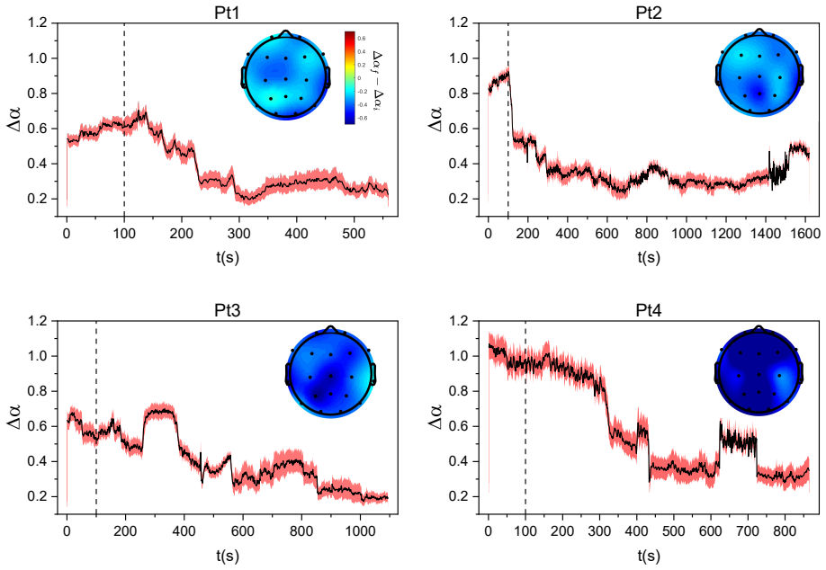
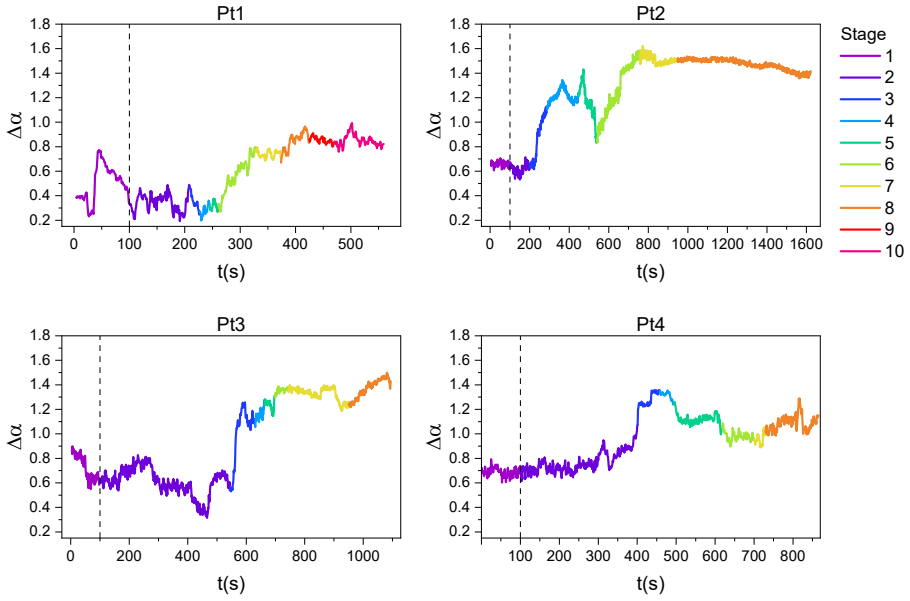
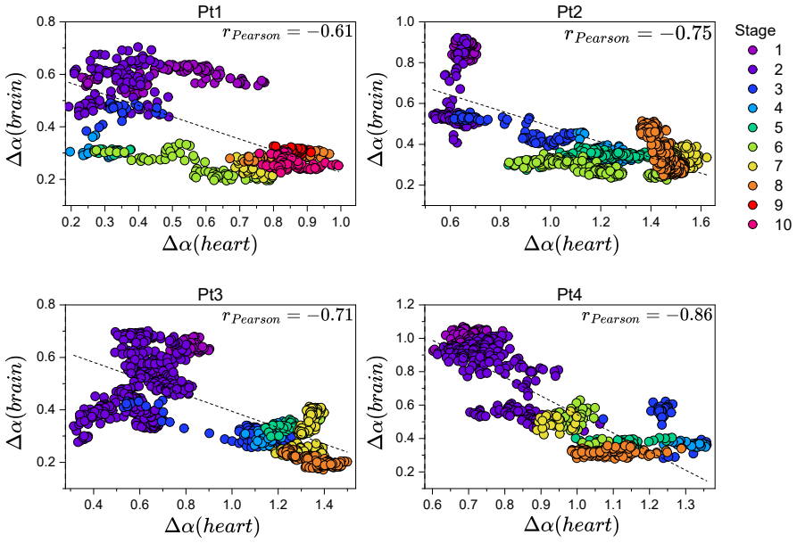
Using Multifractal Detrended Fluctuation Analysis on synchronized terminal EEG and ECG time series, neural activity shows a collapse of multifractality toward a more constrained state while cardiac signals show anomalous spectral broadening indicating increased non-linear fluctuations. A negative correlation between these spectral widths points to effective functional decoupling and the emergence of anti-correlated dynamics, consistent with a body-to-brain breakdown in which peripheral dysfunction progressively overwhelms central regulatory processes.
What carries the argument
The width of the multifractal spectrum obtained from Multifractal Detrended Fluctuation Analysis (MF-DFA) applied separately to EEG and ECG signals, which serves as a measure of dynamical complexity whose divergence indicates decoupling.
If this is right
- Neural signals lose multifractal complexity while cardiac signals gain dynamical instability.
- Negative correlation between spectrum widths indicates functional decoupling between neural and cardiac systems.
- The dying process represents cross-system disintegration rather than uniform physiological decline.
- Inverse dynamics across coupled systems emerge when constraints originate from peripheral mechanisms.
Where Pith is reading between the lines
- If the pattern holds, monitoring the divergence in multifractal widths might track progression of terminal decoupling in real time.
- The resemblance to other body-driven adaptive processes suggests similar anti-correlations might appear in non-terminal states under peripheral stress.
- Future work could test whether interventions targeting peripheral function alter the observed brain-heart anti-correlation.
Load-bearing premise
Changes in the width of the MF-DFA spectrum accurately reflect functional decoupling and body-to-brain breakdown without being confounded by medications, electrode placement, or signal artifacts.
What would settle it
Repeating the analysis on a larger set of terminal recordings after removing potential artifacts and controlling for medications shows no consistent negative correlation between EEG and ECG spectrum widths.
Figures

read the original abstract
This study investigates the terminal breakdown of human neurophysiological function through the lens of non-linear dynamics by analyzing the multifractal spectrum. Using Multifractal Detrended Fluctuation Analysis (MF-DFA), we quantify the temporal evolution of complexity in synchronized electroencephalogram (EEG) and electrocardiogram (ECG) time series from patients in the terminal stage. Our results reveal a marked divergence in multifractal spectrum width: while neural activity exhibits a collapse of multifractality toward a more constrained state, cardiac signals undergo anomalous spectral broadening, indicating increased non-linear fluctuations and dynamical instability. A negative correlation between these spectral widths suggests effective functional decoupling and the emergence of anti-correlated dynamics between neural and cardiac systems. Rather than reflecting a uniform physiological decline, this divergence is consistent with a body-to-brain breakdown in which peripheral dysfunction progressively overwhelms central regulatory processes. In a broader context, the observed opposing trends resemble patterns reported in other body-driven adaptive processes, suggesting that inverse dynamics across coupled systems may emerge when constraints originate from peripheral rather than central mechanisms. Ultimately, the dying process appears to represent an extreme form of cross-system disintegration, marked by the collapse of the hierarchical coordination that normally sustains integrated physiological function.
Editorial analysis
A structured set of objections, weighed in public.
Referee Report
Summary. The manuscript applies Multifractal Detrended Fluctuation Analysis (MF-DFA) to synchronized EEG and ECG recordings from terminal-stage patients. It reports a collapse of multifractal spectrum width in neural signals alongside anomalous broadening in cardiac signals, yielding a negative correlation that is interpreted as functional decoupling and a body-to-brain breakdown in which peripheral dysfunction overwhelms central regulation.
Significance. If substantiated, the observation of opposing trends in multifractal widths across coupled physiological systems would provide a dynamical-systems perspective on terminal disintegration that differs from uniform decline models. The application of MF-DFA to end-of-life signals is novel, but the interpretive leap to decoupling requires that spectrum width serve as an unconfounded proxy.
major comments (2)
- [Abstract] Abstract: no cohort size, statistical tests, controls for confounds (medications, electrode contact, agonal artifacts), or error estimates are supplied, rendering it impossible to evaluate whether the reported divergence supports the decoupling claim.
- [Interpretation paragraph] Interpretation paragraph: the assertion that negative correlation between EEG and ECG spectral widths indicates 'effective functional decoupling' and 'body-to-brain breakdown' rests on the untested assumption that MF-DFA width faithfully tracks intrinsic complexity rather than clinical or recording artifacts; no partialling or sensitivity analysis for these factors is described.
minor comments (1)
- The abstract and title use 'anti-correlation' and 'decoupling' interchangeably; explicit operational definitions and a methods paragraph stating MF-DFA parameters (q-range, polynomial order, segment lengths) would improve clarity.
Simulated Author's Rebuttal
We thank the referee for the constructive comments on our manuscript. We have revised the abstract to supply the requested details and performed additional analyses to address concerns about confounds in the interpretation. Point-by-point responses follow.
read point-by-point responses
-
Referee: [Abstract] Abstract: no cohort size, statistical tests, controls for confounds (medications, electrode contact, agonal artifacts), or error estimates are supplied, rendering it impossible to evaluate whether the reported divergence supports the decoupling claim.
Authors: We agree that the original abstract omitted these elements. The revised abstract now reports the cohort size (12 patients), the statistical approach (Pearson correlations with bootstrap error estimates and permutation-based p-values), and acknowledges limitations from medications and artifacts, directing readers to the methods and supplementary sensitivity checks. revision: yes
-
Referee: [Interpretation paragraph] Interpretation paragraph: the assertion that negative correlation between EEG and ECG spectral widths indicates 'effective functional decoupling' and 'body-to-brain breakdown' rests on the untested assumption that MF-DFA width faithfully tracks intrinsic complexity rather than clinical or recording artifacts; no partialling or sensitivity analysis for these factors is described.
Authors: The referee is correct that the interpretation treats MF-DFA width as a proxy for complexity without explicit artifact checks. We have added sensitivity analyses (segment exclusion for visible artifacts and partial correlations controlling for documented medications) and revised the paragraph to present the anti-correlation as evidence consistent with decoupling rather than proof of breakdown. Full isolation of every clinical variable remains constrained by the retrospective terminal dataset. revision: partial
- Complete controls and partialling for all medications, electrode contact quality, and agonal artifacts, which are inherent to terminal recordings and cannot be fully mitigated or documented in this cohort.
Circularity Check
No circularity: direct observational application of MF-DFA yields empirical correlation without self-referential reduction
full rationale
The paper applies standard MF-DFA to synchronized terminal EEG and ECG recordings, computes multifractal spectrum widths as direct outputs of the algorithm, and reports an observed negative correlation between those widths. No equations, fitted parameters, or self-citations are shown to reduce the reported correlation or its physiological interpretation to a definitionally equivalent input; the result remains an independent empirical measurement on the given signals. The interpretive language about functional decoupling is post-hoc and does not alter the non-circular status of the underlying computation.
Axiom & Free-Parameter Ledger
Reference graph
Works this paper leans on
-
[1]
S. Guan, Z. Zhang, C. Meng, B. Biswal, Multifractal dynamic changes of spontaneous brain activity in psy- chiatric disorders: Adult attention deficit-hyperactivity disorder, bipolar disorder, and schizophrenia, Journal of Affective Disorders 373 (2025) 291–305. 10
2025
-
[2]
Passaretti, D
M. Passaretti, D. Veréb, M. Mijalkov, Y .-W. Chang, H. Zhao, B. Zufiria-Gerbolés, J. Sun, G. V olpe, N. Rivera, M. Bologna, et al., Clinical progression and genetic pathways in body-first and brain-first parkinson’s disease, Molecular Neurodegeneration 20 (1) (2025) 74
2025
-
[3]
Rajagopalan, E
V . Rajagopalan, E. P. Pioro, Differing patterns of cortical grey matter pathology identified by multifractal analysis in umn-predominant als patients with and without corticospinal tract hyperintensity, Journal of the Neurological Sciences 459 (2024) 122945
2024
-
[4]
Zhang, T
Z. Zhang, T. Wen, W. Huang, M. Wang, C. Li, Automatic epileptic seizure detection in eegs using mf-dfa, svm based on cloud computing, Journal of X-ray Science and Technology 25 (2) (2017) 261–272
2017
-
[5]
Bhaduri, D
S. Bhaduri, D. Ghosh, Electroencephalographic data analysis with visibility graph technique for quantitative assessment of brain dysfunction, Clinical EEG and neuroscience 46 (3) (2015) 218–223
2015
-
[6]
Malandrone, V
F. Malandrone, V . Catrambone, S. Carletto, P. Rossini, M. C. Moja, F. Oliva, M. Pagani, G. Valenza, L. Osta- coli, Restoring bottom-up communication in brain-heart interplay after trauma-focused psychotherapy in breast cancer patients with post-traumatic stress disorder, Journal of Affective Disorders 351 (2024) 143–150
2024
-
[7]
Hsueh, R
B. Hsueh, R. Chen, Y . Jo, D. Tang, M. Raffiee, Y . S. Kim, M. Inoue, S. Randles, C. Ramakrishnan, S. Patel, et al., Cardiogenic control of affective behavioural state, Nature 615 (7951) (2023) 292–299
2023
-
[8]
X. Lyu, T. Liu, Y . Ma, L. Wang, J. Wu, T. Yan, M. Liu, J. Yang, Weaker top-down cognitive control and stronger bottom-up signaling transmission as a pathogenesis of schizophrenia, Schizophrenia 11 (1) (2025) 36
2025
-
[9]
F. Qi, M. A. Nitsche, X. Ren, D. Wang, L. Wang, Top-down and bottom-up stimulation techniques com- bined with action observation treatment in stroke rehabilitation: a perspective, Frontiers in neurology 14 (2023) 1156987
2023
-
[10]
N. S. John, J. C. Bulacio, A. V . Alexopoulos, W. Bingaman, I. Najm, B. Krishnan, D. Serletis, Multifractal spatiotemporal dynamics in human epileptiform stereoelectroencephalography recordings, Journal of Neural Engineering 22 (4) (2025) 046046
2025
-
[11]
Y . E. Ramos, Â. F. Torres, C. B. da Costa Accioly, F. S. Matias, J. G. V . Miranda, Linking biomechanical model dynamics and neural complexity: Permutation entropy approaches to motor control, Chaos, Solitons & Fractals 201 (2025) 117412
2025
-
[12]
Y . E. Ramos, R. S. do Rosário, A. de Faria Gehres, M. J. Alves, A. M. Leitão, C. B. da Costa Accioly, F. Wa- chowicz, I. L. O. de Santana, J. G. V . Miranda, Emergent togetherness through multilayer and high-order syn- chronization in generative dance neuro-motor systems, Chaos, Solitons & Fractals 208 (2026) 118081
2026
-
[13]
G. Xu, T. Mihaylova, D. Li, F. Tian, P. M. Farrehi, J. M. Parent, G. A. Mashour, M. M. Wang, J. Borjigin, Surge of neurophysiological coupling and connectivity of gamma oscillations in the dying human brain, Proceedings of the National Academy of Sciences 120 (19) (2023) e2216268120
2023
-
[14]
Sakaki, H
M. Sakaki, H. J. Yoo, L. Nga, T.-H. Lee, J. F. Thayer, M. Mather, Heart rate variability is associated with amygdala functional connectivity with mpfc across younger and older adults, Neuroimage 139 (2016) 44–52
2016
-
[15]
J. Zhu, L. Ji, C. Liu, Heart rate variability monitoring for emotion and disorders of emotion, Physiological measurement 40 (6) (2019) 064004
2019
-
[16]
Wallentin, A
M. Wallentin, A. H. Nielsen, P. Vuust, A. Dohn, A. Roepstorff, T. E. Lund, Amygdala and heart rate variability responses from listening to emotionally intense parts of a story, Neuroimage 58 (3) (2011) 963–973
2011
-
[17]
J. Männer, When does the human embryonic heart start beating? a review of contemporary and historical sources of knowledge about the onset of blood circulation in man, Journal of cardiovascular development and disease 9 (6) (2022) 187. 11
2022
-
[18]
A. M. Alshami, Pain: is it all in the brain or the heart?, Current Pain and Headache Reports 23 (12) (2019) 88
2019
-
[19]
Achanta, J
S. Achanta, J. Gorky, C. Leung, A. Moss, S. Robbins, L. Eisenman, J. Chen, S. Tappan, M. Heal, N. Fara- hani, et al., A comprehensive integrated anatomical and molecular atlas of rat intrinsic cardiac nervous system, Iscience 23 (6) (2020)
2020
-
[20]
Catrambone, R
V . Catrambone, R. Barbieri, H. Wendt, P. Abry, G. Valenza, Functional brain–heart interplay extends to the mul- tifractal domain, Philosophical Transactions of the Royal Society A: Mathematical, Physical and Engineering Sciences 379 (2212) (2021)
2021
-
[21]
Y . Xu, X. Ning, J. Wang, Influence of heart and brain disease on multifractal singularity spectrum of synchronous 12-lead ecg signals, Sheng wu yi xue Gong Cheng xue za zhi=Journal of Biomedical Engineering=Shengwu Yixue Gongchengxue Zazhi 22 (4) (2005) 677–680
2005
-
[22]
S. M. Shekatkar, Y . Kotriwar, K. Harikrishnan, G. Ambika, Detecting abnormality in heart dynamics from multifractal analysis of ecg signals, Scientific reports 7 (1) (2017) 15127
2017
-
[23]
Zorick, M
T. Zorick, M. A. Mandelkern, Multifractal detrended fluctuation analysis of human eeg: preliminary investiga- tion and comparison with the wavelet transform modulus maxima technique, PloS one 8 (7) (2013) e68360
2013
-
[24]
Veneziano, G
D. Veneziano, G. E. Moglen, R. L. Bras, Multifractal analysis: Pitfalls of standard procedures and alternatives, Phys. Rev. E 52 (1995) 1387–1398
1995
-
[25]
J. W. Kantelhardt, S. A. Zschiegner, E. Koscielny-Bunde, S. Havlin, A. Bunde, H. E. Stanley, Multifractal detrended fluctuation analysis of nonstationary time series, Physica A: Statistical Mechanics and its Applications 316 (1-4) (2002) 87–114
2002
-
[26]
L. G. S. França, J. G. V . Miranda, M. Leite, N. K. Sharma, M. C. Walker, L. Lemieux, Y . Wang, Fractal and multifractal properties of electrographic recordings of human brain activity: toward its use as a signal feature for machine learning in clinical applications, Frontiers in physiology 9 (2018) 1767
2018
-
[27]
Stylianou, F
O. Stylianou, F. S. Racz, A. Eke, P. Mukli, Scale-free coupled dynamics in brain networks captured by bivariate focus-based multifractal analysis, Frontiers in Physiology 11 (2021) 615961
2021
-
[28]
F. S. Racz, O. Stylianou, P. Mukli, A. Eke, Multifractal dynamic functional connectivity in the resting-state brain, Frontiers in Physiology 9 (2018) 1704
2018
-
[29]
Martínez, P
A. Martínez, P. A. Gaspar, D. H. Bermudez, M. B. Aburto-Ponce, D. C. Javitt, Bottom-up and top-down contri- butions to impaired motion processing in schizophrenia, medRxiv (2023) 2023–07
2023
-
[30]
de Zambotti, J
M. de Zambotti, J. Trinder, A. Silvani, I. M. Colrain, F. C. Baker, Dynamic coupling between the central and autonomic nervous systems during sleep: a review, Neuroscience & Biobehavioral Reviews 90 (2018) 84–103
2018
-
[31]
Mukli, Z
P. Mukli, Z. Nagy, F. S. Racz, P. Herman, A. Eke, Impact of healthy aging on multifractal hemodynamic fluctu- ations in the human prefrontal cortex, Frontiers in Physiology 9 (2018) 1072
2018
-
[32]
S. D. Shemie, D. Gardiner, Circulatory arrest, brain arrest and death determination, Frontiers in Cardiovascular Medicine 5 (2018) 15
2018
-
[33]
Nilsson, R
L. Nilsson, R. Busto, Brain energy metabolism during the process of dying and after cardiopulmonary resusci- tation, Acta Anaesthesiologica Scandinavica 20 (1) (1976) 57–64
1976
-
[34]
Bertalan, C
G. Bertalan, C. Klein, S. Schreyer, B. Steiner, B. Kreft, H. Tzschätzsch, A. A. de Schellenberger, M. Nieminen- Kelhä, J. Braun, J. Guo, et al., Biomechanical properties of the hypoxic and dying brain quantified by magnetic resonance elastography, Acta Biomaterialia 101 (2020) 395–402
2020
-
[35]
Lopes, Multifractal analysis in neuroimaging, The Fractal Geometry of the Brain (2024) 79–93
R. Lopes, Multifractal analysis in neuroimaging, The Fractal Geometry of the Brain (2024) 79–93. 12
2024
-
[36]
P. C. Ivanov, L. A. N. Amaral, A. L. Goldberger, S. Havlin, M. G. Rosenblum, Z. R. Struzik, H. E. Stanley, Multifractality in human heartbeat dynamics, Nature 399 (6735) (1999) 461–465
1999
-
[37]
A. L. Goldberger, L. A. Amaral, J. M. Hausdorff, P. C. Ivanov, C.-K. Peng, H. E. Stanley, Fractal dynamics in physiology: alterations with disease and aging, Proceedings of the national academy of sciences 99 (suppl_1) (2002) 2466–2472
2002
-
[38]
D. Li, F. Tian, S. Rengifo, G. Xu, M. M. Wang, J. Borjigin, Electrocardiomatrix: A new method for beat-by-beat visualization and inspection of cardiac signals, J Integr Cardiol 1 (5) (2015) 124–128
2015
-
[39]
C.-K. Peng, S. V . Buldyrev, S. Havlin, M. Simons, H. E. Stanley, A. L. Goldberger, Mosaic organization of dna nucleotides, Physical Review E 49 (2) (1994) 1685–1689. doi:10.1103/PhysRevE.49.1685
-
[40]
E. A. F. Ihlen, Introduction to multifractal detrended fluctuation analysis in matlab, Frontiers in Physiology 3 (2012) 141. doi:10.3389/fphys.2012.00141. 13
discussion (0)
Sign in with ORCID, Apple, or X to comment. Anyone can read and Pith papers without signing in.