Effusivity-Controlled Interfacial Thermal Transport Revealed by Nanoscale Optical Thermometry
Pith reviewed 2026-06-26 10:04 UTC · model grok-4.3
The pith
Thermal diffusion along material interfaces is controlled by effusivity contrast rather than bulk diffusivities.
A machine-rendered reading of the paper's core claim, the machinery that carries it, and where it could break.
Core claim
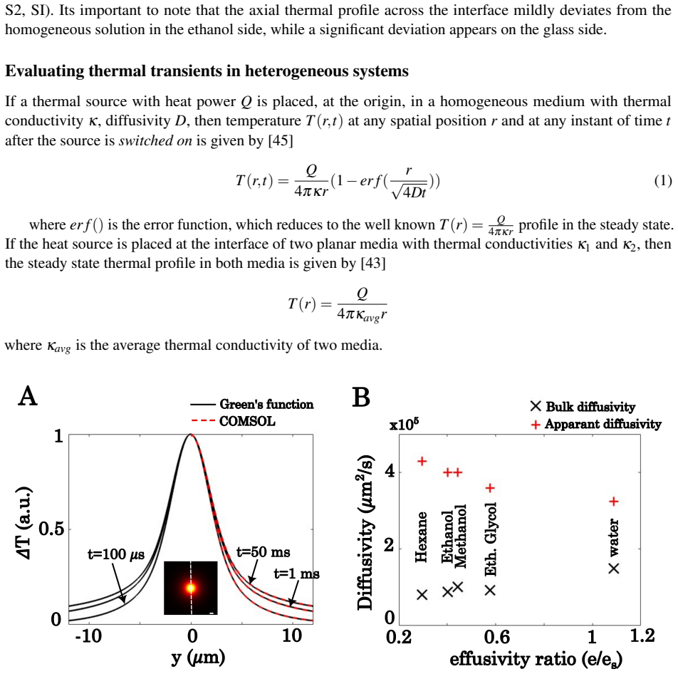
Thermal diffusion along an interface is controlled by their thermal effusivity contrast. An effective interfacial diffusivity is derived that accurately describes the lateral propagation of thermal fields and is validated through finite-element simulations across a broad range of liquid-glass interfaces. Liquids with lower bulk thermal diffusivities exhibit faster interfacial thermal spreading due to their lower effusivities, and the measured diffusivities agree quantitatively with theoretical predictions.
What carries the argument
Effective interfacial diffusivity derived from effusivity contrast, which governs the lateral propagation of thermal fields observed via thermal optical diffraction tomography.
If this is right
- Liquids with lower bulk diffusivities can produce faster interfacial thermal spreading when their effusivities are lower.
- The derived effective diffusivity matches simulation results across varied liquid-glass material pairs.
- Volumetric imaging combined with the effusivity model enables quantitative study of heat flow in heterogeneous systems.
- Interfacial heat transport can be engineered by selecting material pairs according to their effusivity contrast.
Where Pith is reading between the lines
- The same effusivity-controlled description may apply to interfaces beyond the liquid-glass cases simulated and measured.
- The optical tomography approach could be used to map heat flow at additional types of heterogeneous boundaries in real time.
- Engineering applications could select material combinations to accelerate or suppress lateral heat spreading at interfaces.
Load-bearing premise
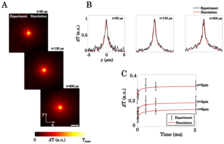
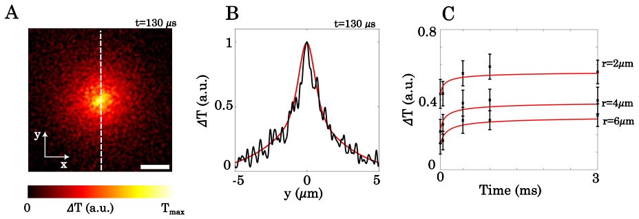
The three-dimensional temperature fields reconstructed from thermally induced refractive index changes accurately reflect the true spatio-temporal evolution of heat without significant contributions from non-thermal optical effects.
What would settle it
Direct measurement of lateral heat propagation speed at a liquid-glass interface that deviates from the value predicted by the effusivity-contrast formula for effective interfacial diffusivity.
Figures

read the original abstract
Quantitative imaging of heat transport with high spatial and temporal resolution is essential for understanding thermal processes in heterogeneous systems, yet direct measurements of transient temperature fields at material interfaces remain challenging. Here, we employ time resolved thermal optical diffraction tomography (thermal ODT), a label free nanoscale optical thermometry technique that reconstructs spatio-temporal evolution of three dimensional temperature fields from thermally induced refractive index changes. We show that thermal diffusion along an interface is controlled by their thermal effusivity contrast. We also derive an effective interfacial diffusivity that accurately describes the lateral propagation of thermal fields and validate the model through finite-element simulations across a broad range of liquid-glass interfaces. Surprisingly, liquids with lower bulk thermal diffusivities exhibit faster interfacial thermal spreading due to their lower effusivities. The measured diffusivities agree quantitatively with theoretical predictions over diverse material combinations. By combining volumetric thermal imaging with a general framework for interfacial heat transport, our work establishes thermal ODT as a powerful platform for investigating nanoscale thermodynamics and engineering heat flow in heterogeneous environments.
Editorial analysis
A structured set of objections, weighed in public.
Referee Report
Summary. The manuscript introduces time-resolved thermal optical diffraction tomography (thermal ODT) for nanoscale imaging of 3D temperature fields at material interfaces. It demonstrates that thermal diffusion along liquid-glass interfaces is governed by the effusivity contrast between the materials. An effective interfacial diffusivity is derived to describe lateral thermal propagation, validated against finite-element simulations for various liquid-glass combinations. The work finds that liquids with lower bulk diffusivities can exhibit faster interfacial spreading due to lower effusivities, with quantitative agreement between measurements and theory.
Significance. If the central assumption holds, this work provides a general framework for interfacial heat transport and establishes thermal ODT as a tool for investigating nanoscale thermodynamics. The quantitative validation with simulations across diverse interfaces is a notable strength, as is the derivation of the effective diffusivity model.
major comments (1)
- [Abstract / reconstruction methods] The claim of quantitative agreement between measured diffusivities and theoretical predictions (abstract) relies on the fidelity of the 3D temperature reconstruction from refractive index changes at the interface. The manuscript should explicitly address potential confounding non-thermal optical effects (such as adsorption-induced RI shifts or scattering at the liquid-glass boundary) and provide evidence that they do not bias the extracted lateral spreading rates, as this is load-bearing for the effusivity-control claim.
minor comments (1)
- Ensure that all simulation parameters and material properties used in the FEM validation are clearly tabulated for reproducibility.
Simulated Author's Rebuttal
We thank the referee for their constructive comment on the potential impact of non-thermal optical effects. We address this point directly below and have revised the manuscript to include an explicit discussion of these concerns.
read point-by-point responses
-
Referee: [Abstract / reconstruction methods] The claim of quantitative agreement between measured diffusivities and theoretical predictions (abstract) relies on the fidelity of the 3D temperature reconstruction from refractive index changes at the interface. The manuscript should explicitly address potential confounding non-thermal optical effects (such as adsorption-induced RI shifts or scattering at the liquid-glass boundary) and provide evidence that they do not bias the extracted lateral spreading rates, as this is load-bearing for the effusivity-control claim.
Authors: We agree that an explicit treatment of possible non-thermal contributions is warranted to support the central claim. In the revised manuscript we have added a dedicated paragraph in the Methods section (new subsection 'Assessment of non-thermal refractive-index contributions') that (i) notes the time scale separation between our nanosecond-scale heating pulses and slower adsorption kinetics, (ii) cites literature values showing that adsorption-induced RI shifts are at least an order of magnitude smaller than the thermo-optic signal under our conditions, and (iii) explains that index-matched liquid-glass pairs used in control experiments suppress scattering at the interface. We further note that any residual non-thermal bias would not be expected to produce the observed quantitative match to the effusivity-derived diffusivity model across six chemically distinct liquid-glass combinations; such agreement would be unlikely if the extracted spreading rates were systematically offset by material-specific optical artifacts. These additions directly address the referee's concern while leaving the reported results and conclusions unchanged. revision: yes
Circularity Check
No circularity: effective diffusivity derived from standard effusivity equations and validated externally
full rationale
The paper derives an effective interfacial diffusivity from effusivity contrast using standard heat transport relations and validates the result against independent finite-element simulations over multiple liquid-glass pairs. No quoted step reduces a prediction to a fitted input by construction, invokes self-citation as load-bearing premise, or renames a known result as new unification. The optical reconstruction method is an experimental input to the measurements, not part of the analytic derivation chain itself. The work is therefore self-contained against external benchmarks.
Axiom & Free-Parameter Ledger
axioms (1)
- domain assumption Thermally induced refractive index changes allow accurate reconstruction of 3D temperature fields via thermal ODT.
Reference graph
Works this paper leans on
-
[1]
& Quidant, R
Ciraulo, B., Garcia-Guirado, J., de Miguel, I., Ortega Arroyo, J. & Quidant, R. Long- range optofluidic control with plasmon heating.Nature Communications12(2021). URL https://www.nature.com/articles/s41467-021-22280-3
2021
-
[2]
URL https://www.nature.com/articles/s41566-025-01731-z?fromPaywallRec=false
Schmidt, F.et al.Three-dimensional optofluidic control using reconfig- urable thermal barriers.Nature Photonics19, 1385–1391 (2025). URL https://www.nature.com/articles/s41566-025-01731-z?fromPaywallRec=false
2025
-
[3]
Chand, R., Rani, C. E., Paul, D. & Kumar, G. V . P. Emergence of directional rotation in an optothermally activated colloidal system.ACS Photonics10, 4006–4013 (2023). URL https://pubs.acs.org/doi/full/10.1021/acsphotonics.3c00890
-
[4]
Donner, J. S., Baffou, G., McCloskey, D. & Quidant, R. Plasmon-assisted optofluidics.ACS Nano5, 5457–5462 (2011). URLhttps://pubs.acs.org/doi/full/10.1021/nn200590u
-
[5]
Jones, S., Andrén, D., Karpinski, P. & Käll, M. Photothermal heating of plasmonic nanoantennas: Influence on trapped particle dynamics and colloid distribution.ACS Photonics5, 2878–2887 (2018). URLhttps://pubs.acs.org/doi/full/10.1021/acsphotonics.8b00231
-
[6]
Dubi, Y ., Un, I. W. & Sivan, Y . Thermal effects – an alternative mechanism for plasmon-assisted photocatalysis.Chemical Science11, 5017–5027 (2020). URL https://pubs.rsc.org/en/content/articlelanding/2020/sc/c9sc06480j
2020
-
[7]
& Polshettiwar, V
Verma, R., Sharma, G. & Polshettiwar, V . The paradox of thermal vs. non-thermal effects in plasmonic photocatalysis.Nature Communications15(2024). URL https://www.nature.com/articles/s41467-024-51916-3
2024
-
[8]
URL https://www.cell.com/joule/fulltext/S2542-4351(25)00233-8
Liu, C.et al.Micro/nanoscale thermometry in photothermal catalysis.Joule9, 102052 (2025). URL https://www.cell.com/joule/fulltext/S2542-4351(25)00233-8
2025
-
[9]
& Quidant, R
Baffou, G., Bordacchini, I., Baldi, A. & Quidant, R. Simple experimental procedures to distinguish photothermal from hot-carrier processes in plasmonics.Light: Science & Applications9(2020). URL https://www.nature.com/articles/s41377-020-00345-0
2020
-
[10]
URL https://www.nature.com/articles/s41467-023-42167-9?fromPaywallRec=false
Biswas, A.et al.Photothermally heated colloidal synthesis of nanoparticles driven by silica-encapsulated plasmonic heat sources.Nature Communications14(2023). URL https://www.nature.com/articles/s41467-023-42167-9?fromPaywallRec=false. 9
2023
-
[11]
URL https://www.nature.com/articles/s41467-022-33074-6
Molinaro, C.et al.Life at high temperature observed in vitro upon laser heating of gold nanoparticles.Nature Communications13(2022). URL https://www.nature.com/articles/s41467-022-33074-6
2022
-
[12]
A., Yeatman, E
Kim, J. A., Yeatman, E. M. & Thompson, A. J. Plasmonic optical fiber for bac- teria manipulation—characterization and visualization of accumulation behavior un- der plasmo-thermal trapping.Biomedical Optics Express12, 3917 (2021). URL https://opg.optica.org/boe/fulltext.cfm?uri=boe-12-7-3917
2021
-
[13]
& Sauter, M
Pieper, K.-W. & Sauter, M. Direct temperature measurement of integrated microelectronic devices by thermally induced leakage currents.Microelectronics Reliability41, 133–136 (2001). URL https://www.sciencedirect.com/science/article/pii/S0026271400002213
2001
-
[14]
URL https://www.nature.com/articles/s41566-019-0486-3
Berto, P.et al.Tunable and free-form planar optics.Nature Photonics13, 649–656 (2019). URL https://www.nature.com/articles/s41566-019-0486-3
2019
-
[15]
Herzog, J. B., Knight, M. W. & Natelson, D. Thermoplasmonics: Quantifying plasmonic heating in single nanowires.Nano Letters14, 499–503 (2014). URL https://pubs.acs.org/doi/10.1021/nl403510u
-
[16]
URLhttps://pubs.acs.org/doi/10.1021/nn2047586
Baffou, G.et al.Thermal imaging of nanostructures by quantitative optical phase analysis.ACS Nano 6, 2452–2458 (2012). URLhttps://pubs.acs.org/doi/10.1021/nn2047586
-
[18]
B., Ciraulo, B., Schmidt, F., Arroyo, J
Vasista, A. B., Ciraulo, B., Schmidt, F., Arroyo, J. O. & Quidant, R. Non–steady state ther- mometry with optical diffraction tomography.Science Advances10, eadk5440 (2024). URL https://www.science.org/doi/10.1126/sciadv.adk5440
-
[19]
Ross, D., Gaitan, M. & Locascio, L. E. Temperature measurement in microfluidic systems us- ing a temperature-dependent fluorescent dye.Analytical Chemistry73, 4117–4123 (2001). URL https://pubs.acs.org/doi/full/10.1021/ac010370l
-
[20]
& Cao, W
Zhou, Y ., Qin, F., Zheng, Y ., Zhang, Z. & Cao, W. Fluorescence intensity ra- tio method for temperature sensing.Optics Letters40, 4544 (2015). URL https://opg.optica.org/ol/abstract.cfm?uri=ol-40-19-4544
2015
-
[21]
P., Kulzer, F
Baffou, G., Kreuzer, M. P., Kulzer, F. & Quidant, R. Temperature mapping near plasmonic nanostructures using fluorescence polarization anisotropy.Optics Express17, 3291 (2009). URL https://opg.optica.org/oe/fulltext.cfm?uri=oe-17-5-3291
2009
-
[22]
URL https://ieeexplore.ieee.org/abstract/document/974795
Kuball, M.et al.Measurement of temperature in active high-power algan/gan hfets using raman spectroscopy.IEEE Electron Device Letters23, 7–9 (2002). URL https://ieeexplore.ieee.org/abstract/document/974795
2002
-
[23]
& Cahill, D
Xie, X. & Cahill, D. G. Thermometry of plasmonic nanostructures by anti- stokes electronic raman scattering.Applied Physics Letters109(2016). URL https://pubs.aip.org/aip/apl/article/109/18/183104/32041/Thermometry-of-plasmonic-nanostructures-by-anti
2016
-
[24]
URL https://pubs.acs.org/doi/10.1021/acsnano.0c06185
Barella, M.et al.In situ photothermal response of single gold nanoparticles through hy- perspectral imaging anti-stokes thermometry.ACS Nano15, 2458–2467 (2020). URL https://pubs.acs.org/doi/10.1021/acsnano.0c06185. 10
-
[25]
URL https://pubs.acs.org/doi/full/10.1021/acsnano.5b05306
Chen, Z.et al.Imaging local heating and thermal diffusion of nanomaterials with plasmonic thermal microscopy.ACS Nano9, 11574–11581 (2015). URL https://pubs.acs.org/doi/full/10.1021/acsnano.5b05306
-
[26]
Heber, A., Selmke, M. & Cichos, F. Thermal diffusivities studied by single-particle photothermal deflection microscopy.ACS Photonics4, 681–687 (2017). URL https://pubs.acs.org/doi/full/10.1021/acsphotonics.7b00044
-
[27]
& Cichos, F
Heber, A., Selmke, M. & Cichos, F. Thermal diffusivity measured using a single plas- monic nanoparticle.Physical Chemistry Chemical Physics17, 20868–20872 (2015). URL https://pubs.rsc.org/en/content/articlelanding/2015/cp/c5cp02920a
2015
-
[28]
H., Braun, J
Olson, D. H., Braun, J. L. & Hopkins, P. E. Spatially resolved ther- moreflectance techniques for thermal conductivity measurements from the nanoscale to the mesoscale.Journal of Applied Physics126(2019). URL https://pubs.aip.org/aip/jap/article/126/15/150901/1062183/Spatially-resolved-thermoreflectance-techniques
2019
-
[29]
URL https://www.nature.com/articles/s41467-017-02652-4
Ziabari, A.et al.Full-field thermal imaging of quasiballistic crosstalk reduction in nanoscale devices.Na- ture Communications9(2018). URL https://www.nature.com/articles/s41467-017-02652-4
2018
-
[30]
URL https://www.sciencedirect.com/science/article/pii/S1359431126012500
Zhang, Z.et al.Spatial variation of thermal properties in thermal interface materials using a fre- quency domain thermoreflectance method.Applied Thermal Engineering298, 130942 (2026). URL https://www.sciencedirect.com/science/article/pii/S1359431126012500
2026
-
[31]
URL https://iopscience.iop.org/article/10.1088/0022-3727/42/14/143001/meta
Farzaneh, M.et al.Ccd-based thermoreflectance microscopy: principles and ap- plications.Journal of Physics D: Applied Physics42, 143001 (2009). URL https://iopscience.iop.org/article/10.1088/0022-3727/42/14/143001/meta
-
[32]
& Schmidt, A
Yang, J., Maragliano, C. & Schmidt, A. J. Thermal property microscopy with fre- quency domain thermoreflectance.Review of Scientific Instruments84(2013). URL https://pubs.aip.org/aip/rsi/article/84/10/104904/360276
2013
-
[34]
URLhttps://doi.org/10.1117/1.JBO.20.11.111208
Park, H.et al.Three-dimensional refractive index tomograms and deformability of individual human red blood cells from cord blood of newborn infants and maternal blood.Journal of Biomedical Optics 20, 111208 (2015). URLhttps://doi.org/10.1117/1.JBO.20.11.111208
-
[35]
URL https://opg.optica.org/boe/fulltext.cfm?uri=boe-6-10-3865
Yoon, J.et al.Label-free characterization of white blood cells by measuring 3d refractive index maps.Biomedical Optics Express6, 3865 (2015). URL https://opg.optica.org/boe/fulltext.cfm?uri=boe-6-10-3865
2015
-
[36]
& Park, Y
Cho, S., Kim, S., Kim, Y . & Park, Y . Optical imaging techniques for the study of malaria.Trends in Biotechnology30, 71–79 (2012)
2012
-
[37]
Kim, K.et al.High-resolution three-dimensional imaging of red blood cells parasitized by plasmodium falciparum and in situ hemozoin crystals using opti- cal diffraction tomography.Journal of Biomedical Optics19, 1 (2013). URL https://www.spiedigitallibrary.org/journals/journal-of-biomedical-optics/volume-19/issue-01/011005/High-resolution-three-dimensiona...
-
[38]
URLhttps://www.pnas.org/doi/full/10.1073/pnas.0806100105
Park, Y .et al.Refractive index maps and membrane dynamics of human red blood cells parasitized by plasmodium falciparum.Proceedings of the National Academy of Sciences105, 13730–13735 (2008). URLhttps://www.pnas.org/doi/full/10.1073/pnas.0806100105
-
[39]
URLhttps://www.nature.com/articles/s41563-022-01202-8
Shin, S.et al.Tomographic measurement of dielectric tensors at optical frequency.Nature Materials 21, 317–324 (2022). URLhttps://www.nature.com/articles/s41563-022-01202-8
2022
-
[40]
The role of thermal effusivity in heat exchange between finite-sized bod- ies.International Journal of Heat and Mass Transfer202, 123721 (2023)
Jain, A. The role of thermal effusivity in heat exchange between finite-sized bod- ies.International Journal of Heat and Mass Transfer202, 123721 (2023). URL https://www.sciencedirect.com/science/article/pii/S0017931022011899
2023
-
[41]
Oh, J.et al.Optical measurements of three-dimensional microscopic temperature distributions around gold nanorods excited by localized surface plasmon resonance.Physical Review Applied11(2019)
2019
-
[42]
URLhttps://opg.optica.org/oe/fulltext.cfm?uri=oe-17-1-266
Sung, Y .et al.Optical diffraction tomography for high resolution live cell imaging.Optics Express17, 266 (2009). URLhttps://opg.optica.org/oe/fulltext.cfm?uri=oe-17-1-266
2009
-
[43]
& Girard, C
Baffou, G., Quidant, R. & Girard, C. Thermoplasmonics modeling: A green’s function approach. Physical Review B82, 165424 (2010)
2010
-
[44]
& Baffou, G
Rogez, B., Marmri, Z., Thibaudau, F. & Baffou, G. Thermoplasmonics of metal layers and nanoholes.APL Photonics6, 101101 (2021). URL https://pubs.aip.org/aip/app/article/6/10/101101/279664/Thermoplasmonics-of-metal-layers-and-nanoholes
2021
-
[45]
Baffou, G.Thermoplasmonics: Heating Metal Nanoparticles Using Light(Cambridge University Press, 2017)
2017
-
[46]
Nikoobakht, B. & El-Sayed, M. A. Preparation and growth mechanism of gold nanorods (nrs) using seed-mediated growth method.Chemistry of Materials15, 1957–1962 (2003). URL https://pubs.acs.org/doi/10.1021/cm020732l
-
[47]
& Grigull, U
Thormählen, I., Straub, J. & Grigull, U. Refractive index of water and its dependence on wavelength, temperature, and density.Journal of Physical and Chemical Reference Data14, 933–945 (1985). 12 Supplementary Information Effusivity-Controlled Interfacial Thermal Transport Revealed by Nanoscale Optical Thermometry S1 Experimental setup Figure S1: Schemati...
1985
discussion (0)
Sign in with ORCID, Apple, or X to comment. Anyone can read and Pith papers without signing in.