Distilling Temporal Coherence into 2D Networks for Transrectal Ultrasound Prostate Video Segmentation
Pith reviewed 2026-07-01 06:16 UTC · model grok-4.3
The pith
Temporal coherence distilled during training lets a 2D network deliver consistent real-time prostate segmentation in TRUS video.
A machine-rendered reading of the paper's core claim, the machinery that carries it, and where it could break.
Core claim
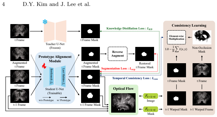
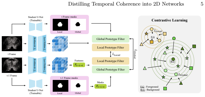
The authors establish that a Temporally Consistent Learning Framework—built around a Confidence-Weighted Temporal Consistency objective derived from optical flow warping residuals and a Dual-scale Prototype Alignment Module—can distill temporal and semantic coherence into an ordinary 2D segmentation network, yielding state-of-the-art accuracy and frame-to-frame consistency at single-frame inference speed on both SUN-SEG and the newly collected TRUS-V benchmark of 2,679 frames.
What carries the argument
The Confidence-Weighted Temporal Consistency objective, which attenuates gradients from regions with high optical-flow warping residuals, together with the Dual-scale Prototype Alignment Module that enforces contrastive alignment of boundary and semantic features.
If this is right
- Single-frame inference runs at real-time speed while inheriting video-level consistency.
- Pseudo-labeling via geometric equivariance reduces reliance on dense per-frame annotations.
- Knowledge distillation from a pretrained teacher further improves the distilled 2D student.
- The same selective weighting of stable anatomy can be applied to other video segmentation tasks where one structure is geometrically reliable.
Where Pith is reading between the lines
- The same stability-based weighting may help temporal models in cardiac or abdominal ultrasound where motion artifacts are common.
- If the optical-flow residual reliably identifies unreliable regions, the weighting scheme could be ported to other consistency losses beyond optical flow.
- Real-time consistent segmentation could support closed-loop guidance in biopsy or ablation procedures that currently tolerate frame-to-frame jitter.
Load-bearing premise
The prostate maintains geometric stability across frames while surrounding tissue and acoustic conditions fluctuate.
What would settle it
If the method shows no measurable gain in temporal-consistency metrics over plain 2D baselines when evaluated on the TRUS-V test set, the distillation claim would be falsified.
Figures


read the original abstract
Real-time video segmentation of the prostate in Transrectal Ultrasound (TRUS) is essential for image-guided interventions. While conventional 2D methods suffer from inter-frame inconsistencies by disregarding temporal context, 3D architectures incur prohibitive latency. To resolve this dilemma, we present a Temporally Consistent Learning Framework that distills temporal coherence into a 2D network during training, preserving single-frame inference efficiency. Our design is driven by a key clinical observation: the prostate exhibits geometric stability, whereas the surrounding acoustic environment fluctuates due to physiological motion and transducer pressure. Because conventional temporal constraints propagate erroneous gradients from these unstable regions, we introduce a Confidence-Weighted Temporal Consistency objective derived from optical flow warping residuals, selectively attenuating contributions from unreliable regions. Complementing this pixel-wise constraint, a Dual-scale Prototype Alignment Module enforces semantic coherence through contrastive optimization of local boundary and global semantic features. Furthermore, to eliminate the need for dense per-frame video annotations, we employ geometric equivariance-based pseudo-labeling with knowledge distillation from a pretrained teacher. Extensive experiments on SUN-SEG and our newly introduced TRUS-V benchmark (2,679 frames) demonstrate state-of-the-art accuracy and temporal consistency at real-time speed. Code and dataset are available at https://github.com/DYDevelop/DTC-TRUS.
Editorial analysis
A structured set of objections, weighed in public.
Referee Report
Summary. The paper proposes a Temporally Consistent Learning Framework that distills temporal coherence into 2D networks for real-time TRUS prostate video segmentation. It introduces a Confidence-Weighted Temporal Consistency objective derived from optical flow warping residuals to selectively attenuate gradients from unstable regions (motivated by the observation that the prostate is geometrically stable while the acoustic environment fluctuates), a Dual-scale Prototype Alignment Module for contrastive semantic coherence at boundary and global scales, and geometric equivariance-based pseudo-labeling with teacher distillation to reduce annotation requirements. Experiments on SUN-SEG and the new TRUS-V benchmark (2,679 frames) claim SOTA accuracy and temporal consistency at real-time inference speed, with code and data released.
Significance. If validated, the framework offers a practical solution for consistent real-time segmentation in image-guided prostate interventions by avoiding 3D latency while incorporating temporal constraints during training. The public release of code and the TRUS-V dataset strengthens reproducibility and enables community follow-up. The selective weighting approach, if supported by data, could improve robustness in variable acoustic conditions without sacrificing target-region accuracy.
major comments (1)
- [Abstract and §3] Abstract and §3 (method description): The Confidence-Weighted Temporal Consistency objective is justified solely by the key clinical observation that prostate geometry remains stable while surrounding regions fluctuate due to motion and pressure. No quantitative support is provided, such as a per-pixel residual histogram, mean warping error comparison, or statistical test showing systematically lower residuals inside the prostate versus background on TRUS-V (or SUN-SEG). If residuals are comparable or higher within the prostate, the weighting scheme lacks empirical grounding and may attenuate gradients in the region the network must segment accurately.
Simulated Author's Rebuttal
We thank the referee for the constructive feedback and recommendation for major revision. We address the single major comment below.
read point-by-point responses
-
Referee: [Abstract and §3] Abstract and §3 (method description): The Confidence-Weighted Temporal Consistency objective is justified solely by the key clinical observation that prostate geometry remains stable while surrounding regions fluctuate due to motion and pressure. No quantitative support is provided, such as a per-pixel residual histogram, mean warping error comparison, or statistical test showing systematically lower residuals inside the prostate versus background on TRUS-V (or SUN-SEG). If residuals are comparable or higher within the prostate, the weighting scheme lacks empirical grounding and may attenuate gradients in the region the network must segment accurately.
Authors: We agree that the manuscript relies on the stated clinical observation without providing quantitative validation of the optical flow warping residuals on the datasets. To directly address this, the revised version will include a new supplementary analysis (with accompanying figure) showing per-pixel residual histograms, mean warping errors, and a statistical comparison between prostate and background regions on TRUS-V. This will empirically support the selective weighting and confirm that residuals are systematically lower inside the prostate. revision: yes
Circularity Check
No significant circularity; derivation relies on external components and empirical validation
full rationale
The paper defines its Confidence-Weighted Temporal Consistency objective from optical-flow warping residuals and its Dual-scale Prototype Alignment from contrastive optimization of features; both draw on independent external techniques rather than fitting the target segmentation metric. Geometric equivariance pseudo-labeling uses a separate pretrained teacher. No equations reduce the claimed temporal consistency to a fitted parameter by construction, no self-citation chains justify uniqueness, and no ansatz is smuggled via prior work by the same authors. Results are reported on SUN-SEG and the new TRUS-V benchmark, keeping the derivation self-contained against external benchmarks.
Axiom & Free-Parameter Ledger
axioms (1)
- domain assumption The prostate exhibits geometric stability, whereas the surrounding acoustic environment fluctuates due to physiological motion and transducer pressure.
Reference graph
Works this paper leans on
-
[1]
In: International conference on medical image computing and computer- assisted intervention
Bortsova, G., Dubost, F., Hogeweg, L., Katramados, I., De Bruijne, M.: Semi- supervised medical image segmentation via learning consistency under transfor- mations. In: International conference on medical image computing and computer- assisted intervention. pp. 810–818. Springer (2019)
2019
-
[2]
In: Proceedings of the IEEE/CVF conference on computer vision and pattern recognition
Chen, X., He, K.: Exploring simple siamese representation learning. In: Proceedings of the IEEE/CVF conference on computer vision and pattern recognition. pp. 15750–15758 (2021)
2021
-
[3]
In: International conference on medical image computing and computer-assisted intervention
Fan, D.P., Ji, G.P., Zhou, T., Chen, G., Fu, H., Shen, J., Shao, L.: Pranet: Parallel reverse attention network for polyp segmentation. In: International conference on medical image computing and computer-assisted intervention. pp. 263–273. Springer (2020)
2020
-
[4]
In: Scandinavian conference on Image analysis
Farnebäck, G.: Two-frame motion estimation based on polynomial expansion. In: Scandinavian conference on Image analysis. pp. 363–370. Springer (2003)
2003
-
[5]
Journal of Medical Imaging6(1), 011003 (2019)
Ghavami, N., Hu, Y., Bonmati, E., Rodell, R., Gibson, E., Moore, C., Barratt, D.: Integration of spatial information in convolutional neural networks for automatic segmentation of intraoperative transrectal ultrasound images. Journal of Medical Imaging6(1), 011003 (2019)
2019
-
[6]
Distilling the Knowledge in a Neural Network
Hinton, G., Vinyals, O., Dean, J.: Distilling the knowledge in a neural network. arXiv preprint arXiv:1503.02531 (2015) 10 D.Y. Kim and J. Lee et al
work page internal anchor Pith review Pith/arXiv arXiv 2015
-
[7]
In: International Conference on Medical Image Computing and Computer-Assisted Intervention
Hu, Q., Yi, Z., Zhou, Y., Peng, F., Liu, M., Li, Q., Wang, Z.: Sali: Short-term align- ment and long-term interaction network for colonoscopy video polyp segmentation. In: International Conference on Medical Image Computing and Computer-Assisted Intervention. pp. 531–541. Springer (2024)
2024
-
[8]
Advances in neural information processing systems28(2015)
Jaderberg, M., Simonyan, K., Zisserman, A., et al.: Spatial transformer networks. Advances in neural information processing systems28(2015)
2015
-
[9]
Ieee Access9, 40496–40510 (2021)
Jha, D., Ali, S., Tomar, N.K., Johansen, H.D., Johansen, D., Rittscher, J., Riegler, M.A., Halvorsen, P.: Real-time polyp detection, localization and segmentation in colonoscopy using deep learning. Ieee Access9, 40496–40510 (2021)
2021
-
[10]
In: Medical Imaging with Deep Learning
Jha, D., Tomar, N.K., Sharma, V., Bagci, U.: Transnetr: transformer-based residual network for polyp segmentation with multi-center out-of-distribution testing. In: Medical Imaging with Deep Learning. pp. 1372–1384. PMLR (2024)
2024
-
[11]
Machine Intelligence Research 19(6), 531–549 (2022)
Ji, G.P., Xiao, G., Chou, Y.C., Fan, D.P., Zhao, K., Chen, G., Van Gool, L.: Video polyp segmentation: A deep learning perspective. Machine Intelligence Research 19(6), 531–549 (2022)
2022
-
[12]
Medical image analysis 57, 186–196 (2019)
Karimi, D., Zeng, Q., Mathur, P., Avinash, A., Mahdavi, S.S., Spadinger, I., Abol- maesumi,P.,Salcudean,S.E.:Accurateandrobustdeeplearning-basedsegmentation of the prostate clinical target volume in ultrasound images. Medical image analysis 57, 186–196 (2019)
2019
-
[13]
In: Proceedings of the European conference on computer vision (ECCV)
Lai, W.S., Huang, J.B., Wang, O., Shechtman, E., Yumer, E., Yang, M.H.: Learning blind video temporal consistency. In: Proceedings of the European conference on computer vision (ECCV). pp. 170–185 (2018)
2018
-
[14]
Medical physics46(7), 3194–3206 (2019)
Lei, Y., Tian, S., He, X., Wang, T., Wang, B., Patel, P., Jani, A.B., Mao, H., Curran, W.J., Liu, T., et al.: Ultrasound prostate segmentation based on multidirectional deeply supervised v-net. Medical physics46(7), 3194–3206 (2019)
2019
-
[15]
Medical Imaging 2019: Ultrasonic Imaging and Tomography 10955, 198–204 (2019)
Lei, Y., Wang, T., Wang, B., He, X., Tian, S., Jani, A.B., Mao, H., Curran, W.J., Patel, P., Liu, T., et al.: Ultrasound prostate segmentation based on 3d v-net with deep supervision. Medical Imaging 2019: Ultrasonic Imaging and Tomography 10955, 198–204 (2019)
2019
-
[16]
In: International Conference on Medical Image Computing and Computer-Assisted Intervention
Lin, J., Dai, Q., Zhu, L., Fu, H., Wang, Q., Li, W., Rao, W., Huang, X., Wang, L.: Shifting more attention to breast lesion segmentation in ultrasound videos. In: International Conference on Medical Image Computing and Computer-Assisted Intervention. pp. 497–507. Springer (2023)
2023
-
[17]
In: 2016 fourth international conference on 3D vision (3DV)
Milletari, F., Navab, N., Ahmadi, S.A.: V-net: Fully convolutional neural networks for volumetric medical image segmentation. In: 2016 fourth international conference on 3D vision (3DV). pp. 565–571. IEEE (2016)
2016
-
[18]
Medical physics47(6), 2413–2426 (2020)
Orlando, N., Gillies, D.J., Gyacskov, I., Romagnoli, C., D’Souza, D., Fenster, A.: Automatic prostate segmentation using deep learning on clinically diverse 3d transrectal ultrasound images. Medical physics47(6), 2413–2426 (2020)
2020
-
[19]
In: Medical Image Computing and Computer-Assisted Intervention–MICCAI 2015
Ronneberger, O., Fischer, P., Brox, T.: U-net: Convolutional networks for biomed- ical image segmentation. In: Medical Image Computing and Computer-Assisted Intervention–MICCAI 2015. pp. 234–241. Springer (2015)
2015
-
[20]
In: International Conference on Medical Image Computing and Computer-Assisted Intervention
Roy, S., Koehler, G., Ulrich, C., Baumgartner, M., Petersen, J., Isensee, F., Jaeger, P.F., Maier-Hein, K.H.: Mednext: transformer-driven scaling of convnets for medical image segmentation. In: International Conference on Medical Image Computing and Computer-Assisted Intervention. pp. 405–415. Springer (2023)
2023
-
[21]
CA: a cancer journal for clinicians73(1), 17–48 (2023)
Siegel, R.L., Miller, K.D., Wagle, N.S., Jemal, A.: Cancer statistics, 2023. CA: a cancer journal for clinicians73(1), 17–48 (2023)
2023
-
[22]
Advances in neural information processing systems30(2017) Distilling Temporal Coherence into 2D Networks 11
Snell, J., Swersky, K., Zemel, R.: Prototypical networks for few-shot learning. Advances in neural information processing systems30(2017) Distilling Temporal Coherence into 2D Networks 11
2017
-
[23]
IEEE Journal of Biomedical and Health Informatics 26(5), 2252–2263 (2021)
Srivastava, A., Jha, D., Chanda, S., Pal, U., Johansen, H.D., Johansen, D., Riegler, M.A., Ali, S., Halvorsen, P.: Msrf-net: A multi-scale residual fusion network for biomedical image segmentation. IEEE Journal of Biomedical and Health Informatics 26(5), 2252–2263 (2021)
2021
-
[24]
arXiv preprint arXiv:2407.00678 (2024)
Wang, H., Wu, H., Wang, Z., Yue, P., Ni, D., Heng, P.A., Wang, Y.: A review of image processing methods in prostate ultrasound. arXiv preprint arXiv:2407.00678 (2024)
-
[25]
In: proceedings of the IEEE/CVF international conference on computer vision
Wang, K., Liew, J.H., Zou, Y., Zhou, D., Feng, J.: Panet: Few-shot image seman- tic segmentation with prototype alignment. In: proceedings of the IEEE/CVF international conference on computer vision. pp. 9197–9206 (2019)
2019
-
[26]
IEEE transactions on medical imaging38(12), 2768–2778 (2019)
Wang, Y., Dou, H., Hu, X., Zhu, L., Yang, X., Xu, M., Qin, J., Heng, P.A., Wang, T., Ni, D.: Deep attentive features for prostate segmentation in 3d transrectal ultrasound. IEEE transactions on medical imaging38(12), 2768–2778 (2019)
2019
-
[27]
arXiv preprint arXiv:2407.05703 (2024)
Xu, H., Yang, Y., Aviles-Rivero, A.I., Yang, G., Qin, J., Zhu, L.: Lgrnet: Local-global reciprocal network for uterine fibroid segmentation in ultrasound videos. arXiv preprint arXiv:2407.05703 (2024)
-
[28]
In: Proceedings of the AAAI Conference on Artificial Intelligence
Yang, X., Yu, L., Wu, L., Wang, Y., Ni, D., Qin, J., Heng, P.A.: Fine-grained recurrent neural networks for automatic prostate segmentation in ultrasound images. In: Proceedings of the AAAI Conference on Artificial Intelligence. vol. 31 (2017)
2017
-
[29]
In: International conference on medical image computing and computer-assisted intervention
Zhang, R., Li, G., Li, Z., Cui, S., Qian, D., Yu, Y.: Adaptive context selection for polyp segmentation. In: International conference on medical image computing and computer-assisted intervention. pp. 253–262. Springer (2020)
2020
-
[30]
In: International conference on medical image computing and computer-assisted intervention
Zhao, X., Wu, Z., Tan, S., Fan, D.J., Li, Z., Wan, X., Li, G.: Semi-supervised spatial temporal attention network for video polyp segmentation. In: International conference on medical image computing and computer-assisted intervention. pp. 456–466. Springer (2022)
2022
-
[31]
IEEE Journal of Biomedical and Health Informatics (2025)
Zhao, Y., Wang, X., Yin, J.: Efficient video polyp segmentation by deformable alignment and local attention. IEEE Journal of Biomedical and Health Informatics (2025)
2025
-
[32]
IEEE transactions on medical imaging39(6), 1856–1867 (2019)
Zhou, Z., Siddiquee, M.M.R., Tajbakhsh, N., Liang, J.: Unet++: Redesigning skip connections to exploit multiscale features in image segmentation. IEEE transactions on medical imaging39(6), 1856–1867 (2019)
2019
discussion (0)
Sign in with ORCID, Apple, or X to comment. Anyone can read and Pith papers without signing in.