CSV-ViT: A Vision Transformer with the Variable-sized Cortical Supervertices for Detection of Alzheimer's Disease Pathologies
Pith reviewed 2026-06-29 18:00 UTC · model grok-4.3
The pith
A Vision Transformer using variable-sized cortical supervertices outperforms prior surface models on MRI classification of Alzheimer's disease, amyloid positivity, and tau positivity.
A machine-rendered reading of the paper's core claim, the machinery that carries it, and where it could break.
Core claim
The paper claims that its cortical surface tokenization, which performs ROI-preserving vertex-based variable-sized patch partitioning into cortical supervertices, combined with a padding-tolerant and mask-aware Vision Transformer, produces higher classification performance than recent surface-based models when applied to T1-weighted MRI for AD diagnosis, amyloid positivity, and tau positivity.
What carries the argument
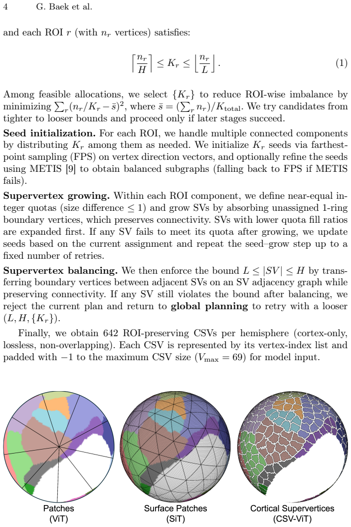
Cortical supervertices (CSVs), defined as ROI-preserving, vertex-based, variable-sized patches obtained by partitioning the cortical surface, together with padding and mask-aware patch embedding that allows the Vision Transformer to process variable patch sizes without boundary duplication or information loss.
If this is right
- The model supports MRI-based prediction of AD-related status prior to PET or CSF confirmation.
- Variable-sized partitioning avoids inclusion of non-cortical regions and duplicate vertices at patch boundaries.
- The framework achieves higher classification performance across AD diagnosis, amyloid positivity, and tau positivity tasks.
- The tokenization enables learning directly from the spherical topology of brain cortical surfaces.
Where Pith is reading between the lines
- The same supervertex tokenization could be tested on other cortical-surface classification problems such as additional neurodegenerative conditions.
- Combining CSV-ViT predictions with PET data in a multi-modal setting might further improve specificity for amyloid and tau status.
- Systematic variation of supervertex size distributions could reveal an optimal granularity for different diagnostic targets.
Load-bearing premise
The variable-sized cortical supervertex partitioning preserves the region of interest without including non-cortical regions such as the medial wall, and the padding plus mask-aware patch embedding does not degrade information or introduce artifacts that affect classification.
What would settle it
A direct replication on the same T1-weighted MRI datasets and tasks in which CSV-ViT fails to exceed the classification accuracy of the compared recent surface-based models on any of the three targets.
Figures

read the original abstract
Confirming Alzheimer's disease (AD) typically relies on positron emission tomography (PET), which remains costly and invasive, motivating the use of structural MRI-based prescreening. Deep learning on non-Euclidean manifolds, particularly brain cortical surfaces, faces significant challenges due to the data's spherical topology. Recent surface models have enabled learning from cortical surface data; however, imposing face-based uniform patches often causes duplicate vertices at patch boundaries. In general, many surface-based models are limited in their awareness of the region of interest (ROI), which can result in non-cortical regions, such as the medial wall, being included. We propose a cortical surface tokenization that performs ROI-preserving, vertex-based, variable-sized patch partitioning. We refer to these cortical surface patches as cortical supervertices (CSVs). Building on this representation, we design the CSV Vision Transformer (CSV-ViT), a variable-size patch-tolerant Vision Transformer that uses padding and a mask-aware patch embedding. We used T1-weighted MRI and evaluated our framework by classifying AD-related status into three categories: AD diagnosis, amyloid positivity, and tau positivity. Across the experiments, CSV-ViT achieved higher classification performance than recent surface-based models. The results suggest that the proposed CSV-ViT may support MRI-based prediction of AD-related status prior to PET or CSF confirmation.
Editorial analysis
A structured set of objections, weighed in public.
Referee Report
Summary. The paper proposes CSV-ViT, a Vision Transformer for cortical surface data from T1-weighted MRI that tokenizes the surface using ROI-preserving, vertex-based, variable-sized cortical supervertices (CSVs). It introduces padding and mask-aware patch embedding to handle variable patch sizes in the ViT, and evaluates the model on three binary classification tasks: AD diagnosis, amyloid positivity, and tau positivity. The central claim is that CSV-ViT outperforms recent surface-based models on these tasks.
Significance. If the performance gains hold under full validation, the work offers a practical advance in non-invasive MRI-based prescreening for AD-related biomarkers, addressing known difficulties with uniform face-based patching and medial-wall inclusion on spherical cortical surfaces. The explicit handling of variable-sized, ROI-preserving supervertices and mask-aware embedding is a targeted technical contribution to manifold learning on brain surfaces.
minor comments (3)
- [Methods] The abstract and method sections would benefit from explicit dataset sizes, train/validation/test splits, and subject-level cross-validation details to allow direct comparison with prior surface-based baselines.
- [Experiments] Figure captions and the experimental results section should report exact AUC, accuracy, and F1 values with confidence intervals or standard deviations across runs rather than qualitative statements of 'higher performance'.
- [Section 3.1] The description of the supervertex partitioning algorithm would be clearer with a short pseudocode block or explicit reference to the number of supervertices per hemisphere and the stopping criterion used.
Simulated Author's Rebuttal
We thank the referee for the positive assessment of our work and the recommendation for minor revision. We appreciate the recognition of the technical contributions regarding ROI-preserving variable-sized cortical supervertices and the mask-aware ViT embedding.
Circularity Check
No significant circularity
full rationale
The manuscript presents an empirical architecture proposal (CSV tokenization plus mask-aware ViT) whose central claim is comparative classification accuracy on three AD-related tasks from T1 MRI. No equations, derivations, or parameter-fitting steps appear that could reduce a claimed prediction to a self-defined input. The method description relies on standard ViT components with added padding/masking; performance is reported via direct experiment rather than any self-referential construction. No load-bearing self-citations or uniqueness theorems are invoked. The derivation chain is therefore self-contained against external benchmarks.
Axiom & Free-Parameter Ledger
Reference graph
Works this paper leans on
-
[1]
Dahan, S., Fawaz, A., Williams, L.Z., Yang, C., et al.: Surface Vision Transformers: Attention-Based Modelling applied to Cortical Analysis. In: Medical Imaging with Deep Learning (MIDL). pp. 282–303 (2022).https://doi.org/10.48550/arXiv. 2203.16414
work page internal anchor Pith review doi:10.48550/arxiv 2022
-
[2]
Sharp, N., Attaiki, S., Crane, K., et al.: DiffusionNet: Discretization agnos- tic learning on surfaces. ACM Trans. Graph.41(3), 27:1–27:16 (2022).https: //doi.org/10.1145/3507905
-
[3]
Dahan,S.,Williams,L.Z.,etal.:TheMultiscaleSurfaceVisionTransformer.arXiv preprintarXiv:2303.11909(2024).https://doi.org/10.48550/arXiv.2303.11909
-
[4]
Medical Image Analysis107, 103793 (2025).https://doi.org/10.1016/j.media.2025.103793
Li, Z., Zhang, J., et al.: SurfGNN: A robust surface-based prediction model with in- terpretability for coactivation maps of spatial and cortical features. Medical Image Analysis107, 103793 (2025).https://doi.org/10.1016/j.media.2025.103793
-
[5]
Cerebral Cortex14(1), 11–22 (2004).https://doi.org/10.1093/ cercor/bhg087
Fischl, B., Van Der Kouwe, A., et al.: Automatically parcellating the human cerebral cortex. Cerebral Cortex14(1), 11–22 (2004).https://doi.org/10.1093/ cercor/bhg087
2004
-
[6]
Johnson, K.A., Sperling, R.A., Gidicsin, C.M., Carmasin, J.S., Maye, J.E., Cole- man, R.E., et al.: Florbetapir (F18-AV-45) PET to assess amyloid burden in Alzheimer’s disease dementia, mild cognitive impairment, and normal aging. Alzheimers Dement.9(5 Suppl), S72–S83 (2013).https://doi.org/10.1016/j. jalz.2012.10.007
work page doi:10.1016/j 2013
-
[7]
Ann Neurol.78(5), 787–800 (2015).https://doi.org/10.1002/ana.24517
Marquié, M., Normandin, M.D., Vanderburg, C.R., Costantino, I.M., Bien, E.A., Rycyna, L.G., et al.: Validating novel tau positron emission tomography tracer [F-18]-AV-1451 (T807) on postmortem brain tissue. Ann Neurol.78(5), 787–800 (2015).https://doi.org/10.1002/ana.24517
-
[8]
An Image is Worth 16x16 Words: Transformers for Image Recognition at Scale
Dosovitskiy, A., Beyer, L., Kolesnikov, A., Weissenborn, D., Zhai, X., Unterthiner, T., et al.: An Image is Worth 16x16 Words: Transformers for Image Recognition at Scale. In: International Conference on Learning Representations (ICLR) (2021). https://doi.org/10.48550/arXiv.2010.11929
work page internal anchor Pith review Pith/arXiv arXiv doi:10.48550/arxiv.2010.11929 2021
-
[9]
Karypis, G., Kumar, V.: A Fast and High Quality Multilevel Scheme for Parti- tioning Irregular Graphs. SIAM J. Sci. Comput.20(1), 359–392 (1998).https: //doi.org/10.1137/S1064827595287997
-
[10]
Neurology74(3), 201–209 (2010)
Petersen, R.C., Aisen, P.S., Beckett, L.A., et al.: Alzheimer’s Disease Neuroimag- ing Initiative (ADNI): clinical characterization. Neurology74(3), 201–209 (2010). https://doi.org/10.1212/WNL.0b013e3181cb3e25
-
[11]
Marcus, D.S., Fotenos, A.F., Csernansky, J.G., Morris, J.C., Buckner, R.L.: Open Access Series of Imaging Studies: Longitudinal MRI Data in Nondemented and Demented Older Adults. J. Cogn. Neurosci.22(12), 2677–2684 (2010).https: //doi.org/10.1162/jocn.2009.21407
-
[12]
Alzheimers Dement.7(3), 270–279 (2011).https: //doi.org/10.1016/j.jalz.2011.03.008 10 G
Albert, M.S., DeKosky, S.T., Dickson, D., et al.: The diagnosis of mild cog- nitive impairment due to Alzheimer’s disease: Recommendations from the Na- tional Institute on Aging–Alzheimer’s Association workgroups on diagnostic guide- lines for Alzheimer’s disease. Alzheimers Dement.7(3), 270–279 (2011).https: //doi.org/10.1016/j.jalz.2011.03.008 10 G. Baek et al
-
[13]
Bondi, M.W., Edmonds, E.C., Jak, A.J., et al.: Neuropsychological criteria for mild cognitive impairment improves diagnostic precision, biomarker associations, and progression rates. J. Alzheimers Dis.42(1), 275–289 (2014).https://doi. org/10.3233/JAD-140276
-
[14]
Alzheimers Dement.10(5), 511–521.e1 (2014).https://doi.org/10
Nettiksimmons, J., DeCarli, C., Landau, S., Beckett, L., Alzheimer’s Disease Neu- roimaging Initiative: Biological heterogeneity in ADNI amnestic mild cognitive impairment. Alzheimers Dement.10(5), 511–521.e1 (2014).https://doi.org/10. 1016/j.jalz.2013.09.003
2014
-
[15]
Alzheimers Dement (Amst)10, 245–252 (2018)
Gordon, B.A., McCullough, A., Mishra, S., et al.: Cross-sectional and longitudi- nal atrophy is preferentially associated with tau rather than amyloidβpositron emission tomography pathology. Alzheimers Dement (Amst)10, 245–252 (2018). https://doi.org/10.1016/j.dadm.2018.02.003
-
[16]
Neurology92(6), e601–e612 (2019).https://doi.org/10.1212/WNL.0000000000006875
Ossenkoppele, R., Smith, R., Ohlsson, T., et al.: Associations between tau, Aβ, and cortical thickness with cognition in Alzheimer disease. Neurology92(6), e601–e612 (2019).https://doi.org/10.1212/WNL.0000000000006875
-
[17]
Alzheimers Dement.17, 1085–1096 (2021).https://doi.org/10.1002/alz.12249
Harrison, T.M., Du, R., Klencklen, G., Baker, S.L., Jagust, W.J.: Distinct effects of beta-amyloid and tau on cortical thickness in cognitively healthy older adults. Alzheimers Dement.17, 1085–1096 (2021).https://doi.org/10.1002/alz.12249
-
[18]
Lew, C.O., Zhou, L., et al.: MRI-based Deep Learning Assessment of Amyloid, Tau, and Neurodegeneration Biomarker Status across the Alzheimer Disease Spectrum. Radiology. 309(1), e222441 (2023).https://doi.org/10.1148/radiol.222441
discussion (0)
Sign in with ORCID, Apple, or X to comment. Anyone can read and Pith papers without signing in.