Unsupervised Susceptibility Distortion Correction of EPI without Calibration Scans via Image Translation-Based Registration
Pith reviewed 2026-06-26 12:33 UTC · model grok-4.3
The pith
SACRED corrects EPI distortions in fMRI without calibration scans by translating BOLD images to T1-weighted contrast for unsupervised registration.
A machine-rendered reading of the paper's core claim, the machinery that carries it, and where it could break.
Core claim
SACRED employs an invertible neural network as the image translation backbone to bridge the contrast gap between BOLD and T1w images while enforcing structural consistency through a modality independent neighborhood descriptor. This design enables the use of a mono-contrast similarity objective to train the registration network in an unsupervised manner without requiring distortion-corrected BOLD images. In addition, we incorporate test-time adaptation (TTA) to further enhance performance on out-of-distribution (OOD) data at inference time. SACRED was evaluated on one in-distribution (ID) dataset and two OOD datasets, and was compared with representative fMRI distortion correction methods.
What carries the argument
Invertible neural network for image translation combined with modality-independent neighborhood descriptor, enabling mono-contrast similarity objective for unsupervised registration across the BOLD-T1w gap.
If this is right
- SACRED outperforms competing methods on both in-distribution and out-of-distribution datasets.
- It exhibits robustness to scanner and population shifts partly enabled by test-time adaptation.
- Distortion correction is possible using only a routinely acquired T1-weighted image and a unidirectional phase-encoding BOLD image.
- No additional calibration scans are required at acquisition time.
Where Pith is reading between the lines
- The same translation-plus-registration pattern could be tested on other contrasts that lack paired ground-truth corrected data.
- Reducing the dependence on calibration scans may shorten total scan time in clinical fMRI protocols.
- Further gains might come from combining the current TTA step with additional domain-adaptation losses not explored in the paper.
Load-bearing premise
The invertible neural network plus modality-independent neighborhood descriptor can produce accurate mono-contrast similarity registration across the BOLD-T1w contrast gap without any distortion-corrected BOLD ground truth during training.
What would settle it
On a held-out dataset supplied with field-map ground truth, the positions of anatomical landmarks after SACRED correction do not match the expected undistorted locations more closely than corrections obtained from conventional calibration-based methods.
Figures


read the original abstract
Functional magnetic resonance imaging (fMRI) utilizes echo-planar imaging (EPI) to capture blood-oxygen-level-dependent (BOLD) signals with high temporal resolution. However, EPI is inherently sensitive to magnetic field inhomogeneities, resulting in susceptibility-induced geometric distortions along the phase-encoding (PE) direction. To correct these distortions, conventional approaches rely on additional calibration scans, such as field maps or reverse PE acquisitions, which are not always available in practice. To overcome this limitation, we propose SACRED, a calibration scan-free susceptibility distortion correction framework that corrects geometric distortions via image translation-based registration using only a routinely acquired anatomical T1-weighted (T1w) image and a unidirectional PE BOLD image. SACRED employs an invertible neural network as the image translation backbone to bridge the contrast gap between BOLD and T1w images while enforcing structural consistency through a modality independent neighborhood descriptor. This design enables the use of a mono-contrast similarity objective to train the registration network in an unsupervised manner without requiring distortion-corrected BOLD images. In addition, we incorporate test-time adaptation (TTA) to further enhance performance on out-of-distribution (OOD) data at inference time. SACRED was evaluated on one in-distribution (ID) dataset and two OOD datasets, and was compared with representative fMRI distortion correction methods. The results demonstrate that SACRED significantly outperforms competing methods on both ID and OOD datasets, exhibiting robustness to scanner and population shifts, partly enabled by TTA. The code will be made publicly available upon acceptance.
Editorial analysis
A structured set of objections, weighed in public.
Referee Report
Summary. The paper proposes SACRED, a calibration-scan-free method for correcting susceptibility-induced geometric distortions in unidirectional-PE EPI BOLD images. It employs an invertible neural network to translate between BOLD and T1w contrasts while using a modality-independent neighborhood descriptor (MIND) to enable unsupervised mono-contrast similarity registration; test-time adaptation (TTA) is added to improve robustness on out-of-distribution data. The method is evaluated on one in-distribution and two out-of-distribution datasets and is reported to significantly outperform representative competing fMRI distortion-correction approaches.
Significance. If the central assumption holds—that the learned translation plus MIND descriptor yields a similarity optimum that corresponds to true geometric undistortion rather than mere contrast matching—this approach would address a practical barrier in fMRI preprocessing by removing the requirement for field maps or reverse-PE scans. The explicit use of TTA for scanner/population shifts and the unsupervised training without distortion-corrected BOLD ground truth would be notable strengths for multi-site applicability.
major comments (2)
- [Abstract] Abstract: the claim that SACRED 'significantly outperforms competing methods on both ID and OOD datasets' is presented without any quantitative metrics, error bars, dataset sizes, statistical tests, or even the names of the competing methods. Because this performance claim is the primary evidence offered for the method's utility, the absence of these numbers prevents assessment of whether the gains are load-bearing or merely incremental.
- [Methods] Methods (image-translation and registration sections): the central modeling assumption—that the invertible-NN translation combined with MIND produces a mono-contrast similarity signal whose optimum aligns with true susceptibility-induced displacements—is not directly tested. No experiment is described that compares the estimated deformation fields against ground-truth distortion maps (field-map or reverse-PE corrected) on held-out data; without such validation it remains possible that the registration is optimizing residual contrast mismatch rather than geometry.
minor comments (1)
- [Abstract] Abstract: the acronym SACRED is introduced without expansion.
Simulated Author's Rebuttal
We thank the referee for the constructive feedback. We address each major comment below and outline planned revisions to strengthen the manuscript.
read point-by-point responses
-
Referee: [Abstract] Abstract: the claim that SACRED 'significantly outperforms competing methods on both ID and OOD datasets' is presented without any quantitative metrics, error bars, dataset sizes, statistical tests, or even the names of the competing methods. Because this performance claim is the primary evidence offered for the method's utility, the absence of these numbers prevents assessment of whether the gains are load-bearing or merely incremental.
Authors: We agree that the abstract would benefit from quantitative support for the performance claims. In the revised manuscript, we will update the abstract to report specific metrics (e.g., mean displacement errors with standard deviations), dataset sizes (number of subjects and volumes), statistical test results (e.g., p-values), and the names of the competing methods (such as TOPUP and fieldmap-based approaches). This will enable readers to evaluate the magnitude and reliability of the reported improvements. revision: yes
-
Referee: [Methods] Methods (image-translation and registration sections): the central modeling assumption—that the invertible-NN translation combined with MIND produces a mono-contrast similarity signal whose optimum aligns with true susceptibility-induced displacements—is not directly tested. No experiment is described that compares the estimated deformation fields against ground-truth distortion maps (field-map or reverse-PE corrected) on held-out data; without such validation it remains possible that the registration is optimizing residual contrast mismatch rather than geometry.
Authors: We acknowledge that a direct comparison of estimated deformation fields to ground-truth distortion maps on held-out data would provide stronger evidence that the similarity optimum corresponds to geometric correction. Our current evaluation relies on indirect validation via improved alignment with T1w images and downstream fMRI task performance on ID and OOD datasets. In the revision, we will add an experiment on available subsets with field maps or reverse-PE data to compare deformation fields against ground truth, thereby addressing this modeling assumption more explicitly. revision: yes
Circularity Check
No circularity: standard unsupervised NN registration with external T1w reference
full rationale
The paper describes an image-translation registration method using an invertible neural network and MIND descriptor for unsupervised training on BOLD-T1w pairs. No equations, derivations, or self-citations are presented that reduce any claimed result to its own inputs by construction. Training relies on a mono-contrast similarity objective and an external anatomical T1w image rather than any fitted parameter or self-referential loop. The approach is self-contained against standard ML registration benchmarks and does not invoke uniqueness theorems or prior author work as load-bearing justification.
Axiom & Free-Parameter Ledger
Reference graph
Works this paper leans on
-
[1]
We propose SACRED, a calibration scan-free fMRI susceptibility-induced distortion correction (SDC) method that requires only routinely acquired T1w images and unidirectional PE BOLD images
-
[2]
We introduce an image translation-based registration framework that leverages an INN and MIND-SSC to bridge the contrast gap between BOLD and T1w images, enabling the use of standard mono-contrast similarity metrics while preserving structural consistency
-
[3]
SACRED demonstrates improved robustness under vari- ous distribution shifts, enabled by TTA
-
[4]
To the best of our knowledge, this study presents the first comprehensive comparative evaluation of nearly all rep- resentative publicly available calibration scan-free fMRI distortion correction methods. II. RELATEDWORK A. Calibration Scan-Free fMRI Susceptibility Distortion Correction Calibration scan-free fMRI SDC has been studied to correct distortion...
-
[5]
In our implementation, we defined a 4-neighborhood con- figurationNconsisting of top, bottom, left, and right neigh- bors
Structural Consistency Loss:To ensure the synthesized pseudo BOLD images maintain the anatomical fidelity of the T1w images, we employed a structural consistency loss, Lstructure, based on the MIND-SSC descriptor [23]. In our implementation, we defined a 4-neighborhood con- figurationNconsisting of top, bottom, left, and right neigh- bors. To capture loca...
-
[6]
This loss enforces that the input image is recoverable from its translated representation
Image Reconstruction Loss:To leverage the invertibility of the image translation network, we incorporate an image reconstruction loss. This loss enforces that the input image is recoverable from its translated representation. We compute theL 1 distance between the original input T1w imagexand the reconstructed imageˆ x, which is obtained via the inverse m...
-
[7]
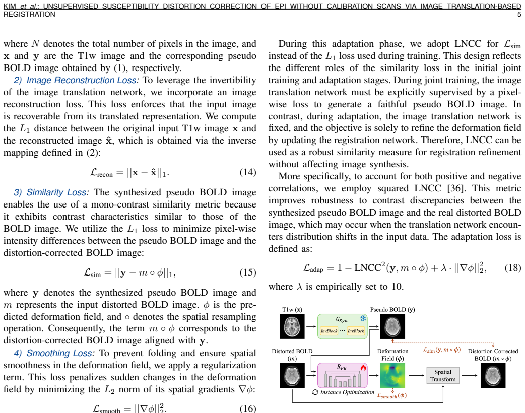
Similarity Loss:The synthesized pseudo BOLD image enables the use of a mono-contrast similarity metric because it exhibits contrast characteristics similar to those of the BOLD image. We utilize theL 1 loss to minimize pixel-wise intensity differences between the pseudo BOLD image and the distortion-corrected BOLD image: Lsim =||y−m◦ϕ|| 1,(15) whereydenot...
-
[8]
This loss penalizes sudden changes in the deformation field by minimizing theL 2 norm of its spatial gradients∇ϕ: Lsmooth =||∇ϕ|| 2 2.(16)
Smoothing Loss:To prevent folding and ensure spatial smoothness in the deformation field, we apply a regularization term. This loss penalizes sudden changes in the deformation field by minimizing theL 2 norm of its spatial gradients∇ϕ: Lsmooth =||∇ϕ|| 2 2.(16)
-
[9]
In our experiments, we empirically set these weights toλ 1 = 0.5,λ 2 = 0.1,λ 3 = 1, andλ 4 = 5
T otal Loss:The final objective function for joint op- timization is a weighted sum of the four loss components discussed above: Ltotal =λ 1 · Lstructure +λ 2 · Lrecon +λ 3 · Lsim +λ 4 · Lsmooth, (17) whereλ 1, λ2, λ3,andλ 4 are hyperparameters that balance the contribution of each term. In our experiments, we empirically set these weights toλ 1 = 0.5,λ 2...
-
[10]
First, SyN-SDC 1, im- plemented in the widely used fMRI preprocessing pipeline fM- RIPrep [15], was included
Conventional Approaches:Two representative non- learning-based methods were selected. First, SyN-SDC 1, im- plemented in the widely used fMRI preprocessing pipeline fM- RIPrep [15], was included. SyN-SDC corrects susceptibility- induced distortions by performing symmetric diffeomorphic registration (SyN) [47] between the distorted BOLD image and an anatom...
-
[11]
First, SynBOLD-DisCo3 [16] was included
Learning-Based Approaches:We further compared our technique with two recent learning-based SDC methods. First, SynBOLD-DisCo3 [16] was included. This method trains a U- Net [26] in a supervised manner to synthesize an undistorted BOLD image using TOPUP-corrected images as ground truth, and subsequently applies TOPUP using the original distorted BOLD image...
-
[12]
Quantitative results are summarized in Table III
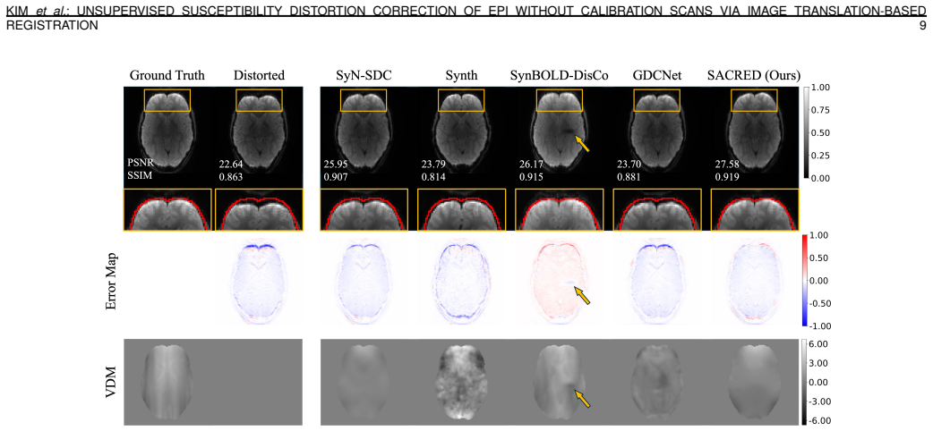
SUDMEX-TMS Dataset:Synth was excluded from this evaluation due to the absence of T2w images in the SUDMEX- TMS dataset. Quantitative results are summarized in Table III. As this dataset was acquired on a different scanner platform, it enables evaluation under scanner-induced distribution shifts. Compared with the uncorrected distorted BOLD images, SACRED ...
-
[13]
QT AB Dataset:Quantitative results on the QTAB dataset are summarized in Table IV. As an OOD dataset acquired on a different scanner platform and comprising subjects from a distinct age range, QTAB enables evaluation under both scanner and population distribution shifts. Compared with the uncorrected distorted BOLD images, SACRED reduced NRMSE by approxim...
-
[14]
Quantitative results are summarized in Table V
Ablation on Loss Functions and T est-Time Adaptation:We conducted ablation studies to evaluate the contributions of the image reconstruction loss (L recon), the structure consistency loss (L structure), and TTA. Quantitative results are summarized in Table V. Starting from the baseline configuration (top row in Ta- ble V), where the model was trained usin...
-
[15]
Specifically, we compared the proposed LNCC 2 loss with theL 1 loss, which was used during initial training and evaluated here as an alternative TTA objective
Analysis of T est-Time Adaptation:We analyzed the effect of the similarity loss function and the number of optimization iterations used during TTA. Specifically, we compared the proposed LNCC 2 loss with theL 1 loss, which was used during initial training and evaluated here as an alternative TTA objective. As shown in Fig. 8, using LNCC 2 consistently out...
-
[16]
Ablation on the Image T ranslation Network Architecture: We conducted an ablation study on the architecture of the image translation network to evaluate its ability to preserve structural consistency during the image translation process. Specifically, we compared the proposed INN-based archi- tecture with a ResNet-based generator [50], [51] and U- Net [26...
-
[17]
Proceedings of the National Academy of Sciences , volume=
S. Ogawa, T. M. Lee, A. R. Kay, and D. W. Tank, “Brain magnetic resonance imaging with contrast dependent on blood oxygenation.”Proceedings of the National Academy of Sciences, vol. 87, no. 24, pp. 9868–9872, Dec. 1990. [Online]. Available: https://pnas.org/doi/full/10.1073/pnas.87.24.9868
-
[18]
Echo-Planar Imaging: Magnetic Resonance Imaging in a Fraction of a Second,
M. K. Stehling, R. Turner, and P. Mansfield, “Echo-Planar Imaging: Magnetic Resonance Imaging in a Fraction of a Second,”Science, vol. 254, no. 5028, pp. 43–50, Oct. 1991. [Online]. Available: https://www.science.org/doi/10.1126/science.1925560
-
[19]
Susceptibility artefacts in NMR imaging,
K. M. L ¨udeke, P. R ¨oschmann, and R. Tischler, “Susceptibility artefacts in NMR imaging,”Magnetic Resonance Imaging, vol. 3, no. 4, pp. 329–343, 1985. [Online]. Available: https://www.sciencedirect.com/ science/article/pii/0730725X85903972
arXiv 1985
-
[20]
Effects of Field-Map Distortion Correction on Resting State Functional Connectivity MRI,
H. Togo, J. Rokicki, K. Yoshinaga, T. Hisatsune, H. Matsuda, N. Haga, and T. Hanakawa, “Effects of Field-Map Distortion Correction on Resting State Functional Connectivity MRI,”Frontiers in Neuroscience, vol. 11, p. 656, Dec. 2017. [Online]. Available: http://journal.frontiersin.org/article/10.3389/fnins.2017.00656/full
-
[21]
Preprocessing Variability in fMRI Predictive Modeling: Effects of Distortion Correction on Functional Connectivity- Based Predictions,
Z. Li, B. Lamichhane, A. Patel, R. Salas, N. Moukaddam, and A. Sabharwal, “Preprocessing Variability in fMRI Predictive Modeling: Effects of Distortion Correction on Functional Connectivity- Based Predictions,” inIEEE-EMBS International Conference on Biomedical and Health Informatics 2025, 2025. [Online]. Available: https://openreview.net/forum?id=5wNyRv1PD8
2025
-
[22]
Advances in functional and structural MR image analysis and implementation as FSL,
S. M. Smith, M. Jenkinson, M. W. Woolrich, C. F. Beckmann, T. E. Behrens, H. Johansen-Berg, P. R. Bannister, M. De Luca, I. Drobnjak, D. E. Flitney, R. K. Niazy, J. Saunders, J. Vickers, Y . Zhang, N. De Stefano, J. M. Brady, and P. M. Matthews, “Advances in functional and structural MR image analysis and implementation as FSL,”NeuroImage, vol. 23, pp. S2...
-
[23]
Available: https://linkinghub.elsevier.com/retrieve/pii/ S1053811904003933
[Online]. Available: https://linkinghub.elsevier.com/retrieve/pii/ S1053811904003933
-
[24]
Correction for geometric distortion in echo planar images from B0field variations,
P. Jezzard and R. S. Balaban, “Correction for geometric distortion in echo planar images from B0field variations,”Magnetic Resonance in Medicine, vol. 34, no. 1, pp. 65–73, Jul. 1995. [Online]. Available: https://onlinelibrary.wiley.com/doi/10.1002/mrm.1910340111 12 IEEE TRANSACTIONS AND JOURNALS TEMPLATE
-
[25]
How to correct susceptibility distortions in spin-echo echo-planar images: application to diffusion tensor imaging,
J. L. Andersson, S. Skare, and J. Ashburner, “How to correct susceptibility distortions in spin-echo echo-planar images: application to diffusion tensor imaging,”NeuroImage, vol. 20, no. 2, pp. 870–888, Oct. 2003. [Online]. Available: https://linkinghub.elsevier.com/retrieve/ pii/S1053811903003367
2003
-
[26]
Distortion correction using topup algorithm by single k-space (task) for echo planar imaging,
S.-H. Hwang, H.-S. Lee, S. H. Choi, and S.-H. Park, “Distortion correction using topup algorithm by single k-space (task) for echo planar imaging,”Scientific Reports, vol. 13, no. 1, p. 18751, 2023
2023
-
[27]
Using synthetic MR images for distortion correction,
D. F. Montez, A. N. Van, R. L. Miller, N. A. Seider, S. Marek, A. Zheng, D. J. Newbold, K. Scheidter, E. Feczko, A. J. Perrone, O. Miranda-Dominguez, E. A. Earl, B. P. Kay, A. K. Jha, A. Sotiras, T. O. Laumann, D. J. Greene, E. M. Gordon, M. D. Tisdall, A. Van Der Kouwe, D. A. Fair, and N. U. Dosenbach, “Using synthetic MR images for distortion correction...
2023
-
[28]
An integrated approach to correction for off-resonance effects and subject movement in diffusion mr imaging,
J. L. Andersson and S. N. Sotiropoulos, “An integrated approach to correction for off-resonance effects and subject movement in diffusion mr imaging,”NeuroImage, vol. 125, pp. 1063–1078, 2016. [Online]. Available: https://www.sciencedirect.com/science/article/pii/ S1053811915009209
2016
-
[29]
Accurate alignment of functional EPI data to anatomical MRI using a physics- based distortion model,
C. Studholme, R. Constable, and J. Duncan, “Accurate alignment of functional EPI data to anatomical MRI using a physics- based distortion model,”IEEE Transactions on Medical Imaging, vol. 19, no. 11, pp. 1115–1127, Nov. 2000. [Online]. Available: http://ieeexplore.ieee.org/document/896788/
2000
-
[30]
A. Gholipour, N. Kehtarnavaz, K. Gopinath, and R. Briggs, “Cross- Validation of Deformable Registration With Field Maps in Functional Magnetic Resonance Brain Imaging,”IEEE Journal of Selected Topics in Signal Processing, vol. 2, no. 6, pp. 854–869, Dec. 2008. [Online]. Available: http://ieeexplore.ieee.org/document/4740308/
arXiv 2008
-
[31]
Unwarping of unidirectionally distorted epi images,
J. Kybic, P. Th ´evenaz, A. Nirkko, and M. Unser, “Unwarping of unidirectionally distorted epi images,”IEEE transactions on medical imaging, vol. 19, no. 2, pp. 80–93, 2000
2000
-
[32]
fMRIPrep: a robust preprocessing pipeline for functional MRI,
O. Esteban, C. J. Markiewicz, R. W. Blair, C. A. Moodie, A. I. Isik, A. Erramuzpe, J. D. Kent, M. Goncalves, E. DuPre, M. Snyder, H. Oya, S. S. Ghosh, J. Wright, J. Durnez, R. A. Poldrack, and K. J. Gorgolewski, “fMRIPrep: a robust preprocessing pipeline for functional MRI,”Nature Methods, vol. 16, no. 1, pp. 111–116, Jan. 2019. [Online]. Available: https...
2019
-
[33]
Distortion correction of functional mri without reverse phase encoding scans or field maps,
T. Yu, L. Y . Cai, S. Torrisi, A. T. Vu, V . L. Morgan, S. E. Goodale, K. Ramadass, S. L. Meisler, J. Lv, A. E. Warrenet al., “Distortion correction of functional mri without reverse phase encoding scans or field maps,”Magnetic resonance imaging, vol. 103, pp. 18–27, 2023
2023
-
[34]
M. M. Jimeno, K. Bachi, G. Gardner, Y . L. Hurd, J. T. Vaughan Jr, and S. Geethanath, “Gdcnet: Calibrationless geometric distortion correction of echo planar imaging data using deep learning,”arXiv preprint arXiv:2402.18777, 2024
arXiv 2024
-
[35]
Navigating distribution shifts in medical image analysis: A survey,
Z. Su, J. Guo, X. Yang, Q. Wang, F. Coenen, and K. Huang, “Navigating distribution shifts in medical image analysis: A survey,”arXiv preprint arXiv:2411.05824, 2024
Pith/arXiv arXiv 2024
-
[36]
Sacred: Susceptibility artifact correction without reverse phase-encoding for epi using deep learning,
W. Kim and S.-H. Park, “Sacred: Susceptibility artifact correction without reverse phase-encoding for epi using deep learning,” in Cape Town - 2026 ISMRM-ISMRT Annual Meeting and Exhibition. Cape Town, South Africa: ISMRM, May 2026, program Number: 661-03-010. [Online]. Available: http://echo.ismrm.org/abstracts/view/ 42bfb763-f4b2-4bf6-aca8-59aa9cba679f
2026
-
[37]
Nice: Non-linear independent components estimation,
L. Dinh, D. Krueger, and Y . Bengio, “Nice: Non-linear independent components estimation,”arXiv preprint arXiv:1410.8516, 2014
Pith/arXiv arXiv 2014
-
[38]
Density estimation using Real NVP,
L. Dinh, J. Sohl-Dickstein, and S. Bengio, “Density estimation using Real NVP,” Feb. 2017, arXiv:1605.08803 [cs]. [Online]. Available: http://arxiv.org/abs/1605.08803
Pith/arXiv arXiv 2017
-
[39]
Glow: Generative Flow with Invertible 1x1 Convolutions,
D. P. Kingma and P. Dhariwal, “Glow: Generative Flow with Invertible 1x1 Convolutions,” Jul. 2018, arXiv:1807.03039 [stat]. [Online]. Available: http://arxiv.org/abs/1807.03039
Pith/arXiv arXiv 2018
-
[40]
Towards realtime multimodal fusion for image-guided inter- ventions using self-similarities,
M. P. Heinrich, M. Jenkinson, B. W. Papie ˙z, S. M. Brady, and J. A. Schnabel, “Towards realtime multimodal fusion for image-guided inter- ventions using self-similarities,” inInternational conference on medical image computing and computer-assisted intervention. Springer, 2013, pp. 187–194
2013
-
[41]
Image registration by maximization of combined mutual information and gradient informa- tion,
J. P. Pluim, J. A. Maintz, and M. A. Viergever, “Image registration by maximization of combined mutual information and gradient informa- tion,” inInternational Conference on Medical Image Computing and Computer-Assisted Intervention. Springer, 2000, pp. 452–461
2000
-
[42]
Mind: Modality independent neighbour- hood descriptor for multi-modal deformable registration,
M. P. Heinrich, M. Jenkinson, M. Bhushan, T. Matin, F. V . Gleeson, M. Brady, and J. A. Schnabel, “Mind: Modality independent neighbour- hood descriptor for multi-modal deformable registration,”Medical image analysis, vol. 16, no. 7, pp. 1423–1435, 2012
2012
-
[43]
U-net: Convolutional networks for biomedical image segmentation,
O. Ronneberger, P. Fischer, and T. Brox, “U-net: Convolutional networks for biomedical image segmentation,” inInternational Conference on Medical image computing and computer-assisted intervention. Springer, 2015, pp. 234–241
2015
-
[44]
V oxelmorph: a learning framework for deformable medical image registration,
G. Balakrishnan, A. Zhao, M. R. Sabuncu, J. Guttag, and A. V . Dalca, “V oxelmorph: a learning framework for deformable medical image registration,”IEEE transactions on medical imaging, vol. 38, no. 8, pp. 1788–1800, 2019
2019
-
[45]
Multimodality image registration by maximization of mutual informa- tion,
F. Maes, A. Collignon, D. Vandermeulen, G. Marchal, and P. Suetens, “Multimodality image registration by maximization of mutual informa- tion,”IEEE transactions on Medical Imaging, vol. 16, no. 2, pp. 187– 198, 2002
2002
-
[46]
Mutual-information- based registration of medical images: a survey,
J. P. Pluim, J. A. Maintz, and M. A. Viergever, “Mutual-information- based registration of medical images: a survey,”IEEE transactions on medical imaging, vol. 22, no. 8, pp. 986–1004, 2003
2003
-
[47]
Unsupervised deformable registration for multi-modal images via dis- entangled representations,
C. Qin, B. Shi, R. Liao, T. Mansi, D. Rueckert, and A. Kamen, “Unsupervised deformable registration for multi-modal images via dis- entangled representations,” inInternational Conference on Information Processing in Medical Imaging. Springer, 2019, pp. 249–261
2019
-
[48]
Z. Chen, J. Wei, and R. Li, “Unsupervised multi-modal medical image registration via discriminator-free image-to-image translation,”arXiv preprint arXiv:2204.13656, 2022
arXiv 2022
-
[49]
Unsupervised multi-modal medical image registration via invertible translation,
M. Guo, “Unsupervised multi-modal medical image registration via invertible translation,” inEuropean Conference on Computer Vision. Springer, 2024, pp. 22–38
2024
-
[50]
Invertible image signal processing,
Y . Xing, Z. Qian, and Q. Chen, “Invertible image signal processing,” inProceedings of the IEEE/CVF conference on computer vision and pattern recognition, 2021, pp. 6287–6296
2021
-
[51]
Densely connected convolutional networks,
G. Huang, Z. Liu, L. Van Der Maaten, and K. Q. Weinberger, “Densely connected convolutional networks,” inProceedings of the IEEE confer- ence on computer vision and pattern recognition, 2017, pp. 4700–4708
2017
-
[52]
A. V . Dalca, G. Balakrishnan, J. Guttag, and M. R. Sabuncu, Unsupervised Learning for Fast Probabilistic Diffeomorphic Registration. Springer International Publishing, 2018, p. 729–738. [Online]. Available: http://dx.doi.org/10.1007/978-3-030-00928-1 82
-
[53]
Multigradi- con: A foundation model for multimodal medical image registration,
B. Demir, L. Tian, H. Greer, R. Kwitt, F.-X. Vialard, R. S. J. Est ´epar, S. Bouix, R. Rushmore, E. Ebrahim, and M. Niethammer, “Multigradi- con: A foundation model for multimodal medical image registration,” in International Workshop on Biomedical Image Registration. Springer, 2024, pp. 3–18
2024
-
[54]
The nimh intramural healthy volunteer dataset: A comprehensive meg, mri, and behavioral resource,
A. C. Nugent, A. G. Thomas, M. Mahoney, A. Gibbons, J. T. Smith, A. J. Charles, J. S. Shaw, J. D. Stout, A. M. Namyst, A. Basavarajet al., “The nimh intramural healthy volunteer dataset: A comprehensive meg, mri, and behavioral resource,”Scientific Data, vol. 9, no. 1, p. 518, 2022
2022
-
[55]
The mexican dataset of a repetitive transcranial mag- netic stimulation clinical trial on cocaine use disorder patients: Sudmex tms,
D. Angeles-Valdez, J. Rasgado-Toledo, V . Villica ˜na, A. Davalos- Guzman, C. Almanza, A. Fajardo-Valdez, R. Alcala-Lozano, and E. A. Garza-Villarreal, “The mexican dataset of a repetitive transcranial mag- netic stimulation clinical trial on cocaine use disorder patients: Sudmex tms,”Scientific Data, vol. 11, no. 1, p. 408, 2024
2024
-
[56]
The queensland twin adolescent brain project, a longitudinal study of ado- lescent brain development,
L. T. Strike, N. K. Hansell, K.-H. Chuang, J. L. Miller, G. I. de Zu- bicaray, P. M. Thompson, K. L. McMahon, and M. J. Wright, “The queensland twin adolescent brain project, a longitudinal study of ado- lescent brain development,”Scientific Data, vol. 10, no. 1, p. 195, 2023
2023
-
[57]
The openneuro resource for sharing of neuro- science data,
C. J. Markiewicz, K. J. Gorgolewski, F. Feingold, R. Blair, Y . O. Halchenko, E. Miller, N. Hardcastle, J. Wexler, O. Esteban, M. Goncavleset al., “The openneuro resource for sharing of neuro- science data,”Elife, vol. 10, p. e71774, 2021
2021
-
[58]
Jenkinson, C
M. Jenkinson, C. F. Beckmann, T. E. Behrens, M. W. Woolrich, and S. M. Smith, “Fsl,”NeuroImage, vol. 62, no. 2, pp. 782– 790, 2012, 20 YEARS OF fMRI. [Online]. Available: https: //www.sciencedirect.com/science/article/pii/S1053811911010603
2012
-
[59]
N4itk: Improved n3 bias correction,
N. J. Tustison, B. B. Avants, P. A. Cook, Y . Zheng, A. Egan, P. A. Yushkevich, and J. C. Gee, “N4itk: Improved n3 bias correction,”IEEE Transactions on Medical Imaging, vol. 29, no. 6, pp. 1310–1320, 2010
2010
-
[60]
Freesurfer,
B. Fischl, “Freesurfer,”NeuroImage, vol. 62, no. 2, pp. 774– 781, 2012, 20 YEARS OF fMRI. [Online]. Available: https: //www.sciencedirect.com/science/article/pii/S1053811912000389
2012
-
[61]
Accurate and robust brain image alignment using boundary-based registration,
D. N. Greve and B. Fischl, “Accurate and robust brain image alignment using boundary-based registration,”Neuroimage, vol. 48, no. 1, pp. 63– 72, 2009
2009
-
[62]
A. Paszke, S. Gross, F. Massa, A. Lerer, J. Bradbury, G. Chanan, T. Killeen, Z. Lin, N. Gimelshein, L. Antiga, A. Desmaison, A. K ¨opf, E. Yang, Z. DeVito, M. Raison, A. Tejani, S. Chilamkurthy, B. Steiner, L. Fang, J. Bai, and S. Chintala, “Pytorch: An imperative style, KIMet al.: UNSUPERVISED SUSCEPTIBILITY DISTORTION CORRECTION OF EPI WITHOUT CALIBRATI...
Pith/arXiv arXiv 2019
-
[63]
Monai: An open-source framework for deep learning in healthcare,
M. J. Cardoso, W. Li, R. Brown, N. Ma, E. Kerfoot, Y . Wang, B. Murrey, A. Myronenko, C. Zhao, D. Yang, V . Nath, Y . He, Z. Xu, A. Hatamizadeh, A. Myronenko, W. Zhu, Y . Liu, M. Zheng, Y . Tang, I. Yang, M. Zephyr, B. Hashemian, S. Alle, M. Z. Darestani, C. Budd, M. Modat, T. Vercauteren, G. Wang, Y . Li, Y . Hu, Y . Fu, B. Gorman, H. Johnson, B. Generea...
Pith/arXiv arXiv 2022
-
[64]
Symmetric diffeomorphic image registration with cross-correlation: evaluating auto- mated labeling of elderly and neurodegenerative brain,
B. B. Avants, C. L. Epstein, M. Grossman, and J. C. Gee, “Symmetric diffeomorphic image registration with cross-correlation: evaluating auto- mated labeling of elderly and neurodegenerative brain,”Medical image analysis, vol. 12, no. 1, pp. 26–41, 2008
2008
-
[65]
Anatomic localization and quantitative analysis of gradient refocused echo-planar fmri susceptibility artifacts,
J. G. Ojemann, E. Akbudak, A. Z. Snyder, R. C. McKinstry, M. E. Raichle, and T. E. Conturo, “Anatomic localization and quantitative analysis of gradient refocused echo-planar fmri susceptibility artifacts,” Neuroimage, vol. 6, no. 3, pp. 156–167, 1997
1997
-
[66]
Au- tomated brain extraction of multisequence mri using artificial neural networks,
F. Isensee, M. Schell, I. Pflueger, G. Brugnara, D. Bonekamp, U. Neu- berger, A. Wick, H.-P. Schlemmer, S. Heiland, W. Wicket al., “Au- tomated brain extraction of multisequence mri using artificial neural networks,”Human brain mapping, vol. 40, no. 17, pp. 4952–4964, 2019
2019
-
[67]
Unpaired Image- to-Image Translation using Cycle-Consistent Adversarial Networks,
J.-Y . Zhu, T. Park, P. Isola, and A. A. Efros, “Unpaired Image- to-Image Translation using Cycle-Consistent Adversarial Networks,” Aug. 2020, arXiv:1703.10593 [cs.CV]. [Online]. Available: http: //arxiv.org/abs/1703.10593
arXiv 2020
-
[68]
Perceptual Losses for Real-Time Style Transfer and Super-Resolution,
J. Johnson, A. Alahi, and L. Fei-Fei, “Perceptual Losses for Real-Time Style Transfer and Super-Resolution,” Mar. 2016, arXiv:1603.08155 [cs.CV]. [Online]. Available: http://arxiv.org/abs/1603.08155
Pith/arXiv arXiv 2016
-
[69]
Medical image translation with deep learning: Advances, datasets and perspectives,
J. Chen, Z. Ye, R. Zhang, H. Li, B. Fang, L.-b. Zhang, and W. Wang, “Medical image translation with deep learning: Advances, datasets and perspectives,”Medical Image Analysis, vol. 103, p. 103605, 2025
2025
discussion (0)
Sign in with ORCID, Apple, or X to comment. Anyone can read and Pith papers without signing in.