Recognition: unknown
Generative Drifting for Conditional Medical Image Generation
Pith reviewed 2026-05-10 02:17 UTC · model grok-4.3
The pith
GDM reformulates conditional 3D medical image generation as a drifting process that jointly optimizes distribution plausibility and patient fidelity in one inference step.
A machine-rendered reading of the paper's core claim, the machinery that carries it, and where it could break.
Core claim
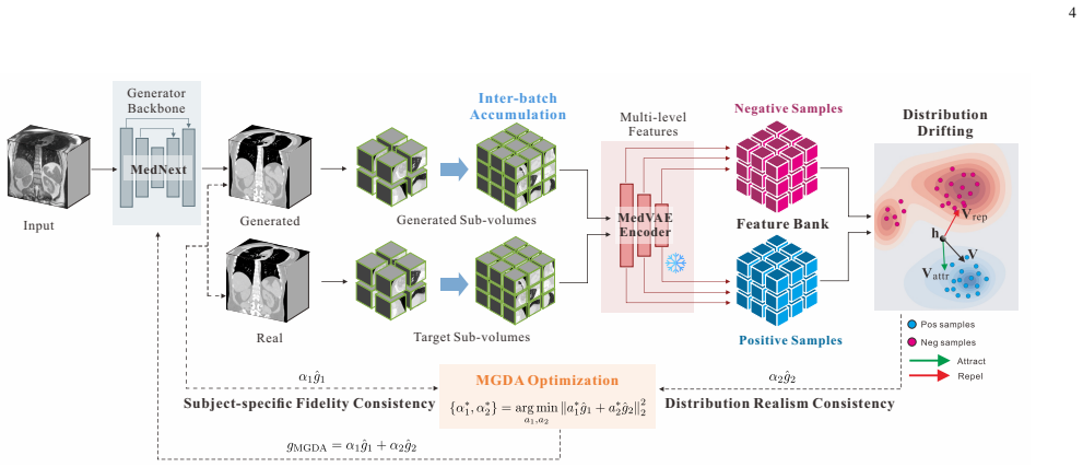
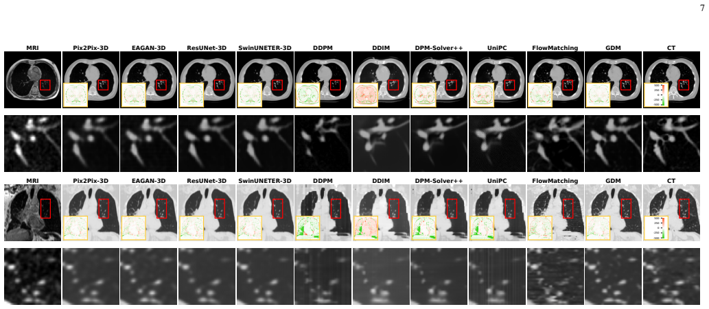
GDM extends drifting to 3D medical imaging by constructing an attractive-repulsive drift that minimizes discrepancy between the generator pushforward and the target distribution, supported by a multi-level feature bank from a medical foundation encoder for global-local-spatial affinity estimation and by gradient coordination in the shared output space to balance distribution-level and fidelity-oriented objectives. On MRI-to-CT synthesis and sparse-view CT reconstruction, the framework consistently outperforms GAN-based, flow-matching-based, SDE-based generative models, and supervised regression methods while improving the joint balance among anatomical fidelity, quantitative reliability, and
What carries the argument
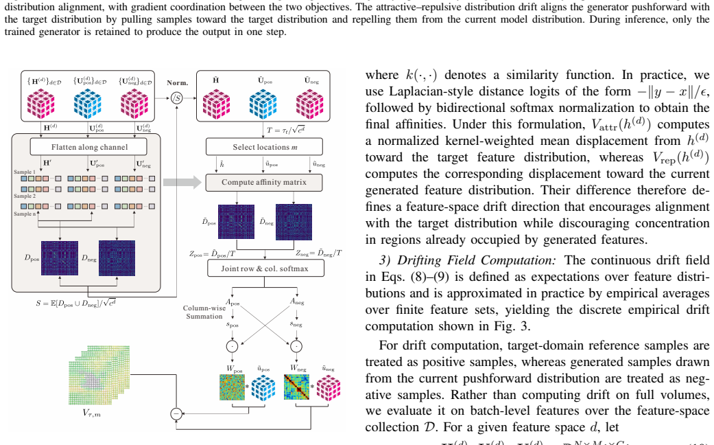
Attractive-repulsive drift supported by a multi-level feature bank extracted from a medical foundation encoder, which supplies reliable affinity estimates and drifting fields across complementary global, local, and spatial representations.
If this is right
- The model retains single-step inference while still promoting both distribution-level plausibility and patient-specific fidelity.
- Gradient coordination in the shared output space improves optimization stability when distribution and fidelity objectives compete.
- Performance gains hold across two representative clinical tasks: MRI-to-CT synthesis and sparse-view CT reconstruction.
- The framework is compatible with a range of backbone architectures because the drifting mechanism operates on top of any generator that produces volumetric outputs.
Where Pith is reading between the lines
- The same drifting construction could be tested on other paired 3D modalities such as PET-CT or ultrasound-CT without requiring new loss terms.
- Because the feature bank is drawn from a pretrained foundation encoder, the method may reduce the volume of paired training data needed for new clinical sites.
- If the drift fields prove interpretable, they could be inspected post hoc to identify which anatomical regions drive the largest corrections during generation.
Load-bearing premise
The multi-level feature bank from the medical foundation encoder supplies reliable affinity estimates and drifting fields that remain stable when learning on 3D volumetric data.
What would settle it
A controlled experiment on a held-out 3D medical synthesis task in which GDM produces lower anatomical fidelity scores or visibly unstable drift fields compared with a strong regression baseline would falsify the central claim.
Figures


read the original abstract
Conditional medical image generation plays an important role in many clinically relevant imaging tasks. However, existing methods still face a fundamental challenge in balancing inference efficiency, patient-specific fidelity, and distribution-level plausibility, particularly in high-dimensional 3D medical imaging. In this work, we propose GDM, a generative drifting framework that reformulates deterministic medical image prediction as a multi-objective learning problem to jointly promote distribution-level plausibility and patient-specific fidelity while retaining one-step inference. GDM extends drifting to 3D medical imaging through an attractive-repulsive drift that minimizes the discrepancy between the generator pushforward and the target distribution. To enable stable drifting-based learning in 3D volumetric data, GDM constructs a multi-level feature bank from a medical foundation encoder to support reliable affinity estimation and drifting field computation across complementary global, local, and spatial representations. In addition, a gradient coordination strategy in the shared output space improves optimization balance under competing distribution-level and fidelity-oriented objectives. We evaluate the proposed framework on two representative tasks, MRI-to-CT synthesis and sparse-view CT reconstruction. Experimental results show that GDM consistently outperforms a wide range of baselines, including GAN-based, flow-matching-based, and SDE-based generative models, as well as supervised regression methods, while improving the balance among anatomical fidelity, quantitative reliability, perceptual realism, and inference efficiency. These findings suggest that GDM provides a practical and effective framework for conditional 3D medical image generation.
Editorial analysis
A structured set of objections, weighed in public.
Referee Report
Summary. The manuscript proposes Generative Drifting Model (GDM), a framework that reformulates conditional 3D medical image generation (e.g., MRI-to-CT synthesis and sparse-view CT reconstruction) as a multi-objective problem. It introduces an attractive-repulsive drift to minimize pushforward-to-target discrepancy, supported by a multi-level feature bank extracted from a medical foundation encoder for affinity estimation across global/local/spatial representations, plus a gradient coordination strategy in shared output space. The work claims one-step inference with consistent outperformance over GAN-, flow-matching-, SDE-based, and supervised regression baselines while improving balance among anatomical fidelity, quantitative reliability, perceptual realism, and efficiency.
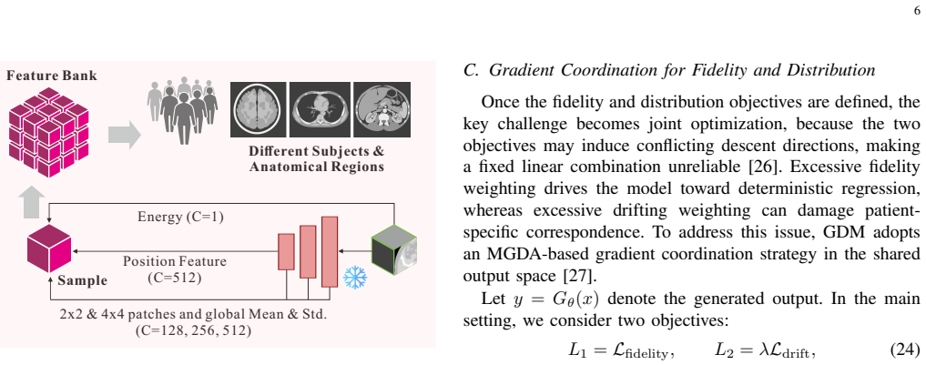
Significance. If the reported gains prove robust and reproducible, GDM could meaningfully advance practical conditional generation for high-dimensional medical volumes by combining distribution-level plausibility with patient-specific fidelity under efficient inference. The extension of drifting via foundation-model features is a targeted contribution, but its significance hinges on whether the empirical advantages are supported by detailed metrics, ablations, and stability checks rather than qualitative assertions alone.
major comments (1)
- [Methods (multi-level feature bank and drifting field computation)] The central claim of stable 3D drifting and consistent outperformance rests on the multi-level feature bank (global, local, and spatial representations from the medical foundation encoder) producing reliable affinities for the attractive-repulsive drift field. The manuscript provides no ablation studies, sensitivity analysis to imaging artifacts (e.g., intensity inhomogeneity), or quantitative verification that this component outperforms simpler feature extractors; without such evidence the load-bearing assumption remains untested and the performance claims cannot be fully evaluated.
minor comments (2)
- [Abstract] The abstract asserts 'consistent outperformance' and 'improved balance' without any numerical metrics, error bars, or dataset sizes; a brief quantitative summary should be added for clarity even though the full results section presumably contains them.
- [Methods] Notation for the drifting field, affinity estimation, and gradient coordination should be introduced with explicit equations and variable definitions in the methods to aid reproducibility.
Simulated Author's Rebuttal
We thank the referee for the thoughtful review and for recognizing the potential of GDM to advance conditional 3D medical image generation. The feedback on the multi-level feature bank is valuable and highlights an area where additional evidence can strengthen the manuscript. We address the major comment below and will revise the paper accordingly to improve clarity and empirical support.
read point-by-point responses
-
Referee: [Methods (multi-level feature bank and drifting field computation)] The central claim of stable 3D drifting and consistent outperformance rests on the multi-level feature bank (global, local, and spatial representations from the medical foundation encoder) producing reliable affinities for the attractive-repulsive drift field. The manuscript provides no ablation studies, sensitivity analysis to imaging artifacts (e.g., intensity inhomogeneity), or quantitative verification that this component outperforms simpler feature extractors; without such evidence the load-bearing assumption remains untested and the performance claims cannot be fully evaluated.
Authors: We agree that dedicated validation of the multi-level feature bank would strengthen the manuscript. The current version emphasizes the overall framework, end-to-end results on MRI-to-CT and sparse-view CT tasks, and comparisons against GAN, flow-matching, SDE, and regression baselines, but does not include isolated ablations of the feature bank or explicit sensitivity tests to artifacts such as intensity inhomogeneity. In the revised manuscript we will add (i) ablation studies replacing the multi-level bank with simpler single-level or non-foundation encoders while keeping all other components fixed, (ii) quantitative metrics (e.g., affinity correlation, drift-field stability, and downstream generation quality) demonstrating the benefit of the multi-level design, and (iii) sensitivity analyses on subsets of the data exhibiting intensity inhomogeneity and other common artifacts. These additions will be presented with tables and figures to allow direct evaluation of the component's contribution. revision: yes
Circularity Check
No circularity: derivation chain is self-contained as an independent extension
full rationale
The abstract and description present GDM as a reformulation of deterministic prediction into multi-objective learning with an attractive-repulsive drift minimizing pushforward-to-target discrepancy, enabled by a multi-level feature bank from a medical foundation encoder plus gradient coordination. No equations, fitted parameters, or self-citations are shown that reduce any claimed result (e.g., outperformance or stability) to quantities defined by the paper's own inputs by construction. The central components are introduced as extensions rather than tautological renamings or self-referential fits, and experimental claims stand apart from the framework definition. This is the normal case of a non-circular methodological proposal.
Axiom & Free-Parameter Ledger
Forward citations
Cited by 1 Pith paper
-
One-Step Generative Modeling via Wasserstein Gradient Flows
W-Flow achieves state-of-the-art one-step ImageNet 256x256 generation at 1.29 FID by training a static neural network to follow a Wasserstein gradient flow that minimizes Sinkhorn divergence, delivering roughly 100x f...
Reference graph
Works this paper leans on
-
[1]
Diffboost: Enhancing medical image segmen- tation via text-guided diffusion model,
Z. Zhang, L. Yao, B. Wang, D. Jha, G. Durak, E. Keles, A. Medetal- ibeyoglu, and U. Bagci, “Diffboost: Enhancing medical image segmen- tation via text-guided diffusion model,”IEEE Transactions on Medical Imaging, vol. 44, no. 9, pp. 3670–3682, 2024
2024
-
[2]
Unsupervised medical image translation with adversarial diffusion models,
M. ¨Ozbey, O. Dalmaz, S. U. Dar, H. A. Bedel, S ¸. ¨Ozturk, A. G ¨ung¨or, and T. Cukur, “Unsupervised medical image translation with adversarial diffusion models,”IEEE Transactions on Medical Imaging, vol. 42, no. 12, pp. 3524–3539, 2023
2023
-
[3]
Cross- conditioned diffusion model for medical image to image translation,
Z. Xing, S. Yang, S. Chen, T. Ye, Y . Yang, J. Qin, and L. Zhu, “Cross- conditioned diffusion model for medical image to image translation,” in International Conference on Medical Image Computing and Computer- Assisted Intervention. Springer, 2024, pp. 201–211
2024
-
[4]
CT-sdm: A sampling diffu- sion model for sparse-view CT reconstruction across various sampling rates,
L. Yang, J. Huang, G. Yang, and D. Zhang, “CT-sdm: A sampling diffu- sion model for sparse-view CT reconstruction across various sampling rates,”IEEE Transactions on Medical Imaging, 2025. 11
2025
-
[5]
Regression is all you need for medical image translation,
S. Rassmann, D. K ¨ugler, C. Ewert, and M. Reuter, “Regression is all you need for medical image translation,”IEEE Transactions on Medical Imaging, 2026
2026
-
[6]
Tomographic foundation Model—FORCE: Flow-oriented reconstruction conditioning engine,
W. Xia, C. Niu, and G. Wang, “Tomographic foundation Model—FORCE: Flow-oriented reconstruction conditioning engine,” IEEE Transactions on Medical Imaging, 2026
2026
-
[7]
A new deep convolutional neural network design with efficient learning capability: Application to CT image synthesis from MRI,
A. Bahrami, A. Karimian, E. Fatemizadeh, H. Arabi, and H. Zaidi, “A new deep convolutional neural network design with efficient learning capability: Application to CT image synthesis from MRI,”Medical Physics, vol. 47, no. 10, pp. 5158–5171, 2020
2020
-
[8]
Low-dose CT with a residual encoder-decoder convolutional neural network,
H. Chen, Y . Zhang, M. K. Kalra, F. Lin, Y . Chen, P. Liao, J. Zhou, and G. Wang, “Low-dose CT with a residual encoder-decoder convolutional neural network,”IEEE Transactions on Medical Imaging, vol. 36, no. 12, pp. 2524–2535, 2017
2017
-
[9]
MRI-only based synthetic CT generation using dense cycle consistent generative adversarial networks,
Y . Lei, J. Harms, T. Wang, Y . Liu, H.-K. Shu, A. B. Jani, W. J. Curran, H. Mao, T. Liu, and X. Yang, “MRI-only based synthetic CT generation using dense cycle consistent generative adversarial networks,”Medical Physics, vol. 46, no. 8, pp. 3565–3581, 2019
2019
-
[10]
Yang Song, Liyue Shen, Lei Xing, and Stefano Ermon
Y . Song, L. Shen, L. Xing, and S. Ermon, “Solving inverse problems in medical imaging with score-based generative models,”arXiv preprint arXiv:2111.08005, 2021
-
[11]
Dual-domain collaborative diffusion sampling for multi-source stationary computed tomography reconstruction,
Z. Li, D. Chang, Z. Zhang, F. Luo, Q. Liu, J. Zhang, G. Yang, and W. Wu, “Dual-domain collaborative diffusion sampling for multi-source stationary computed tomography reconstruction,”IEEE Transactions on Medical Imaging, vol. 43, no. 10, pp. 3398–3411, 2024
2024
-
[12]
Physics-informed score-based diffusion model for limited-angle reconstruction of cardiac computed tomography,
S. Han, Y . Xu, D. Wang, B. Morovati, L. Zhou, J. S. Maltz, G. Wang, and H. Yu, “Physics-informed score-based diffusion model for limited-angle reconstruction of cardiac computed tomography,”IEEE Transactions on Medical Imaging, vol. 44, no. 9, pp. 3629–3640, 2024
2024
-
[13]
Two-and-a-half order score-based model for solving 3D ill-posed inverse problems,
Z. Li, Y . Wang, J. Zhang, W. Wu, and H. Yu, “Two-and-a-half order score-based model for solving 3D ill-posed inverse problems,”Comput- ers in Biology and Medicine, vol. 168, p. 107819, 2024
2024
-
[14]
Generative Modeling via Drifting
M. Deng, H. Li, T. Li, Y . Du, and K. He, “Generative modeling via drifting,”arXiv preprint arXiv:2602.04770, 2026
work page internal anchor Pith review arXiv 2026
-
[15]
Deep con- volutional neural network for inverse problems in imaging,
K. H. Jin, M. T. McCann, E. Froustey, and M. Unser, “Deep con- volutional neural network for inverse problems in imaging,”IEEE Transactions on Image Processing, vol. 26, no. 9, pp. 4509–4522, 2017
2017
-
[16]
Image-to-image translation with conditional adversarial networks,
P. Isola, J.-Y . Zhu, T. Zhou, and A. A. Efros, “Image-to-image translation with conditional adversarial networks,” inProceedings of the IEEE Conference on Computer Vision and Pattern Recognition, 2017, pp. 1125–1134
2017
-
[17]
Swin unetr: Swin transformers for semantic segmentation of brain tumors in MRI images,
A. Hatamizadeh, V . Nath, Y . Tang, D. Yang, H. R. Roth, and D. Xu, “Swin unetr: Swin transformers for semantic segmentation of brain tumors in MRI images,” inInternational MICCAI Brainlesion Workshop. Springer, 2021, pp. 272–284
2021
-
[18]
A review of deep learning CT reconstruction from incomplete projection data,
T. Wang, W. Xia, J. Lu, and Y . Zhang, “A review of deep learning CT reconstruction from incomplete projection data,”IEEE Transactions on Radiation and Plasma Medical Sciences, vol. 8, no. 2, pp. 138–152, 2023
2023
-
[19]
Ea- GANs: edge-aware generative adversarial networks for cross-modality MR image synthesis,
B. Yu, L. Zhou, L. Wang, Y . Shi, J. Fripp, and P. Bourgeat, “Ea- GANs: edge-aware generative adversarial networks for cross-modality MR image synthesis,”IEEE Transactions on Medical Imaging, vol. 38, no. 7, pp. 1750–1762, 2019
2019
-
[20]
Generative adversarial network in medical imaging: A review,
X. Yi, E. Walia, and P. Babyn, “Generative adversarial network in medical imaging: A review,”Medical Image Analysis, vol. 58, p. 101552, 2019
2019
-
[21]
Which training methods for GANs do actually converge?
L. Mescheder, A. Geiger, and S. Nowozin, “Which training methods for GANs do actually converge?” inInternational Conference on Machine Learning. PMLR, 2018, pp. 3481–3490
2018
-
[22]
Flow Matching for Generative Modeling
Y . Lipman, R. T. Chen, H. Ben-Hamu, M. Nickel, and M. Le, “Flow matching for generative modeling,”arXiv preprint arXiv:2210.02747, 2022
work page internal anchor Pith review Pith/arXiv arXiv 2022
-
[23]
Solving 3D inverse problems using pre-trained 2D diffusion models,
H. Chung, D. Ryu, M. T. McCann, M. L. Klasky, and J. C. Ye, “Solving 3D inverse problems using pre-trained 2D diffusion models,” inProceedings of the IEEE/CVF Conference on Computer Vision and Pattern Recognition, 2023, pp. 22 542–22 551
2023
-
[24]
Fast and robust fixed-point algorithms for independent component analysis,
A. Hyvarinen, “Fast and robust fixed-point algorithms for independent component analysis,”IEEE Transactions on Neural Networks, vol. 10, no. 3, pp. 626–634, 1999
1999
-
[25]
MedV AE: Efficient automated interpretation of medical images with large-scale generalizable autoencoders,
M. Varma, A. Kumar, R. V . der Sluijs, S. Ostmeier, L. Blankemeier, P. J. M. Chambon, C. Bluethgen, J. Prince, C. Langlotz, and A. S. Chaudhari, “MedV AE: Efficient automated interpretation of medical images with large-scale generalizable autoencoders,” inMedical Imaging with Deep Learning, 2025
2025
-
[26]
Conflict-averse gradient descent for multi-task learning,
B. Liu, X. Liu, X. Jin, P. Stone, and Q. Liu, “Conflict-averse gradient descent for multi-task learning,”Advances in neural information pro- cessing systems, vol. 34, pp. 18 878–18 890, 2021
2021
-
[27]
Multi-task learning as multi-objective opti- mization,
O. Sener and V . Koltun, “Multi-task learning as multi-objective opti- mization,”Advances in Neural Information Processing Systems, vol. 31, 2018
2018
-
[28]
SynthRAD2025 grand challenge dataset: Generating synthetic CTs for radiotherapy from head to abdomen,
A. Thummerer, E. van der Bijl, A. J. Galapon, F. Kamp, M. Savenije, C. Muijs, S. Aluwini, R. J. Steenbakkers, S. Beuel, M. P. Intvenet al., “SynthRAD2025 grand challenge dataset: Generating synthetic CTs for radiotherapy from head to abdomen,”Medical Physics, vol. 52, no. 7, p. e17981, 2025
2025
-
[29]
Weighted res-unet for high-quality retina vessel segmentation,
X. Xiao, S. Lian, Z. Luo, and S. Li, “Weighted res-unet for high-quality retina vessel segmentation,” in2018 9th International Conference on Information Technology in Medicine and Education (ITME). IEEE, 2018, pp. 327–331
2018
-
[30]
Denoising diffusion probabilistic models,
J. Ho, A. Jain, and P. Abbeel, “Denoising diffusion probabilistic models,” Advances in Neural Information Processing Systems, vol. 33, pp. 6840– 6851, 2020
2020
-
[31]
Denoising Diffusion Implicit Models
J. Song, C. Meng, and S. Ermon, “Denoising diffusion implicit models,” arXiv preprint arXiv:2010.02502, 2020
work page internal anchor Pith review Pith/arXiv arXiv 2010
-
[32]
Dpm-solver++: Fast solver for guided sampling of diffusion probabilistic models,
C. Lu, Y . Zhou, F. Bao, J. Chen, C. Li, and J. Zhu, “Dpm-solver++: Fast solver for guided sampling of diffusion probabilistic models,”Machine Intelligence Research, vol. 22, no. 4, pp. 730–751, 2025
2025
-
[33]
Unipc: A unified predictor- corrector framework for fast sampling of diffusion models,
W. Zhao, L. Bai, Y . Rao, J. Zhou, and J. Lu, “Unipc: A unified predictor- corrector framework for fast sampling of diffusion models,”Advances in Neural Information Processing Systems, vol. 36, pp. 49 842–49 869, 2023
2023
-
[34]
Totalsegmen- tator: robust segmentation of 104 anatomic structures in CT images,
J. Wasserthal, H.-C. Breit, M. T. Meyer, M. Pradella, D. Hinck, A. W. Sauter, T. Heye, D. T. Boll, J. Cyriac, S. Yanget al., “Totalsegmen- tator: robust segmentation of 104 anatomic structures in CT images,” Radiology: Artificial Intelligence, vol. 5, no. 5, p. e230024, 2023
2023
-
[35]
The noise power spectrum of CT images,
M. F. Kijewski and P. F. Judy, “The noise power spectrum of CT images,” Physics in Medicine & Biology, vol. 32, no. 5, pp. 565–575, 1987
1987
-
[36]
Low-dose CT image and projection dataset,
T. R. Moen, B. Chen, D. R. Holmes III, X. Duan, Z. Yu, L. Yu, S. Leng, J. G. Fletcher, and C. H. McCollough, “Low-dose CT image and projection dataset,”Medical Physics, vol. 48, no. 2, pp. 902–911, 2021
2021
-
[37]
Single-slice rebinning method for helical cone-beam CT,
F. Noo, M. Defrise, and R. Clackdoyle, “Single-slice rebinning method for helical cone-beam CT,”Physics in Medicine & Biology, vol. 44, no. 2, pp. 561–570, 1999
1999
-
[38]
Mednext: transformer-driven scaling of convnets for medical image segmentation,
S. Roy, G. Koehler, C. Ulrich, M. Baumgartner, J. Petersen, F. Isensee, P. F. Jaeger, and K. H. Maier-Hein, “Mednext: transformer-driven scaling of convnets for medical image segmentation,” inInternational Confer- ence on Medical Image Computing and Computer-assisted Intervention. Springer, 2023, pp. 405–415
2023
discussion (0)
Sign in with ORCID, Apple, or X to comment. Anyone can read and Pith papers without signing in.